. 2022 Sep;118(3):258-277.
doi: 10.1111/mmi.14966. Epub 2022 Aug 15.

Role of novel polysaccharide layers in assembly of the exosporium, the outermost protein layer of the Bacillus anthracis spore

Affiliations

Role of novel polysaccharide layers in assembly of the exosporium, the outermost protein layer of the Bacillus anthracis spore

Dörte Lehmann et al. Mol Microbiol. 2022 Sep.

Abstract

A fundamental question in cell biology is how cells assemble their outer layers. The bacterial endospore is a well-established model for cell layer assembly. However, the assembly of the exosporium, a complex protein shell comprising the outermost layer in the pathogen Bacillus anthracis, remains poorly understood. Exosporium assembly begins with the deposition of proteins at one side of the spore surface, followed by the progressive encirclement of the spore. We seek to resolve a major open question: the mechanism directing exosporium assembly to the spore, and then into a closed shell. We hypothesized that material directly underneath the exosporium (the interspace) directs exosporium assembly to the spore and drives encirclement. In support of this, we show that the interspace possesses at least two distinct layers of polysaccharide. Secondly, we show that putative polysaccharide biosynthetic genes are required for exosporium encirclement, suggesting a direct role for the interspace. These results not only significantly clarify the mechanism of assembly of the exosporium, an especially widespread bacterial outer layer, but also suggest a novel mechanism in which polysaccharide layers drive the assembly of a protein shell.

Keywords: Bacillus anthracis; exosporium; interspace; polysaccharide; spore.


Figures

Fig 1.
Fig 1.. Previous (A) and new model (B) of B. anthracis spore formation.
A) It is known that the cap assembles early during sporulation before completion of the forespore engulfment (I) on the mother cell side of the septum. However, when visualized by TEM a gap can be observed between the cap and forespore membrane. This gap likely contains some coat proteins and interspace material of unknown composition (striped layer, I). After engulfment (II) a thin layer of coat and interspace material forms around the forespore. At the same time the assembly of the exosporium (green) starts on the edge of the cap, on top of the interspace. Simultaneously, the interspace material underneath the cap region widens (III) and densely staining coat features around the forespore can be detected by TEM. As exosporium assembly continues to enclose the entire forespore, the interspace widens along the spore, and coat and exosporium assembly complete (IV-V), and the spore is released (VI). B) Cap and inner polysaccharide appear prior to engulfment (I). Inner polysaccharide assembles around the forespore, and the coat forms under the inner polysaccharide. Exosporium assembly initiates at the cap’s leading edge (II). Exosporium assembly continues while outer polysaccharide accumulates at the mother cell-proximal pole (III-IV). Outer polysaccharide distributes laterally around the forespore, the mother cell lyses and the spore is released (V-VI).
Fig 2.
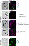
Fig 2.. Epifluorescence microscopic detection of interspace polysaccharide using WGA.
CotE mutant and wild-type spores were visualized either by bright-field microscopy (A, C, E, G, I) or by epifluorescence microscopy, after staining an N-acetylglucosamine-comprising polysaccharide with WGA (magenta; B, D, F, H, J; WGA staining is pictured as ellipses (cartoon) on the right side of the panels) and anti-BclA BA-MAB5 monoclonal antibody (green; B, J; green ellipses to the right of the panels) to counterstain the exosporium. As controls, we assessed WGA binding in the presence of chitin hydrolysate (D, chitin hydrolases block WGA binding to the spore), 500 mM GlcNAc monomer (F, the affinity of WGA is stronger towards the spore than GlcNAc monomer), or 1M NaCl (H, 1 M NaCl does not affect WGA staining).
Fig 3.
Fig 3.. Epifluorescence microscopic visualization of outer polysaccharide.
Wild type spores (A-C), or bclA (D, E) mutant spores were visualized by phase contrast (A, D), or epifluorescence microscopy (B, C, E). The images in panels C-E are enlarged relative to A and B. Spores were stained with SJA (red; B, C) and counterstained using polyclonal anti-BclA antibody (to detect the exosporium surface, green; B, C) or anti-BxpB/ExsFA antibodies (to detect the exosporium basal layer, green; E).
Fig 4.
Fig 4.. Localization of outer polysaccharide relative to the exosporium using ellipsoid localization microscopy (ELM).
Spores were visualized by epifluorescence microscopy (A). Images were put into the ellipsoid model, while decreasing the inferred point-spread function to remove instrumental blurring (B), then the radius of a sphere of equal volume to the fitted ellipsoid of 42 spores was analysed (C). bclA mutant spores were stained with SJA (magenta, A), and counterstained using anti-BxpB/ExsFA antibodies (green, A). The error bars indicate the standard error of the mean.
Fig 5.
Fig 5.. Epifluorescence microscopic visualization of outer polysaccharide in cotO mutant spores.
CotO mutant spores were visualized by phase contrast (A, C), or epifluorescence microscopy (B, D). Spores were stained with SJA (red; B), or WGA (red; no WGA staining is detectable in cotO mutant spores) and counterstained using polyclonal anti-BclA antibody (green; B, D).
Fig 6.
Fig 6.. Light and transmission electron microscopic analysis of cotE cotO mutant and wild type spores.
Spores were visualized using epifluorescence microscopy with polyclonal anti-BclA antibodies (green; A, D) and WGA (magenta, A, D (no WGA staining detected in wild type spores)), phase contrast microscopy (B, E), and thin-section TEM (C, F (wild type image is a representative image taken from an independent experiment)). Exosporium (Exo), coat (Ct) and cortex (Cx) are indicated in C and F. A piece of unassembled exosporium stuck to the coat (which has separated from the cortex) is visible in the lower spore in C (these features are common in cotE mutant spores, Giorno et al., 2007). The bar in C and F indicates 200 nm.
Fig 7.
Fig 7.. Epifluorescence microscopic detection of polysaccharide in ysxE mutant spores.
YsxE mutant and wild type spores were visualized by phase contrast (A, C, E, G, I, K) or epifluorescence (B, D, F, H, J, L) microscopy. Spores were stained with WGA (magenta, B, H; no WGA staining detectable in wild type spores), SJA (magenta, D, J) or both (WGA in magenta and SJA in green; F, L (no WGA staining detectable in wild type spores)). Spores in B, D, H, and J were also stained with anti-BclA polyclonal antibodies (green).
Fig 8.
Fig 8.. Epifluorescence microscopic analysis of inner polysaccharide deposition in wild type sporangia during sporulation.
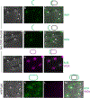
Sporangia were harvested from hours t2 through t7 of sporulation, fixed using methanol, and visualized by phase-contrast (top row) or epifluorescence (bottom row) microscopy. Cells were stained with Hoechst 33342 (to detect DNA, blue), WGA (to detect inner polysaccharide, magenta) and anti-BclA monoclonal antibodies (to detect the exosporium surface, green, A) or anti-BxpB antibodies (to detect the exosporium basal layer, green, B)
Fig 9.
Fig 9.. Epifluorescence microscopic analysis of inner polysaccharide deposition in ysxE mutant sporangia during sporulation.
Sporangia were harvested from hours t2 through t8 of sporulation, fixed with methanol, visualized either by phase-contrast microscopy (top row) or epifluorescence (bottom row). Cells were stained with Hoechst 33342 (to detect DNA, blue), polyclonal anti-BclA antibodies (to detect the exosporium, green) and WGA (to detect inner polysaccharide, magenta; once the exosporium encircles the entire spore (t6) WGA staining pattern is variable, indicated by the dashed line in the cartoon above the panels)
Fig 10.
Fig 10.. Epifluorescence microscopic analysis of inner polysaccharide deposition during sporulation in wild type (A) and exsY mutant (B) sporangia.
Sporangia were harvested from hours t2 through t7, fixed with methanol, and visualized by phase-contrast (top row) or epifluorescence (bottom row) microscopy. Cells were stained with Hoechst 33342 (to detect DNA, blue), anti-CotY/ExsY antibodies (to detect the cap and the exosporium, green) and WGA (to detect the inner polysaccharide, magenta).
Fig 11.
Fig 11.. Epifluorescence microscopic visualization of inner polysaccharide in bas1093 mutant sporangia.
Sporangia were visualized by phase contrast (A, D, G, J) or epifluorescence microscopy (B, C, E, F, H, I, K, L, M). Monoclonal anti-BclA antibody (green; B, C, E, F, I, K, M) or WGA (magenta; H, I, L) was applied. Panels C, F, and M show merged phase contrast and immunofluorescence images. Arrow denotes detached cap-like BclA staining.
Fig 12.
Fig 12.. TEM analysis of bas1093 mutant sporangia and released spores.
Spores and sporangia were visualized using thin-section TEM with Ruthenium red staining. White arrows indicate misassembled coat and/or exosporium. Scale bars indicate 400nm (A) and 200nm (B, C). MCE=mother cell envelope; Cr=core; Cx=cortex. A representative wild type TEM image is shown in Fig. 6
Fig 13.
Fig 13.. Epifluorescence microscopic visualization of inner polysaccharide in bas0371 mutant spores.
Bas0371 mutant and wild type spores were visualized by phase contrast (A, C, E, G, I, K) or epifluorescence microscopy (B, D, F, H, J, L). Polyclonal anti-BclA antibodies (green; B, D, F) and either WGA (magenta; D) or SJA (magenta; F) were applied. Arrows indicate spores encircled partially by or lacking an exosporium.

References

    1. Albers SV, and Meyer BH (2011) The archaeal cell envelope. Nat Rev Microbiol 9: 414–426. - PubMed
    1. Arrieta-Ortiz ML, Hafemeister C, Bate AR, Chu T, Greenfield A, Shuster B, et al. (2015) An experimentally supported model of the Bacillus subtilis global transcriptional regulatory network. Mol Syst Biol 11: 839. - PMC - PubMed
    1. Arroyo J, Farkas V, Sanz AB, and Cabib E (2016) ‘Strengthening the fungal cell wall through chitin-glucan cross-links: effects on morphogenesis and cell integrity’. Cell Microbiol 18: 1239–1250. - PubMed
    1. Bailey-Smith K, Todd SJ, Southworth TW, Proctor J, and Moir A (2005) The ExsA protein of Bacillus cereus is required for assembly of coat and exosporium onto the spore surface. J Bacteriol 187: 3800–3806. - PMC - PubMed
    1. Bartels J, Blüher A, López Castellanos S, Richter M, Günther G, and Mascher T (2019) The Bacillus subtilis endospore crust: protein interaction network, architecture and glycosylation state of a potential glycoprotein layer. Molecular Microbiology 112(5): 1576–1592. - PubMed

Publication types

MeSH terms