. 2023 Feb;18(2):357-363.
doi: 10.4103/1673-5374.346545.

PANoptosis-like cell death in ischemia/reperfusion injury of retinal neurons

Affiliations

PANoptosis-like cell death in ischemia/reperfusion injury of retinal neurons

Wei-Tao Yan et al. Neural Regen Res. 2023 Feb.

Abstract

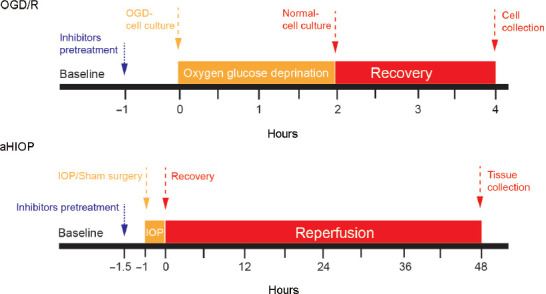
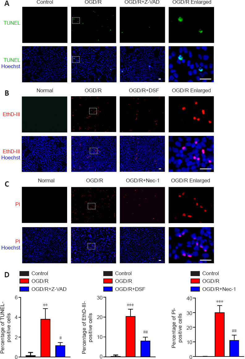
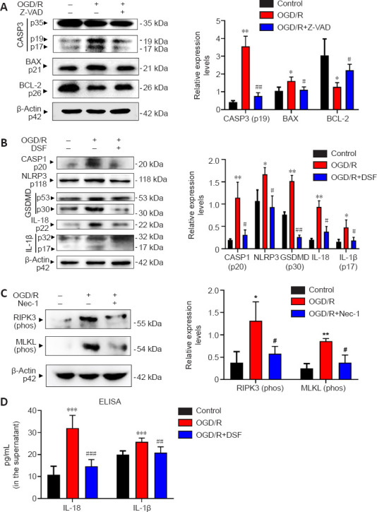
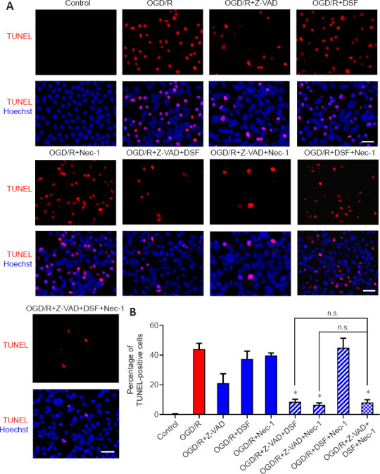
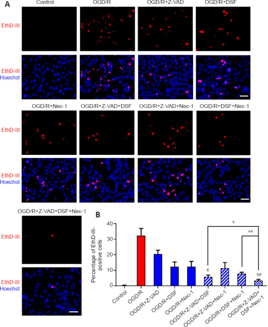
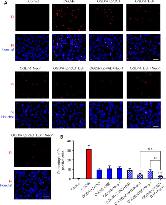
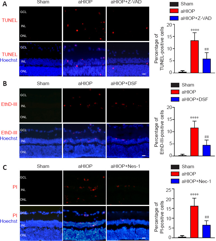
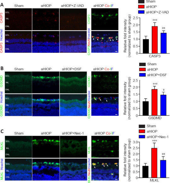
PANoptosis is a newly identified type of regulated cell death that consists of pyroptosis, apoptosis, and necroptosis, which simultaneously occur during the pathophysiological process of infectious and inflammatory diseases. Although our previous literature mining study suggested that PANoptosis might occur in neuronal ischemia/reperfusion injury, little experimental research has been reported on the existence of PANoptosis. In this study, we used in vivo and in vitro retinal neuronal models of ischemia/reperfusion injury to investigate whether PANoptosis-like cell death (simultaneous occurrence of pyroptosis, apoptosis, and necroptosis) exists in retinal neuronal ischemia/reperfusion injury. Our results showed that ischemia/reperfusion injury induced changes in morphological features and protein levels that indicate PANoptosis-like cell death in retinal neurons both in vitro and in vivo. Ischemia/reperfusion injury also significantly upregulated caspase-1, caspase-8, and NLRP3 expression, which are important components of the PANoptosome. These results indicate the existence of PANoptosis-like cell death in ischemia/reperfusion injury of retinal neurons and provide preliminary experimental evidence for future study of this new type of regulated cell death.

Keywords: NOD-like receptor protein 3 (NLRP3); PANoptosis; apoptosis; gasdermin-D (GSDMD); ischemia/reperfusion; mixed lineage kinase domain-like protein (MLKL); necroptosis; pyroptosis; receptor-interacting protein kinase 3 (RIPK3); retinal neuron.


Conflict of interest statement

None

Figures

Figure 1
Figure 1
Schematic of the experimental timeline of OGD/R and aHIOP modeling. In the OGD/R model (top panel), R28 cells were pretreated with inhibitors (or DMSO for control) for 1 hour before establishing the OGD model. Then, the initial culture medium was replaced with glucose-free medium. The cell culture containers were placed in the OGD treatment device, which maintained a hypoxic condition (O2 < 1%). OGD treatment lasted for 2 hours. After OGD treatment, the cells were returned to the initial culture condition for 2 hours. The normal control group was maintained in the normal culture condition for the same length of time. In the aHIOP model (bottom panel), inhibitors (or DMSO for control) were injected into the vitreous cavity 30 minutes before HIOP/sham surgery. To induce ocular hypertension, normal saline was injected into the anterior chamber to form artificial intraocular pressure (IOP). IOP was slowly increased to 110 mmHg and maintained for 60 minutes, then gradually returned to normal level and maintained for 48 hours before retinal tissue collection. In the sham group, a sterilized needle was inserted into the anterior chamber without elevating the IOP at the same time point. aHIOP: Acute high intraocular pressure; DMSO: dimethyl sulphoxide; OGD/R: oxygen-glucose deprivation/recovery.
Figure 2
Figure 2
OGD/R-induced R28 cells exhibit pyroptosis, apoptosis, and necroptosis. (A) Terminal-deoxynucleotidyl transferase mediated nick end labeling (TUNEL) staining for R28 cells. R28 cells were treated with OGD/R and Z-VAD. (B) Ethidium Homodimer III (EthD-III) staining for R28 cells. R28 cells were treated with OGD/R and DSF. (C) Propidium iodide (PI) staining for R28 cells. R28 cells were treated with OGD/R and Nec-1. Scale bars: 50 μm. (D) Percentage of TUNEL-, EthD-III-, PI-positive cells (mean ± SD, n = 3). **P < 0.01, ***P < 0.001, vs. Control; #P < 0.05, ##P < 0.01, vs. OGD/R (Student’s t-test). All cell experiments were performed at least three times independently. DSF: Disulfiram; Nec-1: necrostatin-1; OGD/R: oxygen-glucose deprivation/recovery; Z-VAD: Z-VAD-FMK.
Figure 3
Figure 3
Treatment with OGD/R upregulates key proteins of pyroptosis, apoptosis and necroptosis in R28 cells. (A) Western blotting of caspase-3, BAX and Bcl-2 in R28 cells after treatment with OGD/R and Z-VAD inhibitor. (B) Western blotting of caspase-1, NLRP3, GSDMD, IL-18, IL-1β in R28 cells after treatment with OGD/R and DSF inhibitor. (C) Western blotting of phosphorylated RIPK3 and phosphorylated MLKL in R28 cells after treatment with OGD/R and Nec-1 inhibitor. Quantifications of protein expression are shown to the right of each western blot image. (D) ELISA of the expressions of mature IL-1β and IL-18 in the R28 cell culture supernatant after treatment with OGD/R and DSF inhibitor. Data are expressed as the mean ± SD (n = 3). *P < 0.05, **P < 0.01 vs. Control; #P < 0.05, ##P < 0.01, vs. OGD/R (Student’s t-test). All cell experiments were performed at least three times independently. BAX: Bcl-2-associated X protein; CASP: caspase; DSF: disulfiram; GSDMD: Gasdermin-D; IL: interleukin; MLKL: mixed lineage kinase domain-like; Nec-1: necrostatin-1; NLRP3: NACHT, LRR, and PYD domains-containing protein 3; OGD/R: oxygen-glucose deprivation/recovery; RIPK: receptor-interacting protein kinase; Z-VAD: Z-VAD-FMK.
Figure 4
Figure 4
Combination of RCD inhibitors decreases the OGD/R-induced TUNEL-positive cells. (A) TUNEL staining for R28 cells treated with combinations of RCD inhibitors following OGD/R treatment. Scale bars: 50 μm. Red: TUNEL-positive cells. (B) Percentage of TUNEL-positive cells (mean ± SD, n = 3). *P < 0.05, vs. OGD/R + Z-VAD (Student’s t-test). DSF: Disulfiram; n.s.: not significant; Nec-1: necrostatin-1; OGD/R: oxygen-glucose deprivation/recovery; RCD: regulated cell death; TUNEL: terminal-deoxynucleotidyl transferase mediated nick end labeling; Z-VAD: Z-VAD-FMK.
Figure 5
Figure 5
Combination of RCD inhibitors decreases the OGD/R-induced EthD-III-positive cells. (A) EthD-III staining for R28 cells treated with combinations of RCD inhibitors following OGD/R treatment. Red: EthD-III-positive cells. Scale bars: 50 μm. (B) Percentage of EthD-III-positive cells (mean ± SD, n = 3). *P < 0.05, **P < 0.01, vs. OGD/R + Z-VAD + DSF + Nec-1; #P < 0.05, ##P < 0.01, vs. OGD/R + DSF (Student’s t-test). DSF: Disulfiram; EthD-III: Ethidium Homodimer III; Nec-1: necrostatin-1; OGD/R: oxygen-glucose deprivation/recovery; RCD: regulated cell death; Z-VAD: Z-VAD-FMK.
Figure 6
Figure 6
Combination of RCD inhibitors decreases the OGD/R-induced PI-positive cells. (A) PI staining for R28 cells treated with OGD/R and Z-VAD, DSF, and Nec-1. Red: PI-positive cells. Scale bars: 50 μm. (B) Percentage of PI-positive cells (mean ± SD, n = 3). **P < 0.01, vs. OGD/R + Z-VAD + DSF + Nec-1; ##P < 0.01, ###P < 0.001 vs. OGD/R+Nec-1 (Student’s t-test). All cell experiments were performed at least three times independently. DSF: Disulfiram; n.s.: not significant; Nec-1: necrostatin-1; OGD/R: oxygen-glucose deprivation/recovery; PI: propidium iodide; RCD: regulated cell death; Z-VAD: Z-VAD-FMK.
Figure 7
Figure 7
aHIOP induces pyroptosis, apoptosis, and necroptosis in vivo in rat retina. (A) TUNEL staining for rat retinal sections. The rats were treated with aHIOP and Z-VAD. (B) EthD-III staining for rat retinal sections. The rats were treated with aHIOP and DSF. (C) PI staining for rat retinal sections. The sections were treated with aHIOP and Nec-1. Scale bars: 50 μm. Percentage of TUNEL-, EthD-III-, and PI-positive cells shown to the right of images (mean ± SD, n = 5 rats per group). ***P < 0.001, vs. Sham; ##P < 0.01, vs. aHIOP (Student’s t-test). aHIOP: Acute high intraocular pressure; DSF: disulfiram; GCL: ganglion cell layer; INL: inner nuclear layer; Nec-1: necrostatin-1; ONL: outer nuclear layer; Z-VAD: Z-VAD-FMK.
Figure 8
Figure 8
aHIOP induces high expression of pyroptosis, apoptosis, and necroptosis-related proteins in rat retina. Immunofluorescence staining of CASP3 (A), GSDMD (B) and MLKL (C) of rat retinal tissue sections. NeuN staining was used to indicate ganglion cells in the retina. Scale bars: 50 μm. Quantification of pyroptosis, apoptosis, and necroptosis-related protein expressions shown on the right of images (mean ± SD, n = 5 rats per group). Relative fold intensity of immunofluorescence staining was normalized relative to the intensity of the sham group. **P < 0.001, vs. Sham; #P < 0.05, ##P < 0.01, vs. OGD/R (Student’s t-test). aHIOP: Acute high intraocular pressure; CASP: caspase; Co-IF: co-immunofluorescence; DSF: disulfiram; GCL: ganglion cell layer; GSDMD: gasdermin-D; INL: inner nuclear layer; Nec-1: necrostatin-1; MLKL: mixed lineage kinase domain-like; ONL: outer nuclear layer; Z-VAD: Z-VAD-FMK.

References

    1. Banoth B, Tuladhar S, Karki R, Sharma BR, Briard B, Kesavardhana S, Burton A, Kanneganti TD. ZBP1 promotes fungi-induced inflammasome activation and pyroptosis, apoptosis, and necroptosis (PANoptosis) J Biol Chem. 2020;295:18276–18283. - PMC - PubMed
    1. Basuroy S, Bhattacharya S, Leffler CW, Parfenova H. Nox4 NADPH oxidase mediates oxidative stress and apoptosis caused by TNF-alpha in cerebral vascular endothelial cells. Am J Physiol Cell Physiol. 2009;296:C422–432. - PMC - PubMed
    1. Briard B, Malireddi RKS, Kanneganti TD. Role of inflammasomes/pyroptosis and PANoptosis during fungal infection. PLoS Pathog. 2021;17:e1009358. - PMC - PubMed
    1. Chen H, Deng Y, Gan X, Li Y, Huang W, Lu L, Wei L, Su L, Luo J, Zou B, Hong Y, Cao Y, Liu Y, Chi W. NLRP12 collaborates with NLRP3 and NLRC4 to promote pyroptosis inducing ganglion cell death of acute glaucoma. Mol Neurodegener. 2020;15:26. - PMC - PubMed
    1. Chen S, Yan J, Deng HX, Long LL, Hu YJ, Wang M, Shang L, Chen D, Huang JF, Xiong K. Inhibition of calpain on oxygen glucose deprivation-induced RGC-5 necroptosis. J Huazhong Univ Sci Technolog Med Sci. 2016;36:639–645. - PubMed

LinkOut - more resources