

A comparison of Covid-19 early detection between convolutional neural networks and radiologists
- PMID: 35900673
- PMCID: PMC9330942
- DOI: 10.1186/s13244-022-01250-3
A comparison of Covid-19 early detection between convolutional neural networks and radiologists
Erratum in
-
Correction: A comparison of Covid-19 early detection between convolutional neural networks and radiologists.Insights Imaging. 2022 Oct 20;13(1):172. doi: 10.1186/s13244-022-01314-4. Insights Imaging. 2022. PMID: 36264395 Free PMC article. No abstract available.
Abstract
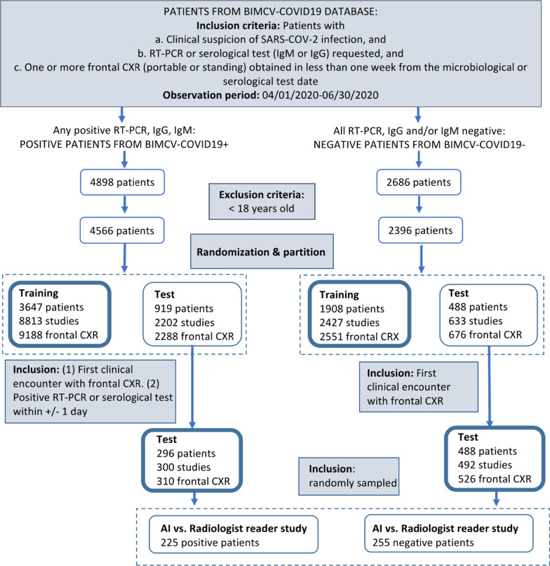
Background: The role of chest radiography in COVID-19 disease has changed since the beginning of the pandemic from a diagnostic tool when microbiological resources were scarce to a different one focused on detecting and monitoring COVID-19 lung involvement. Using chest radiographs, early detection of the disease is still helpful in resource-poor environments. However, the sensitivity of a chest radiograph for diagnosing COVID-19 is modest, even for expert radiologists. In this paper, the performance of a deep learning algorithm on the first clinical encounter is evaluated and compared with a group of radiologists with different years of experience.
Methods: The algorithm uses an ensemble of four deep convolutional networks, Ensemble4Covid, trained to detect COVID-19 on frontal chest radiographs. The algorithm was tested using images from the first clinical encounter of positive and negative cases. Its performance was compared with five radiologists on a smaller test subset of patients. The algorithm's performance was also validated using the public dataset COVIDx.
Results: Compared to the consensus of five radiologists, the Ensemble4Covid model achieved an AUC of 0.85, whereas the radiologists achieved an AUC of 0.71. Compared with other state-of-the-art models, the performance of a single model of our ensemble achieved nonsignificant differences in the public dataset COVIDx.
Conclusion: The results show that the use of images from the first clinical encounter significantly drops the detection performance of COVID-19. The performance of our Ensemble4Covid under these challenging conditions is considerably higher compared to a consensus of five radiologists. Artificial intelligence can be used for the fast diagnosis of COVID-19.
Keywords: Covid-19; Deep learning; Radiology.
© 2022. The Author(s).
Conflict of interest statement
The authors declare that they have no competing interests.
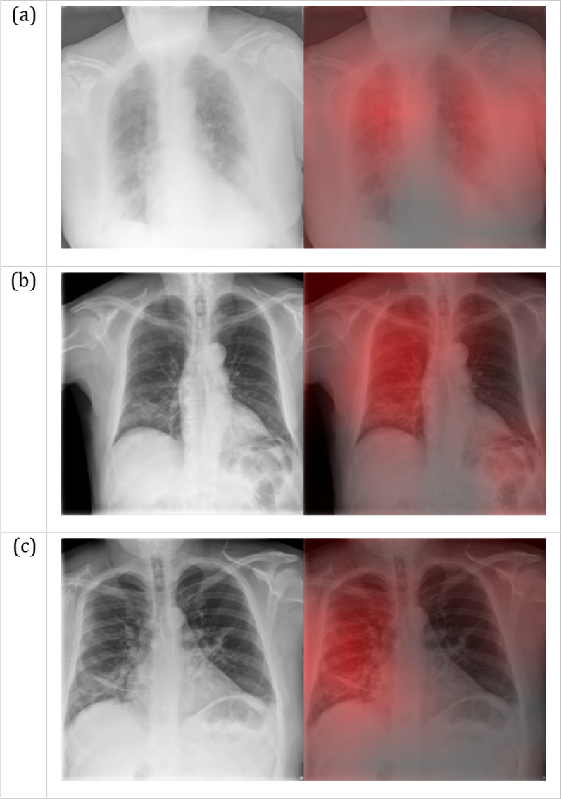
Figures


References
-
- World Health Organization, Coronavirus Disease (Covid 19) (2020) https://www.who.int/health-topics/coronavirus#tab=tab_1. Accessed May 14, 2022
