. 2022 Sep 15;132(18):e154571.
doi: 10.1172/JCI154571.

Small-molecule eRF3a degraders rescue CFTR nonsense mutations by promoting premature termination codon readthrough

Affiliations

Small-molecule eRF3a degraders rescue CFTR nonsense mutations by promoting premature termination codon readthrough

Rhianna E Lee et al. J Clin Invest. .

Abstract

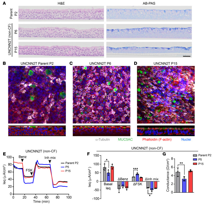
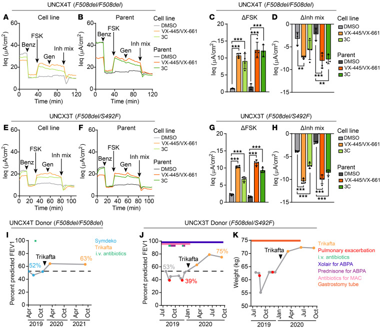
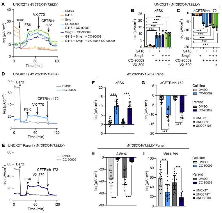
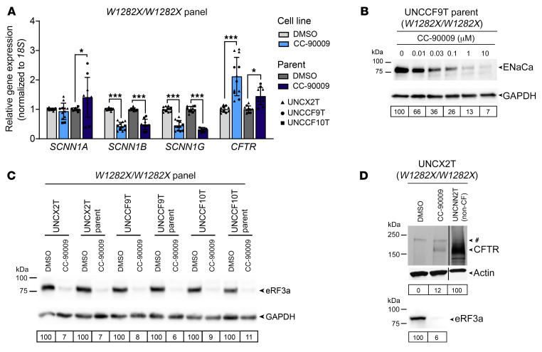
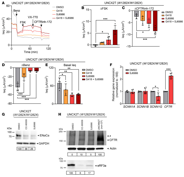
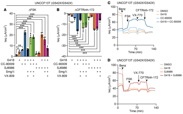
The vast majority of people with cystic fibrosis (CF) are now eligible for CF transmembrane regulator (CFTR) modulator therapy. The remaining individuals with CF harbor premature termination codons (PTCs) or rare CFTR variants with limited treatment options. Although the clinical modulator response can be reliably predicted using primary airway epithelial cells, primary cells carrying rare CFTR variants are scarce. To overcome this obstacle, cell lines can be created by overexpression of mouse Bmi-1 and human TERT (hTERT). Using this approach, we developed 2 non-CF and 6 CF airway epithelial cell lines, 3 of which were homozygous for the W1282X PTC variant. The Bmi-1/hTERT cell lines recapitulated primary cell morphology and ion transport function. The 2 F508del-CFTR cell lines responded robustly to CFTR modulators, which was mirrored in the parent primary cells and in the cell donors' clinical response. Cereblon E3 ligase modulators targeting eukaryotic release factor 3a (eRF3a) rescued W1282X-CFTR function to approximately 20% of WT levels and, when paired with G418, rescued G542X-CFTR function to approximately 50% of WT levels. Intriguingly, eRF3a degraders also diminished epithelial sodium channel (ENaC) function. These studies demonstrate that Bmi-1/hTERT cell lines faithfully mirrored primary cell responses to CFTR modulators and illustrate a therapeutic approach to rescue CFTR nonsense mutations.

Keywords: Genetic diseases; Ion channels; Pulmonology; Therapeutics; Translation.


Figures

Figure 1
Figure 1. Nasal cell lines model primary cell morphology and ion transport function.
(A) H&E and AB-PAS staining of UNCNN2T (non-CF) P2 parent cells and cell line at P6 and P15. Scale bar: 50 μm. (BD) Whole-mount immunostaining of UNCNN2T P2 parent cells (B) and cell line at P6 (C) and P15 (D). α-Tubulin (white), MUC5AC (green), phalloidin (F-actin, red), and Hoechst (nuclei, blue). Scale bar: 25 μm. (EG) TECC-24 measurements of UNCNN2T P2 parent cells and cell line at P5 and P15. (E) TECC-24 tracing representing 3–4 replicates. Acute addition of 6 μM benzamil (Benz), 10 μM FSK, and an inhibitor mixture (Inh mix) consisting of CFTRinh-172, GlyH-101, and bumetanide (each at 20 μM), is indicated by arrows. (F) Basal Ieq and change in Ieq (ΔIeq) in response to benzamil, FSK, and the inhibitor mixture. n = 3–4. (G) Baseline conductance values. n = 3–4. All data were analyzed using an ordinary linear model and are presented as the mean ± SD. *P < 0.05, **P < 0.01, and ***P < 0.001.
Figure 2
Figure 2. Nasal cell lines predict primary cell and clinical response to CFTR modulators.
(A and B) TECC-24 tracings of the UNCX4T nasal cell line (F508del/F508del) (A) and parent primary cells (B) treated with 0.1% DMSO and VX-445 and VX-661 (each at 5 μM), or a triple corrector combination (3C) containing VX-809/3151/4172 (each at 5 μM). Tracings are representative of 3–4 replicates. Acute addition of the potentiator 10 μM genistein is indicated by arrows. (C and D) ΔIeq of UNCX4T and parent cells in response to FSK (C) and the inhibitor mixture (D). n = 3–4. (E and F) TECC-24 tracings of the UNCX3T nasal cell line (F508del/S492F) (E) and parent primary cells (F) pretreated with DMSO, VX-445 and VX-661, or 3C. Tracings are representative of 3–4 replicates. (G and H) ΔIeq of UNCX3T and parent cells in response to FSK (G) and the inhibitor mixture (H). n = 3–4. (I) Change in the percentage of predicted FEV1 before and after Trikafta initiation in the UNCX4T donor (I) and the UNCX3T donor (J). Blue data points indicate FEV1 measured during SYMDEKO therapy, orange data points indicate FEV1 measured during Trikafta therapy, and red data points indicate FEV1 measured during a CF exacerbation. The treatment course for the UNCX4T and UNCX3T donors is indicated above the respective FEV1 plots and includes the timeline of i.v. antibiotics (green), XOLAIR for ABPA (dark blue), prednisone for ABPA (purple), antibiotics for treatment of Mycobacterium avium complex (MAC) (pink). (K) Change in weight in kilograms of the UNCX3T cell donor after Trikafta initiation. Gastrostomy tube use and subsequent removal are indicated by an orange bar. All data were analyzed using an ordinary linear model and are presented as the mean ± SD. Post hoc comparisons were performed using the general linear hypothesis test. **P < 0.01 and ***P < 0.001.
Figure 3
Figure 3. The eRF3a degrader CC-90009 rescues W1282X-CFTR in a panel of cell lines and parent primary cells.
(AC) TECC-24 measurements of the UNCX2T cell line (W1282X/W1282X) treated with 0.1% DMSO, 200 μM G418, 0.3 μM SMG1i, 0.1 μM CC-90009, and 3 μM VX-809, alone or in combination as indicated. Acute addition of the potentiator 10 μM VX-770 is indicated by an arrow. (A) TECC-24 tracing representing 3–4 replicates. (B and C) ΔIeq in response to FSK (B) and CFTRinh-172 (C). Data were analyzed using ordinary linear models. n = 3–4. (DI) TECC-24 measurements of a panel of W1282X/W1282X cell lines and parent cells treated with 0.1% DMSO or 0.1 μM CC-90009. (D and E) TECC-24 tracing of the UNCX2T cell line (D) and parent cells (E). Tracings are representative of the W1282X/W1282X panel containing 3 cell donors with 6 replicates per donor. (FI) ΔIeq in response to FSK (F) and CFTRinh-172 (G), benazmil (H), and basal Ieq (I). Data were analyzed using a linear mixed-effects model with the donor as a random effect factor. n = 6 per donor. Post hoc comparisons were performed using the general linear hypothesis test. All data are presented as the mean ± SD. ***P < 0.001.
Figure 4
Figure 4. CC-90009 reduces ENaC expression and rescues CFTR by promoting readthrough and full-length CFTR production.
(A) Relative SCNN1A, SCNN1B, SCNN1G, and CFTR mRNA levels by RT-qPCR in a panel of W1282X/W1282X cell lines and parent cells. Data were analyzed using a linear mixed-effects model with the donor as a random effect factor and are presented as the mean ± SD. n = 2–6 for each cell line. Post hoc comparisons were performed using the general linear hypothesis test. *P < 0.05 and ***P < 0.001. (B) Western blot for ENaCa in UNCCF9T parent cells treated with escalating doses of CC-90009. ENaCa expression normalized to GAPDH and relative to the 0.1% DMSO control is quantified below. Cells were grown in Vertex ALI media to increase the level of ENaC expression for detectability by Western blotting. (C) Western blot for eRF3a in a panel of W1282X/W1282X cell lines and parent cells treated with 0.1% DMSO or 0.1 μM CC-90009. eRF3a expression normalized to GAPDH and relative to the paired DMSO control is quantified below. (D) CFTR IP–Western blot (top) of the UNCX2T cell line (W1282X/W1282X) treated with 0.1% DMSO or 0.1 μM CC-90009 or the UNCNN2T non-CF cell line. A nonspecific band was observed in all samples and is indicated by a pound sign. CFTR expression normalized to actin and relative to the UNCNN2T non-CF control is quantified below. The same blot was reprobed for eRF3a (bottom). eRF3a expression normalized to actin and relative to the DMSO control is quantified below. All samples were run on the same blot and imaged at the same intensity level.
Figure 5
Figure 5. Another eRF3a degrader, SJ6986, rescues CFTR and decreases ENaC expression.
(AE) TECC-24 measurements of the UNCX2T cell line (W1282X/W1282X) treated with 0.1% DMSO, 200 μM G418, and 0.2 μM SJ6986, alone or in combination. (A) TECC-24 tracing representing 4 replicates. (BD) ΔIeq in response to FSK (B), CFTRinh-172 (C), and benzamil (D). (E) Basal Ieq. n = 4. (F) Relative SCNN1A, SCNN1B, SCNN1G, and CFTR mRNA levels by RT-qPCR in the UNCX2T cell line treated with 0.1% DMSO or 0.2 μM SJ6986. n = 4–6. (G) Western blot for ENaCa in the UNCX2T cell line treated with 0.1% DMSO, 0.1 μM CC-90009, or 0.2 μM SJ6986. ENaCa expression normalized to GAPDH and relative to the DMSO control is quantified below. Cells were grown in Vertex ALI media to increase the level of ENaC expression for detectability by Western blotting. (H) CFTR IP–Western blot (top) of the UNCX2T cell line treated with 0.1% DMSO, 0.1 μM CC-90009, or 0.2 μM SJ6986 or the untreated UNCNN2T non-CF cell line. The final lane (UNCNN2T) also appears in Figure 4D. A nonspecific band observed in all samples is indicated by a pound sign. CFTR expression normalized to actin and relative to the UNCNN2T non-CF control is quantified below. The same blot was reprobed for eRF3a (bottom). eRF3a expression normalized to actin and relative to the DMSO control is quantified below. All data were analyzed using ordinary linear models and are presented as the mean ± SD. *P < 0.05, **P < 0.01, and ***P < 0.001.
Figure 6
Figure 6. eRF3a degraders synergize with G418 to rescue G542X-CFTR function.
(AD) TECC-24 measurements of the UNCCF13T cell line (G542X/G542X) treated with 0.1% DMSO or combinations of 200 μM G418, 0.1 μM CC-90009, 0.2 μM SJ6986, 0.3 μM Smg1i, and 3 μM VX-809. (A and B) Change in Ieq in response to FSK (A) and CFTRinh-172 (B). (C and D) TECC-24 tracings representing 4 replicates. All data were analyzed using ordinary linear models and are presented as the mean ± SD. *P < 0.05, **P < 0.01, and ***P < 0.001.

References

    1. Cutting GR. Cystic fibrosis genetics: from molecular understanding to clinical application. Nat Rev Genet. 2015;16(1):45–56. - PMC - PubMed
    1. Ratjen F, et al. Cystic fibrosis. Nat Rev Dis Primers. 2015;1:15010. doi: 10.1038/nrdp.2015.10. - DOI - PMC - PubMed
    1. Ramsey BW, et al. A CFTR potentiator in patients with cystic fibrosis and the G551D mutation. N Engl J Med. 2011;365(18):1663–1672. doi: 10.1056/NEJMoa1105185. - DOI - PMC - PubMed
    1. Rowe SM, et al. Tezacaftor-ivacaftor in residual-function heterozygotes with cystic fibrosis. N Engl J Med. 2017;377(21):2024–2035. doi: 10.1056/NEJMoa1709847. - DOI - PMC - PubMed
    1. Taylor-Cousar JL, et al. Tezacaftor-ivacaftor in patients with cystic fibrosis homozygous for Phe508del. N Engl J Med. 2017;377(21):2013–2023. doi: 10.1056/NEJMoa1709846. - DOI - PubMed

Publication types

Substances