. 2023 Mar;19(3):873-885.
doi: 10.1080/15548627.2022.2107314. Epub 2022 Aug 8.

USP22 suppresses the NLRP3 inflammasome by degrading NLRP3 via ATG5-dependent autophagy

Affiliations

USP22 suppresses the NLRP3 inflammasome by degrading NLRP3 via ATG5-dependent autophagy

Qianqian Di et al. Autophagy. 2023 Mar.

Abstract

The NLRP3 inflammasome is involved in a diverse range of inflammatory diseases. The activation of inflammasomes must be tightly regulated to prevent excessive inflammation, and the protein ubiquitination system is reported to be one of the ways in which inflammasome activation is regulated. However, the deubiquitination regulatory mechanisms of inflammasome activation remain elusive. Here, we demonstrated that USP22 (ubiquitin specific peptidase 22) promotes NLRP3 degradation and inhibits NLRP3 inflammasome activation. USP22 deficiency or in vivo silencing significantly increases alum-induced peritonitis and lipopolysaccharide-induced systemic inflammation. Mechanistically, USP22 inhibits NLRP3 inflammasome activation via the promotion of ATG5-mediated macroautophagy/autophagy. USP22 stabilizes ATG5 via decreasing K27- and K48-linked ubiquitination of ATG5 at the Lys118 site. Taken together, these findings reveal the role USP22 plays in the regulation of NLRP3 inflammasome activation and suggest a potential therapeutic target to treat NLRP3 inflammasome-related diseases.Abbreviations: ATG5: autophagy related 5; ATP: adenosine triphosphate; CASP1: caspase 1; IL18: interleukin 18; IL1B/IL-1β: interleukin 1 beta; LPS: lipopolysaccharide; NLRC4: NLR family, CARD domain containing 4; NLRP3: NLR family, pyrin domain containing 3; PYCARD/ASC: PYD and CARD domain containing; TNF/TNF-α: tumor necrosis factor; USP22: ubiquitin specific peptidase 22.

Keywords: Autophagy; NLRP3; autophagy related 5; inflammasome; ubiquitin specific peptidase 22.


Conflict of interest statement

All the authors declare that there are no conflicts of interest.

Figures

Figure 1.
Figure 1.
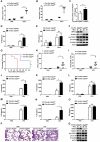
USP22 silencing promotes NLRP3 inflammasome activation. (a) Immunoblots of IL1B, pro-CASP1, and cleaved CASP1 in the supernatants or cell lysates of USP22-silenced mouse primary macrophages or PMA-pretreated human THP-1 cells, primed with LPS, and then stimulated with nigericin (Nig.). (b) ELISA analysis of IL1B in supernates (SNs) from mouse primary macrophages and human THP-1 cells. (c) ELISA analysis of IL18 in SNs from mouse primary macrophages and human THP-1 cells. (d) USP22-silenced or control THP-1 macrophages were treated with palmitate at the indicated concentrations for 24 h. Immunoblots show the NLRP3, IL1B, pro-CASP1 and cleaved CASP1 in the cell lysates. And (e) IL1B in SNs from THP-1 cells silenced of USP22 were analyzed by ELISA. (f) qPCR analysis of the expression of Usp22 in peritoneal exudate cells (PECs) recovered 6 h after alum injection in Usp22-silenced mice and control mice. (g) Immunoblot analysis of NLRP3 expression in peritoneal exudate cells recovered from Usp22-silenced mice and control mice 6 h after alum injection and quantization of NLRP3. (h and i) Flow cytometry analysis of the inflammatory cell subset in peritoneal exudate cells recovered 6 h after alum injection (n = 6). (J) ELISA analysis of IL1B secretion in the lavage fluid from Usp22-silenced mice or control mice (n = 6–8). Data shown are representative of three separate experiments (A, D, and G) or are means ± SEM of three independent experiments (B, C and E). * p < 0.05, ** p < 0.01.
Figure 2.
Figure 2.
Knockout of Usp22 promotes activation of the NLRP3 inflammasome. (a) Cre-Esr Usp22fl/+ and Cre-Esr usp22fl/fl primary macrophages were stimulated with LPS for 16 h and activated with nigericin for 30 min or alum for 6 h. Immunoblot analysis of the expression of indicated proteins. (b) Cre-Esr Usp22fl/+ and Cre-Esr usp22fl/fl BMDMs were stimulated with LPS for 16 h and activated with nigericin for 30 min or alum for 6 h. Immunoblot analysis of the expression of the indicated proteins. (c) ELISA analysis of IL1B in the supernatants of usp22-knockout mouse primary macrophages. (d) ELISA analysis of IL1B in the supernatants of usp22-knockout BMDMs. (e) ELISA analysis of IL18 in the supernatants of usp22-knockout mouse primary macrophages. (f) ELISA analysis of IL18 in the supernatants of usp22-knockout BMDMs. (g) Cre-Esr Usp22fl/+ and Cre-Esr usp22fl/fl primary macrophages were pretreated with MCC950, then stimulated with LPS and nigericin. ELISA analysis of IL1B in the supernatants. (h) ELISA analysis of TNF in the supernatants of usp22-knockout mouse primary macrophages. (i) Cre-Esr Usp22fl/+ and Cre-Esr usp22fl/fl BMDMs were stimulated with LPS for 16 h, then transfected with flagellin (0.5 μg/mL) for 1 h; ELISA analysis of IL1B in the supernatants. (j) Cre-Esr Usp22fl/+ and Cre-Esr usp22fl/fl BMDMs were stimulated with LPS for 16 h, then transfected with poly(dA:dT) (2 μg/mL) for 1 h; ELISA analysis of IL1B in the supernatants. Data shown are representative of three separate experiments (A, B) or are means ± SEM of three independent experiments (C-J). * p < 0.05, ** p < 0.01, *** p < 0.001. ns, not significant.
Figure 3.
Figure 3.
Usp22 knockout exacerbates LPS-induced systemic inflammation and alum-induced peritoneal inflammation. (a-f) 8-week-old Cre-Esr Usp22fl/+ and Cre-Esr usp22fl/fl female mice (n = 6/group) were intraperitoneally injected with alum (1 mg/mouse); 6 h later, the mice were euthanized for the following analyses. (a and b) Flow cytometry analysis of the inflammatory cell subset in peritoneal exudate cells recovered 6 h after alum injection. (c) Protein concentration in the peritoneal lavage fluids. (d and e) ELISA analysis of IL1B and TNF secretion in the peritoneal lavage fluids. (f) Immunoblot analysis with the indicated antibodies of peritoneal exudate cells isolated from Cre-Esr Usp22fl/+ and Cre-Esr usp22fl/fl mice 6 h after alum injection. (g) 8-week-old Cre-Esr Usp22fl/+ and Cre-Esr usp22fl/fl female mice (n = 10/group) were intraperitoneally injected with LPS (20 mg/kg), and survival of the mice was monitored for 48 h. (h-q) 8-week-old Cre-Esr Usp22fl/+ and Cre-Esr usp22fl/fl female mice (n = 6/group) were intraperitoneally injected with LPS (10 mg/kg). (h and i) Flow cytometry analysis of the inflammatory cell subset in peritoneal exudate cells recovered 4 h after LPS injection. (j-l) ELISA analysis of IL1B, IL18, and TNF secretion in the peritoneal lavage fluids. (m-o) ELISA analysis of IL1B, IL18, and TNF secretion in the serum. (p) H&E staining of left-lung samples. (q) Immunoblot analysis with the indicated antibodies of peritoneal exudate cells recovered from Cre-Esr Usp22fl/+ and Cre-Esr usp22fl/fl mice 4 h after LPS injection. Data shown are means ± SEM (A-E and G-O). * p < 0.05, ** p < 0.01, *** p < 0.001. ns, not significant.
Figure 4.
Figure 4.
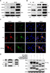
USP22 interacts with NLRP3 at the LRR domain. (a) Co-IP analysis of the interaction between NLRP3 and USP22 in mouse peritoneal macrophages following immunoprecipitation with anti-USP22. (b) Co-IP analysis of the interaction between NLRP3 and USP22 in mouse peritoneal macrophages following immunoprecipitation with anti-NLRP3. (c) Immunofluorescence staining for USP22 (green) and NLRP3 (red) in mouse peritoneal macrophages. Scale bars represent 10 μm. (d) 293 T cells were co-transfected with MYC-USP22 and Flag-NLRP3 or its truncated mutants, immunoprecipitated with anti-Flag, and immunoblotted using the indicated antibodies. Data shown are representative of three separate experiments.
Figure 5.
Figure 5.
USP22 post-translationally regulates NLRP3 stability. (a) 293 T cells were co-transfected with HA-NLRP3 and various concentrations of Flag-USP22, then immunoblot assays were performed using the indicated antibodies. (b) 293 T cells were co-transfected with HA-NLRP3 and Flag-USP22 and incubated with CHX for the indicated times. HA-tag and Flag-tag antibodies were used to detect the expression of relevant proteins. The quantification of relative NLRP3 levels is shown in the lower panel. (c) 293 T cells were co-transfected with HA-NLRP3 and Flag-USP22 for 24 h, incubated with the lysosome inhibitor CQ or proteasome inhibitor MG132 for 4 h, then immunoblot analyses were performed using the indicated antibodies. (d) Cre-Esr Usp22fl/+ and Cre-Esr usp22fl/fl primary macrophages were incubated in EBSS conditions with or without CQ for 3 h, the immunoblotted with the indicated antibodies. (e) Cre-Esr Usp22fl/+ and Cre-Esr usp22fl/fl primary macrophages were treated with rapamycin at the indicated concentration for 12 h, the immunoblotted with the indicated antibodies. (f) Cre-Esr Usp22fl/+ and Cre-Esr usp22fl/fl primary macrophages were incubated in EBSS conditions with or without CQ for 3 h, and immunofluorescence staining for LC3B (green) and DAPI (blue). Scale bars represent 10 μm. (g) Cre-Esr Usp22fl/+ and Cre-Esr usp22fl/fl primary macrophages were treated with rapamycin (250 nM) for 12 h, and immunofluorescence staining for LC3B (green) and DAPI (blue). Scale bars represent 10 μm. (h) Cre-Esr Usp22fl/+ and Cre-Esr usp22fl/fl primary macrophages were stimulated with LPS for 16 h, and with or without rapamycin (250 nM) for 12 h, the immunoblotted with the indicated antibodies. (i) The production of IL1B was analyzed by ELISA, Data shown is mean ± SEM. * p < 0.05, ** p < 0.01, *** p < 0.001. (J) 293 T cells were co-transfected with HA-NLRP3, Flag-USP22, and Flag-USP22-C185S, then immunoblotted with the indicated antibodies. Data shown are representative of three separate experiments.
Figure 6.
Figure 6.
USP22 interacts with and stabilizes ATG5. (a) Immunoblot analysis of the expression of ATG5, ATG7, and SQSTM1/p62 in the cell lysates of Usp22-silenced mouse peritoneal macrophages with or without LPS stimulation. (b) Co-IP analysis of the interaction between NLRP3 and ATG5 in mouse peritoneal macrophages following immunoprecipitation with anti-NLRP3. (c) Immunofluorescence staining for USP22 (green) and ATG5 (red) in mouse peritoneal macrophages. Scale bars represent 10 μm. (d) 293 T cells were transfected with Flag-ATG5 and MYC-USP22, immunoprecipitated with anti-Flag, and then immunoblotted using the indicated antibodies. (e) Immunoblots of IL1B, pro-CASP1, and cleaved CASP1 in the supernatants or cell lysates of Atg5-silenced mouse primary macrophages, primed with LPS, and then stimulated with nigericin (Nig.). (F) IL1B in SNs from macrophages silenced of Atg5 were analyzed by ELISA. (g) 293 T cells were transfected with HA-NLRP3 and Flag-ATG5, then immunoblotted with the indicated antibodies. Data shown are representative of three separate experiments (A-E, G) or are means ± SEM of three independent experiments (F). ** p < 0.01.
Figure 7.
Figure 7.
USP22 promotes the degradation of NLRP3 via ATG5-mediated autophagy. (a) 293 T cells were co-transfected with expression plasmids for Flag-ATG5, various concentrations of MYC-USP22; then, immunoblot analysis of the expression of Flag-ATG5 and MYC-USP22 in 293 T cells was performed. (b) 293 T cells were co-transfected with Flag-ATG5 and MYC-USP22 for 24 h, incubated with the lysosome inhibitor CQ or the proteasome inhibitor MG132 for 4 h, then immunoblot analyses using the indicated antibodies were performed. (c) 293 T cells were co-transfected with Flag-ATG5 and MYC-USP22 and incubated with CHX for the indicated times. MYC-tag and Flag-tag antibodies were used to detect the expression of relevant proteins. (d) Immunoblot analysis of lysates from 293 T cells transfected with HA-tagged ubiquitin (HA-Ub) and Flag-ATG5, with or without MYC-USP22, followed by IP with anti-Flag M2 beads, then probed with anti-HA tag. (e) Immunoblot analysis of lysates from 293 T cells transfected with HA-tagged ubiquitin (HA-Ub), HA-tagged K6-linked ubiquitin (HA-K6-Ub), HA-tagged K11-linked ubiquitin (HA-K11-Ub), HA-tagged K27-linked ubiquitin (HA-K27-Ub), HA-tagged K29-linked ubiquitin (HA-K29-Ub), HA-tagged K33-linked ubiquitin (HA-K33-Ub), HA-tagged K48-linked ubiquitin (HA-K48-Ub), or HA-tagged K63-linked ubiquitin (HA-K63-Ub) and Flag-ATG5, with or without MYC-USP22, followed by IP with anti-Flag M2 beads, then probed with anti-HA. (f) Conserved lysine residues of ATG5 in the indicated species. (g) Immunoblot analysis of lysates from 293 T cells transfected with HA-tagged ubiquitin, MYC-USP22, Flag-ATG5 or indicated mutant Flag-ATG5, followed by IP with anti-Flag M2 beads, then probed with anti-HA. (h) Atg5 knockout or control 293 T cells were co-transfected with HA-NLRP3 and Flag-USP22, then immunoblotted with the indicated antibodies. Data shown are representative of three separate experiments. (i) A model showing how USP22 negatively regulates NLRP3 inflammasome via ATG5-mediated autophagy.

References

    1. Strowig T, Henao-Mejia J, Elinav E, et al. Inflammasomes in health and disease. Nature. 2012;481(7381):278–286. - PubMed
    1. Rathinam VA, Fitzgerald KA.. Inflammasome complexes: emerging mechanisms and effector functions. Cell. 2016;165(4):792–800. - PMC - PubMed
    1. Swanson KV, Deng M, Ting JP. The NLRP3 inflammasome: molecular activation and regulation to therapeutics. Nat Rev Immunol. 2019;19(8):477–489. - PMC - PubMed
    1. Lamkanfi M, Dixit VM, Madhani HD. The inflammasomes. PLoS Pathog. 2009;5(12):e1000510. - PMC - PubMed
    1. Davis BK, Wen H, Ting JP. The inflammasome NLRs in immunity, inflammation, and associated diseases. Annu Rev Immunol. 2011;29(1):707–735. - PMC - PubMed

Publication types