. 2022 Jul 28;17(7):e0272231.
doi: 10.1371/journal.pone.0272231. eCollection 2022.

N-acetyl cysteine prevents arecoline-inhibited C2C12 myoblast differentiation through ERK1/2 phosphorylation

Affiliations

N-acetyl cysteine prevents arecoline-inhibited C2C12 myoblast differentiation through ERK1/2 phosphorylation

Yi-Xuan Li et al. PLoS One. .

Abstract

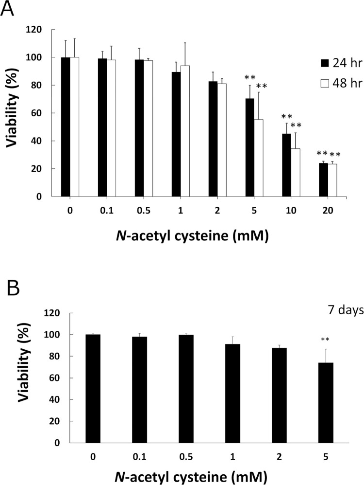
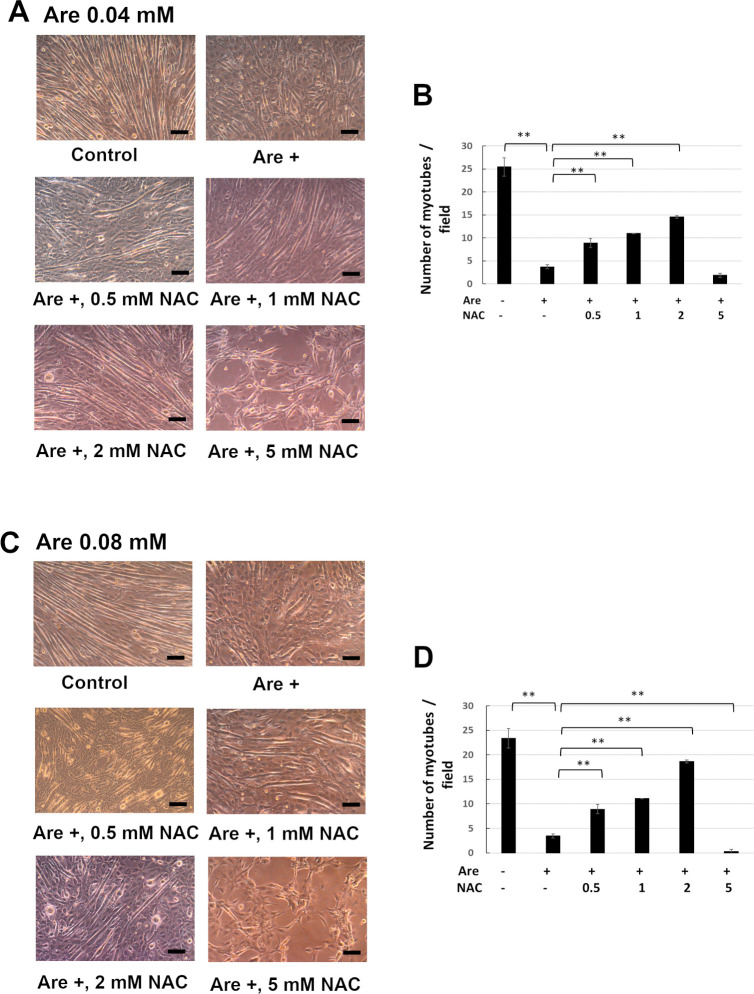
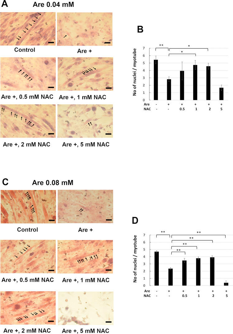
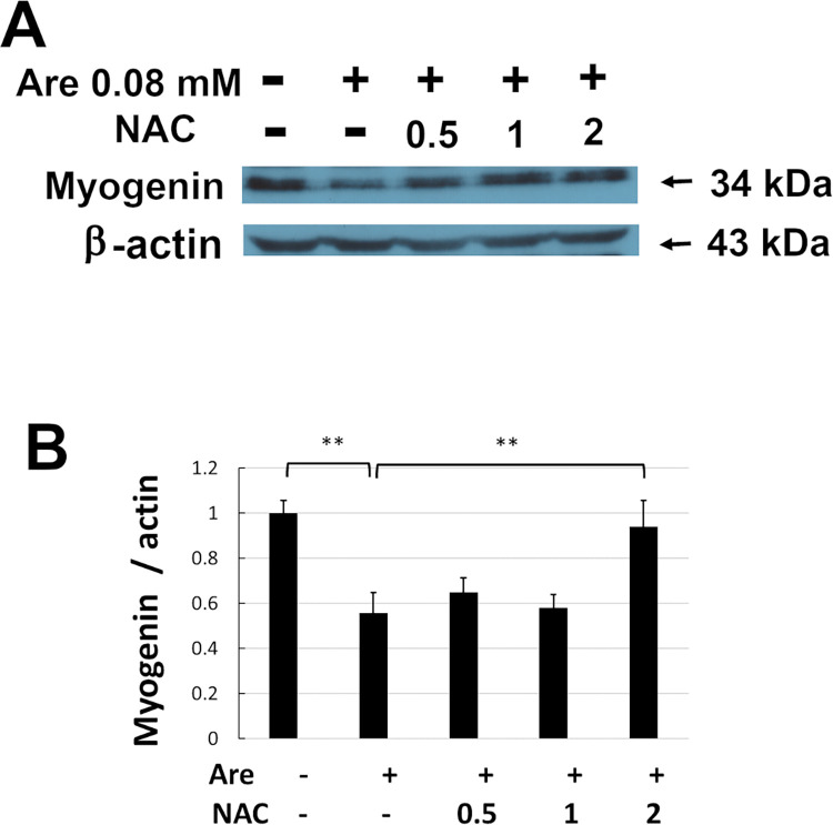
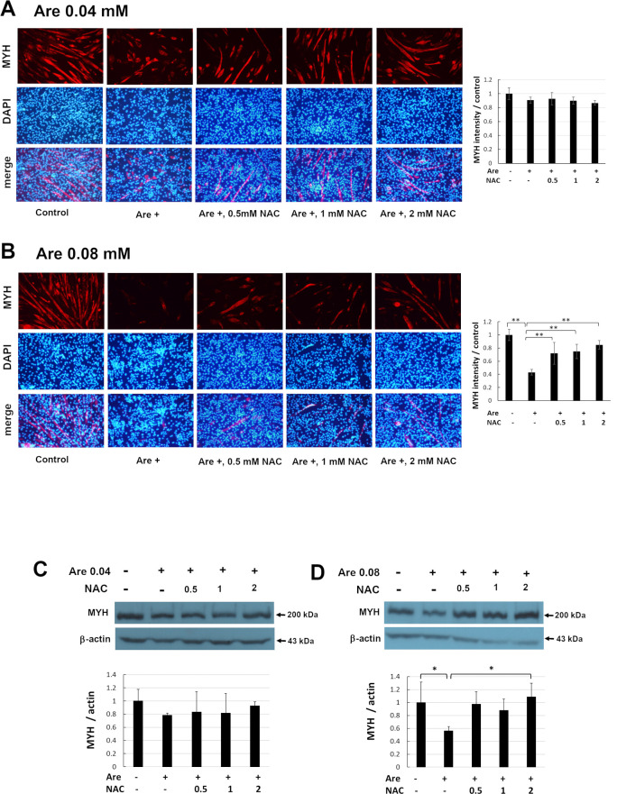
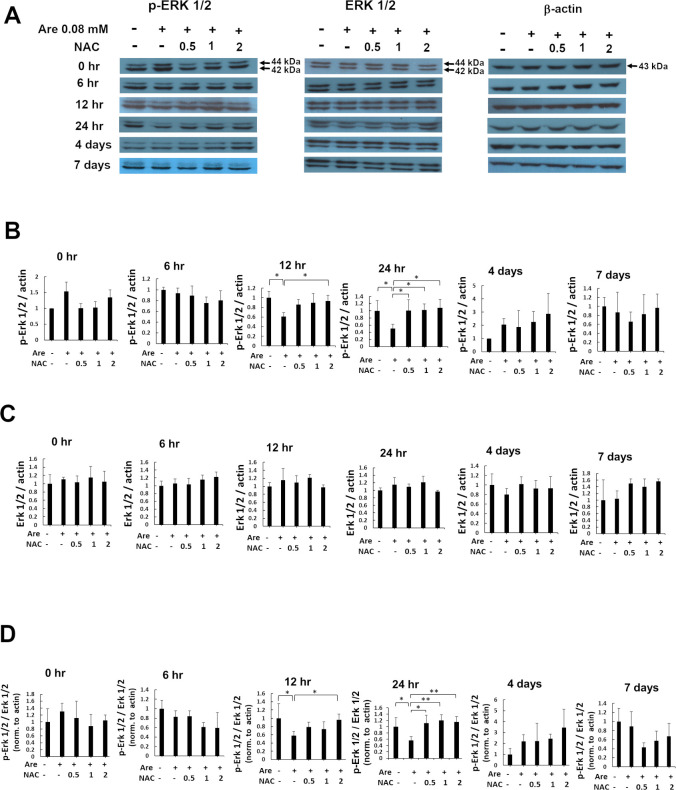
Arecoline is known to induce reactive oxygen species (ROS). Our previous studies showed that arecoline inhibited myogenic differentiation and acetylcholine receptor cluster formation of C2C12 myoblasts. N-acetyl-cysteine (NAC) is a known ROS scavenger. We hypothesize that NAC scavenges the excess ROS caused by arecoline. In this article we examined the effect of NAC on the inhibited myoblast differentiation by arecoline and related mechanisms. We found that NAC less than 2 mM is non-cytotoxic to C2C12 by viability analysis. We further demonstrated that NAC attenuated the decreased number of myotubes and nuclei in each myotube compared to arecoline treatment by H & E staining. We also showed that NAC prevented the decreased expression level of the myogenic markers, myogenin and MYH caused by arecoline, using immunocytochemistry and western blotting. Finally, we found that NAC restored the decreased expression level of p-ERK1/2 by arecoline. In conclusion, our results indicate that NAC attenuates the damage of the arecoline-inhibited C2C12 myoblast differentiation by the activation/phosphorylation of ERK. This is the first report to demonstrate that NAC has beneficial effects on skeletal muscle myogenesis through ERK1/2 upon arecoline treatment. Since defects of skeletal muscle associates with several diseases, NAC can be a potent drug candidate in diseases related to defects in skeletal muscle myogenesis.


Conflict of interest statement

The authors have declared that no competing interests exist.

Figures

Fig 1
Fig 1. Effect of NAC on cell proliferation in C2C12 myoblasts.
Cells were exposed to 0–20 mM arecoline for 24 h (solid bars) or 48 h (open bars) (A) and 7 days (B). The viability of cells was measured with CellTiter 96 Aqueous One Solution Reagent (Promega) in duplicate. The means of duplicate from three independent experiments were used for further analysis. The values of ANOVA at 24 h, 48 h and 7 days were (F7,16 = 56.44, P = 4.28E-10), (F7,16 = 26.97, P = 1.03E-07) and (F5,12 = 7.93, P = 1.66E-3), respectively (mean ± SD; **p < 0.01, as compared to a no-treatment control, Tukey HSD test).
Fig 2
Fig 2. The effects of NAC on the decreased number of myotube formations caused by arecoline.
C2C12 cells were cultured in differentiation medium with 0–5 mM NAC and 0.04 mM (A, B) or 0.08 mM (C, D) arecoline for 7 days. The myotubes were photographed by phase contrast microscopy (A, C). Scale bar, 100 μm. The numbers of myotubes from 30 random fields were counted (B, D). The values of ANOVA with 0.04 and 0.08 mM were (F5,12 = 241.90, P = 1.30E-11) and (F5,12 = 379.27, P = 8.99E-13) (mean ± SD; **p < 0.01, as compared to cells that were treated only with arecoline, Tukey HSD test).
Fig 3
Fig 3. Effect of NAC on the arecoline-inhibited multinucleated myotube formation.
C2C12 cells were cultured in differentiation medium with 0–5 mM NAC and 0.04 mM (A, B) or 0.08 mM (C, D) arecoline for 7 days and H&E staining. The myotubes were photographed by light microscopy (A, C). Arrows indicate the nuclei in the multinucleated myotubes. Scale bar, 100 μm. The numbers of nuclei per myotube from 30 random fields were shown (B, D). The values of ANOVA with 0.04 and 0.08 mM were (F5,12 = 14.51, P = 9.87E-05) and (F5,12 = 154.72, P = 1.83E-10). (mean ± SD; *p < 0.05; **p < 0.01, as compared to cells treated with arecoline only, Tukey HSD test).
Fig 4
Fig 4. Effect of NAC on decreased myogenin expression caused by arecoline in C2C12 cells.
The C2C12 cells were cultured in differentiation medium with 0–2 mM NAC and 0.08 mM arecoline for 7 days. (A) The expression of myogenin protein was detected by Western blotting. β-actin served as a loading control. (B) The band intensities of myogenin were quantified and normalized to the no-treatment control. Data from three independent experiments were used. The values of ANOVA were (F4,10 = 22.02, P = 6.05E-05)(mean ± SD; **p < 0.01 as compared to cells treated with arecoline only, Tukey HSD test).
Fig 5
Fig 5. Effect of NAC on decreased MYH expression caused by arecoline in C2C12 cells.
The C2C12 cells were cultured in differentiation medium with 0–2 mM NAC and 0.04 mM (A, C) or 0.08 mM (B, D) arecoline for 7 days. The distribution of MYH was examined by immunofluorescence under a fluorescence microscope (A and B, left, upper panel). Scale bar represents 100 μm at 100x magnification. Nuclei in the same field were counterstained with DAPI (A and B, left, middle panel). The images of MYH and DAPI were merged (A and B, left, lower panel). The intensities of MYH were quantified and normalized to the no-treatment control from random images of three independent experiments (Fig 5A and 5B, right panels). The values of ANOVA with 0.04 and 0.08 mM were (F4,10 = 2.73, P = 0.09) and (F4,10 = 14.49, P = 3.63E-04). The expression of MYH protein was detected by Western blotting (C and D, upper panel). β-actin served as a loading control. The band intensities of MYH from three independent experiments were quantified and normalized to no-treatment control (C and D, lower panel). The values of ANOVA with 0.04 and 0.08 mM were (F4,10 = 0.63, P = 0.65) and (F4,10 = 5.15, P = 0.02) (mean ± SD; *p < 0.05; **p < 0.01, as compared to cells treated only with arecoline, Tukey HSD test).
Fig 6
Fig 6. The effect of NAC on decreased ERK1/2 Tyr-204 phosphorylation caused by arecoline during myogenic differentiation in C2C12 cells.
(A) The expression of Tyr-204 phosphorylated ERK1/2, ERK1/2 and β-actin proteins were detected by Western blotting at 0, 6, 12, 24 h, 4 and 7 days of myogenic differentiation in C2C12 cells treated with 0.08 mM arecoline and 0–2 mM NAC. p44:ERK1, p42:ERK2. β-actin served as the loading control. (B) The relative expression of p-ERK1/2 protein to β-actin were quantified and normalized to the no-treatment control. The values of ANOVA at 12 and 24h were (F4,10 = 5.03, P = 0.02) and (F4,10 = 5.07, P = 0.02). (C) The relative expression of ERK1/2 protein to β-actin were quantified and normalized to the no-treatment control. (D) The ratio of p-ERK1/2 to ERK1/2 from figure B and C were relative to β-actin and normalized to the no-treatment control. The values of ANOVA at 12 and 24h were (F4,10 = 5.54, P = 0.01) and (F4,10 = 7.94, P = 3.78E-03). Data from three independent experiments were presented (mean ± SD; *p < 0.05; **p < 0.01, as compared to cells treated with arecoline, Tukey HSD test).

Similar articles

Cited by

References

    1. Kamei Y, Miura S, Suzuki M, Kai Y, Mizukami J, Taniguchi T, et al.. Skeletal muscle FOXO1 (FKHR) transgenic mice have less skeletal muscle mass, down-regulated Type I (slow twitch/red muscle) fiber genes, and impaired glycemic control. J Biol Chem, 2004. 279(39): p. 41114–23. doi: 10.1074/jbc.M400674200 . - DOI - PubMed
    1. Morgan J and Partridge T. Skeletal muscle in health and disease. Dis Model Mech, 2020. 13(2): p. dmm042192. doi: 10.1242/dmm.042192 ; PubMed Central PMCID: PMC7044447. - DOI - PMC - PubMed
    1. Truskey GA. Development and application of human skeletal muscle microphysiological systems. Lab Chip, 2018. 18(20): p. 3061–73. doi: 10.1039/c8lc00553b ; PubMed Central PMCID: PMC6177290. - DOI - PMC - PubMed
    1. Lehka L and Rędowicz MJ. Mechanisms regulating myoblast fusion: A multilevel interplay. Semin Cell Dev Biol, 2020. 104: p. 81–92. doi: 10.1016/j.semcdb.2020.02.004 . - DOI - PubMed
    1. Comai G and Tajbakhsh S. Molecular and cellular regulation of skeletal myogenesis. Curr Top Dev Biol, 2014. 110: p. 1–73. doi: 10.1016/B978-0-12-405943-6.00001-4 . - DOI - PubMed

Publication types