. 2022 Aug 19;11(8):2741-2755.
doi: 10.1021/acssynbio.2c00147. Epub 2022 Jul 28.

Building the Plant SynBio Toolbox through Combinatorial Analysis of DNA Regulatory Elements

Affiliations

Building the Plant SynBio Toolbox through Combinatorial Analysis of DNA Regulatory Elements

Alexander C Pfotenhauer et al. ACS Synth Biol. .

Abstract

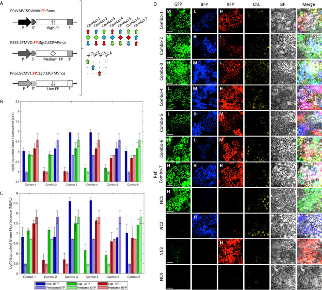
While the installation of complex genetic circuits in microorganisms is relatively routine, the synthetic biology toolbox is severely limited in plants. Of particular concern is the absence of combinatorial analysis of regulatory elements, the long design-build-test cycles associated with transgenic plant analysis, and a lack of naming standardization for cloning parts. Here, we use previously described plant regulatory elements to design, build, and test 91 transgene cassettes for relative expression strength. Constructs were transiently transfected into Nicotiana benthamiana leaves and expression of a fluorescent reporter was measured from plant canopies, leaves, and protoplasts isolated from transfected plants. As anticipated, a dynamic level of expression was achieved from the library, ranging from near undetectable for the weakest cassette to a ∼200-fold increase for the strongest. Analysis of expression levels in plant canopies, individual leaves, and protoplasts were correlated, indicating that any of the methods could be used to evaluate regulatory elements in plants. Through this effort, a well-curated 37-member part library of plant regulatory elements was characterized, providing the necessary data to standardize construct design for precision metabolic engineering in plants.

Keywords: flow cytometry; fluorometry; genetic regulatory elements; single-cell analysis; synthetic biology; transgene expression.


Conflict of interest statement

The authors declare no competing financial interest.

Figures

Figure 1
Figure 1
Comparative study of plant genetic regulatory elements. (A) Schematic representation of DNA constructs and regulatory elements used to modulate gene expression in plant cells. Indicated in the image are promoters (P), 5′ untranslated regions (5′UTR), 3′ untranslated regions (3′UTR), and coding sequences for fluorescent protein reporters (reporters). The nucleotide sequences for the regulatory elements and descriptions are provided in Table S1. (B) Schematic representation of the experimental approach used to test genetic elements in plant cells. The approach involves (1) canopy Agrobacterium tumefaciens-mediated infiltration of Nicotiana benthamiana with DNA constructs, (2) scanning fluorometric analysis of leaf tissue, (3) protoplast isolation from the same tissue, and (4) single cell analysis by flow cytometry.
Figure 2
Figure 2
Comparative analysis of promoters. Each construct contains a different promoter (Table S2), while keeping the 5′UTR (TMVΩ leader), reporter gene (GFP), and 3′UTR (35S CaMV polyA) consistent. Graphs represent scanning fluorometry and flow cytometry data obtained using intact leaf tissue and protoplasts isolated from the same tissue. Scanning fluorometry (excitation 475 nm, emission 509 nm) data is expressed as log10 of CPS (counts per second) values, while flow cytometry (excitation 488 nm, emission 510/10 bandpass filter) data is expressed as log10 of MEFL (molecules of equivalent fluorescein) values. Negative (plants transformed with untransformed Agrobacterium tumefaciens) and positive (plants transformed with the P2x35S:5TMVΩ:GFP:335S construct) controls are indicated with blue and red lines across the graphs, respectively. Data is represented as the mean ± standard deviation (SD) of at least three transformed plants per construct.
Figure 3
Figure 3
Comparative analysis of 3′UTRs. Each construct contains a different 3′UTR (Table S2), while keeping the promoter (CaMV 2x35S), 5′UTR (TMVΩ leader) and reporter gene (GFP) consistent. Graphs represent scanning fluorometry and flow cytometry data obtained using intact leaf tissue and protoplasts isolated from the same tissue. Scanning fluorometry (excitation 475 nm, emission 509 nm) data is expressed as log10 of CPS (counts per second) values, while flow cytometry (excitation 488 nm, emission 510/10 bandpass filter) data is expressed as log10 of MEFL (molecules of equivalent fluorescein) values. Negative (plants transformed with untransformed Agrobacterium tumefaciens) and positive (plants transformed with the P2x35S:5TMVΩ:GFP:335S construct) controls are indicated with blue and red lines across the graphs, respectively. Data is represented as the mean ± standard deviation (SD) of at least three transformed plants per construct.
Figure 4
Figure 4
Comparative analysis of promoter-5′UTR fusions in combination with 3′UTRs of different activities. Each construct contains a different promoter-5′UTR (Table S2). While the reporter gene (GFP) was kept consistent, two different 3′UTRs (nos 3′UTR and gctt3CPMV-nos fusion) were interchanged downstream from each promoter-5′UTR fusion. Graphs represent scanning fluorometry and flow cytometry data obtained using intact leaf tissue and protoplasts isolated from the same tissue. Scanning fluorometry (excitation 475 nm, emission 509 nm) data is expressed as log10 of CPS (counts per second) values, while flow cytometry (excitation 488 nm, emission 510/10 bandpass filter) data is expressed as log10 of MEFL (molecules of equivalent fluorescein) values. Negative (plants transformed with untransformed Agrobacterium tumefaciens) and positive (plants transformed with the P2x35S:5TMVΩ:GFP:335S construct) controls are indicated with blue and red lines across the graphs, respectively. Data is represented as the mean ± standard deviation (SD) of at least three transformed plants per construct.
Figure 5
Figure 5
Comparative analysis 5′UTRs in combination with promoters and 3′UTRs of different activities. Per promoter group (nos, 35S and 2x35S, in A-C, respectively), the library of 5′UTRs (Table S2) was tested along with two 3′UTRs (nos 3′UTR and gctt3CPMV-nos fusion) while keeping the reporter gene (GFP) consistent. The promoter nos was also tested with or without three 5′UTRs (TMVΩ, PVXΩ and 5SO) along with a 4 bp (ttcg) variant of 3CPMV-nos 3′UTR (ttcg3CPMV-nos). Graphs represent scanning fluoromety and flow cytometry data obtained using intact leaf tissue and protoplasts isolated from the same tissue. Scanning fluorometry (excitation 475 nm, emission 509 nm) data is expressed as log10 of CPS (counts per second) values, while flow cytometry (excitation 488 nm, emission 510/10 bandpass filter) data is expressed as log10 of MEFL (molecules of equivalent fluorescein) values. Negative (plants transformed with untransformed Agrobacterium tumefaciens) and positive (plants transformed with the P2x35S:5TMVΩ:GFP:335S construct) controls are indicated with blue and red lines across the graphs, respectively. Data is represented as the mean ± standard deviation (SD) of at least three transformed plants per construct.
Figure 6
Figure 6
Comparison of scanning fluorometry and flow cytometry data for all combinations of genetic regulatory elements. (A, B) Graphs represent scanning fluorometry and flow cytometry data obtained using intact leaf tissue and protoplasts isolated from the same tissue. Scanning fluorometry (excitation 475 nm, emission 509 nm) data is expressed as log10 of CPS (counts per second) values, while flow cytometry (excitation 488 nm, emission 510/10 bandpass filter) data is expressed as log10 of MEFL (molecules of equivalent fluorescein) values. Negative (plants transformed with untransformed Agrobacterium tumefaciens) and positive (plants transformed with the P2x35S:5TMVΩ:GFP:335S construct) controls are indicated with blue and red lines across the graphs, respectively. Data is represented as the mean ± standard deviation (SD) of at least three transformed plants per construct. (C) Graph showing the correlation between scanning fluorometry measurements and flow cytometry data. The R2 correlation value of ∼0.72 is shown in the graph.
Figure 7
Figure 7
Combining transgene cassettes to achieve predicted fluorescence levels within a three-gene pathway. (A) High (PCsVMV-5CsVMV:FP:3nos, H), medium (P35S:5TMVΩ:FP:3gctt3CPMVnos, M), and low (Pnos:5CMV1:FP:3gctt3CPMVnos, L) regulatory element cassettes (Table S2) were designed to control the expression of either GFP, BFP, or RFP reporters in all possible combinations. Combination 1 (Combo-1): H RFP, M GFP, and L BFP; combination 2 (Combo-2): H BFP, M GFP, and L RFP; combination 3 (Combo-3): H GFP, M BFP, and L RFP; combination 4 (Combo-4): H RFP, M BFP, and L GFP; combination 5 (Combo-5): H BFP, M RFP, and L GFP; combination 6 (Combo-6): H GFP, M RFP, and L BFP. (B, C) Graphs represent scanning fluorometry and flow cytometry data obtained using intact leaf tissue and protoplasts isolated from the same tissue. Scanning fluorometry (excitation 475, 550, and 400 nm, emission 509, 574, and 455 nm) data is expressed as log10 of CPS (counts per second) values, while flow cytometry (excitation 488, 561, and 405, emission 510/10, 585/16, and 440/50 bandpass filters) data is expressed as log10 of MEFL (molecules of equivalent fluorescein) values for GFP, RFP, and BFP respectively.. Negative (plants transformed with untransformed Agrobacterium tumefaciens) and positive (plants transformed with the P2x35S:5TMVΩ:GFP:335S construct) controls are indicated with blue and red lines across the graphs, respectively. Data is represented as the mean ± standard deviation (SD) of at least three transformed plants per construct. The fluorescence levels of combinations 1–6 are compared with predicted single cassette fluorescence levels (light green, blue, and red). (D) Confocal images showing the same epidermal N. benthamiana leaf cells transformed with the indicated construct combinations, (Combo-1–6); reference combination 7 (Combo-7): H GFP, H RFP, and H BFP; negative control 1 (NC1): H GFP only; negative control 2 (NC2): H BFP only; negative control 3 (NC3): H RFP only; and negative control 4 (NC4): leaves transformed with wild-type A. tumefaciens. Chlorophyll (Chl), bright-field (BF), and merged images are indicated. Scale bars = 50 μm.
Figure 8
Figure 8
Analysis of the activity of genetic modules in canopies by a fluorescence-inducing laser projector (FILP). (A) Graphs represent fluorometric data obtained using intact leaf tissue. Scanning fluorometry (excitation 475 nm, emission 509 nm) data is expressed as log10 of CPS (counts per second) values, while FILP (Fluorescence-Inducing Laser Projector) (excitation 465, 525 nm, emission 525, 680 nm filters) data is expressed as log10 of pixel intensity. Data of the graph correlating both methods is also shown (R2: ∼0.89). Data is represented as the mean ± standard deviation (SD) of at least three transformed plants per construct. Plants expressing single high (H1–3: PCsVMV-5CsVMV:GFP:3nos, P35S:5RbcS2B:GFP:3nos, and P35S:5PVX:GFP:3nos), medium (M1–3:P35S:5RbcS2B:GFP:3gctt3CPMVnos, PM24MMV:5AIMV:GFP:3nos, and PUBQ11-5UBQ11-link:GFP:3nos), and low (L1–3: P35S:5TMV:GFP:3gctt3CPMVnos, PH4:5H4:GFP:3nos, and Pnos:5CMV1:GFP:3gctt3CPMVnos) expression cassettes (Table S2) have been analyzed using the two methods. (B) FILP images showing N. benthamiana plants expressing high (H), medium (M), and low (L) expression cassettes along with wild-type controls (W). GFP (green), chlorophyll (red), bright-filed (gray), and merge images are shown. Scale bar: 10 cm.

Similar articles

Cited by

References

    1. Smith P.; Gregory P. J. Climate change and sustainable food production. Proc. Nutr. Soc. 2013, 72, 21–28. 10.1017/S0029665112002832. - DOI - PubMed
    1. Bailey-Serres J.; Parker J. E.; Ainsworth E. A.; Oldroyd G. E. D.; Schroeder J. I. Genetic strategies for improving crop yields. Nature 2019, 575, 109–118. 10.1038/s41586-019-1679-0. - DOI - PMC - PubMed
    1. Occhialini A.; Lin M. T.; Andralojc P. J.; Hanson M. R.; Parry M. A. Transgenic tobacco plants with improved cyanobacterial Rubisco expression but no extra assembly factors grow at near wild-type rates if provided with elevated CO2. Plant J. 2016, 85, 148–160. 10.1111/tpj.13098. - DOI - PMC - PubMed
    1. Fitch M. M. M.; Manshardt R. M.; Gonsalves D.; Slightom J. L.; Sanford J. C. Virus resistant papaya plants derived from tissues bombarded with the coat protein gene of papaya ringspot virus. Nat. Biotechnol. 1992, 10, 1466–1472. 10.1038/nbt1192-1466. - DOI
    1. Castiglioni P.; Warner D.; Bensen R. J.; Anstrom D. C.; Harrison J.; Stoecker M.; Abad M.; Kumar G.; Salvador S.; D’Ordine R.; Navarro S.; Back S.; Fernandes M.; Targolli J.; Dasgupta S.; Bonin C.; Luethy M. H.; Heard J. E. Bacterial RNA chaperones confer abiotic stress tolerance in plants and improved grain yield in maize under water-limited conditions. Plant Physiol. 2008, 147, 446–455. 10.1104/pp.108.118828. - DOI - PMC - PubMed

Publication types

MeSH terms