. 2022 Jul 28;18(7):e1010321.
doi: 10.1371/journal.pgen.1010321. eCollection 2022 Jul.

Repression by the H-NS/YmoA histone-like protein complex enables IscR dependent regulation of the Yersinia T3SS

Affiliations

Repression by the H-NS/YmoA histone-like protein complex enables IscR dependent regulation of the Yersinia T3SS

David Balderas et al. PLoS Genet. .

Abstract

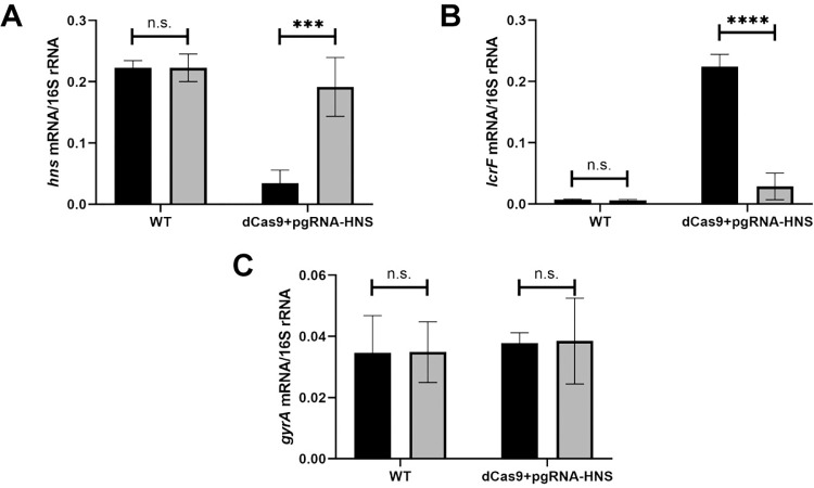
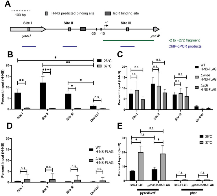
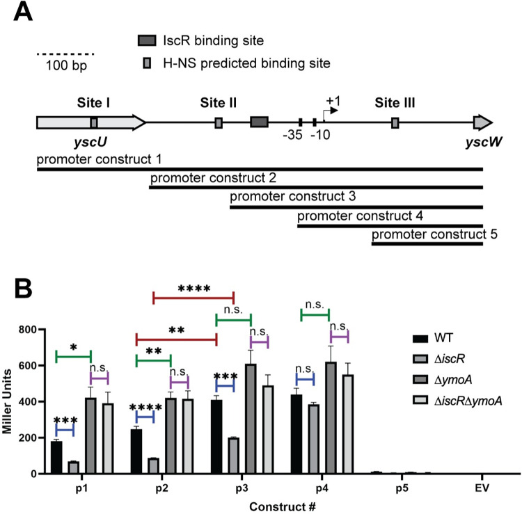
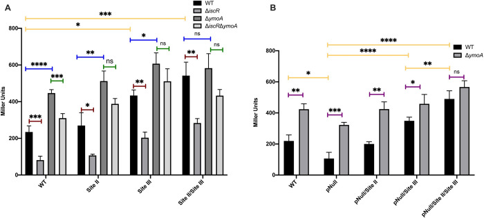
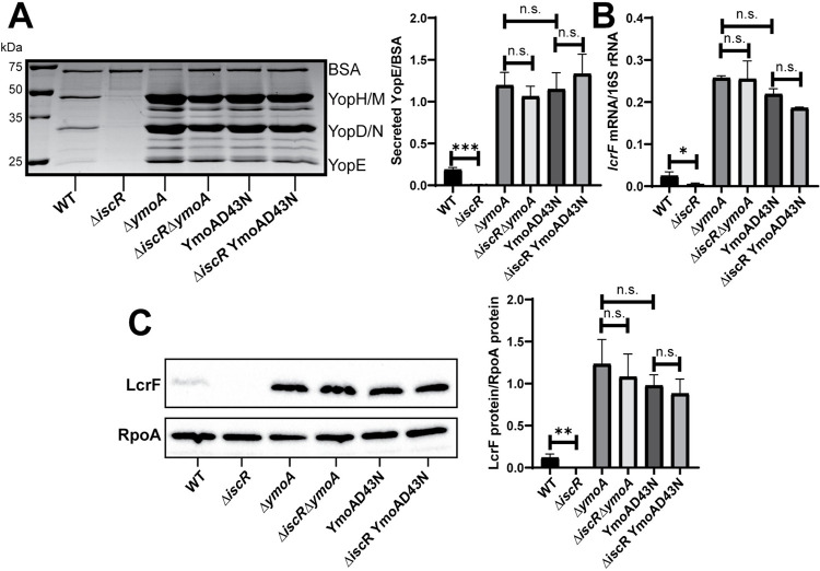
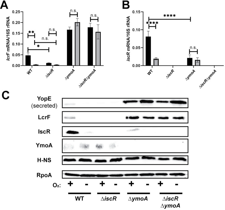
The type III secretion system (T3SS) is an appendage used by many bacterial pathogens, such as pathogenic Yersinia, to subvert host defenses. However, because the T3SS is energetically costly and immunogenic, it must be tightly regulated in response to environmental cues to enable survival in the host. Here we show that expression of the Yersinia Ysc T3SS master regulator, LcrF, is orchestrated by the opposing activities of the repressive H-NS/YmoA histone-like protein complex and induction by the iron and oxygen-regulated IscR transcription factor. While deletion of iscR or ymoA has been shown to decrease and increase LcrF expression and type III secretion, respectively, the role of H-NS in this system has not been definitively established because hns is an essential gene in Yersinia. Using CRISPRi knockdown of hns, we show that hns depletion causes derepression of lcrF. Furthermore, we find that while YmoA is dispensable for H-NS binding to the lcrF promoter, YmoA binding to H-NS is important for H-NS repressive activity. We bioinformatically identified three H-NS binding regions within the lcrF promoter and demonstrate binding of H-NS to these sites in vivo using chromatin immunoprecipitation. Using promoter truncation and binding site mutation analysis, we show that two of these H-NS binding regions are important for H-NS/YmoA-mediated repression of the lcrF promoter. Surprisingly, we find that IscR is dispensable for lcrF transcription in the absence of H-NS/YmoA. Indeed, IscR-dependent regulation of LcrF and type III secretion in response to changes in oxygen, such as those Yersinia is predicted to experience during host infection, only occurs in the presence of an H-NS/YmoA complex. These data suggest that, in the presence of host tissue cues that drive sufficient IscR expression, IscR can act as a roadblock to H-NS/YmoA-dependent repression of RNA polymerase at the lcrF promoter to turn on T3SS expression.


Conflict of interest statement

The authors have declared that no competing interests exist.

Figures

Fig 1
Fig 1. Knockdown of H-NS leads to derepression of LcrF.
Y. pseudotuberculosis strains were grown aerobically in low calcium LB in the absence (grey bars) or presence (black bars) of anhydrotetracycline for 3 hrs at 26°C to induce expression of hns guide RNA and dCas9 and then transferred to 37°C (T3SS inducing conditions) for 1.5 hrs. RNA was analyzed by RT-qPCR for hns (A), lcrF (B), or gyrA (C) mRNA level normalized to 16S rRNA. The average of three biological replicates are shown ± standard deviation. Statistical analysis was performed using an unpaired Student’s t-test (***p < .001, ****p < .0001, and n.s. non-significant).
Fig 2
Fig 2. H-NS and IscR enrichment at the yscW-lcrF promoter.
(A) FIMO-MEME suite tools were used to identify H-NS binding sites in the yscW-lcrF promoter. Shown are the three putative sites identified (p<10−3), referred to as Sites I, II, and III. The previously characterized transcriptional start site (TSS) is shown by the arrow [16], and the known IscR binding site is shown by the grey rectangle [39]. The -2 to +272 DNA fragment (relative to the +1 TSS) previously shown to bind H-NS in vitro [16] is shown, as well as the qPCR products detected following H-NS chromatin immunoprecipitation. (B) The relative enrichment (percent input) of Site I, Site II, and Site III promoter DNA and a negative control promoter (DN756_21750) was analyzed by anti-FLAG ChIP-qPCR in Yersinia expressing H-NS-FLAG. ChIP-qPCR was performed with bacteria grown aerobically at 26°C (black bars) or 37°C (grey bars) in low calcium LB for 3 hrs. (C) ChIP-qPCR was performed with the H-NS-FLAG allele in WT, ΔymoA, or ΔiscR mutant background at 26°C (D) ChIP-qPCR was performed with the H-NS-FLAG allele in WT or ΔiscR mutant background at 37°C. (E) ChIP-qPCR was performed with the IscR-FLAG allele in the WT or ΔymoA mutant background at 26°C (black bars) or 37°C (grey bars). The hpt control promoter, which IscR is not predicted to bind, was used as a negative control. The average of at least three biological replicates ± standard deviation is shown and statistical analysis was performed using Two-way ANOVA (*p < .05, **p < .01, ***p < .001, ****p < .0001 and n.s. non-significant).
Fig 3
Fig 3. The identified H-NS binding Sites II and III are important for regulation of yscW-lcrF promoter activity.
(A) Schematic of PyscW-lcrF::lacZ fusions. Five constructs (p1-p5) were used to assess which regions of pyscW-lcrF allows for H-NS-YmoA repression and IscR activation. (B) Yersinia harboring the various pyscW-lcrF::lacZ plasmids were grown aerobically under T3SS-inducing conditions (low calcium LB at 37°C) for 1.5 hrs and assayed for β-galactosidase (Miller units). The average of at least three biological replicates are shown ± standard deviation. Statistical analysis was performed using an unpaired Student’s t-test (*p < .05, **p < .01, ***p < .001, ****p < .0001, and n.s. non-significant).
Fig 4
Fig 4. The AT-rich Sites II and III bound by H-NS are necessary for YmoA-dependent repression as well as IscR potentiation of yscW-lcrF promoter activity.
(A) The pyscW-lcrF::lacZ reporter 2 construct from Fig 3 was used as a template to generate pyscW-lcrF::lacZ promoter fusions with H-NS Site II and III mutations, and the mutated promoters introduced into the WT, ΔiscR, ΔymoA, and ΔiscRymoA Y. pseudotuberculosis genetic backgrounds. (B) Additionally, pyscW-lcrF::lacZ reporter 2 constructs carrying WT or mutated Site II and/or III were further mutated for the IscR binding site (pNull) and introduced into WT and ΔymoA Y. pseudotuberculosis. All these strains were grown aerobically under T3SS-inducing conditions (low calcium LB at 37°C) for 1.5 hrs and assayed for β-galactosidase (Miller units). The different promoter constructs are listed on the x-axis. The average of at least three biological replicates are shown ± standard deviation. Statistical analysis was performed using (A-B) a one-way ANOVA with Bonferroni’s multiple comparisons test on either (yellow bars) all WT genetic backgrounds carrying different reporter constructs or (blue, green, or red bars) each individual reporter construct expressed in WT, ΔiscR, ΔymoA, or ΔiscRymoA; or (B) an unpaired t test (purple bars). (*p < .05, **p < .01, ***p < .001, ****p < .0001, and n.s. non-significant).
Fig 5
Fig 5. IscR is dispensable for type III secretion in the ΔymoA mutant background.
Yersinia strains were grown aerobically under T3SS-inducing conditions (low calcium at 37°C). (A) Precipitated secreted proteins were visualized by SDS-PAGE followed by Coomassie blue staining. Bovine serum albumin (BSA) was used as a loading control (left panel). Densitometry was used to measure the relative amount of secreted YopE T3SS effector protein versus BSA control. The average of four independent replicates ± standard deviation is shown (right panel). (B) RNA was extracted and RT-qPCR was used to measure relative levels of lcrF mRNA normalized to 16S rRNA. The average of at least three biological replicates are shown ± standard deviation. (C) LcrF protein levels were determined by Western blotting (left panel) and densitometry (right panel) relative to the RpoA loading control. Shown is the average of four independent replicates ± standard deviation. Statistical analysis was performed using an unpaired Student’s t-test (*p < .05, **p < .01, ***p < .001, and n.s. non-significant).
Fig 6
Fig 6. Oxygen-dependent control of lcrF requires YmoA.
Yersinia strains were cultured under T3SS inducing conditions under aerobic (black bars) or anaerobic (grey bars) conditions. Levels of lcrF (A) and iscR (B) mRNA levels were measured by RT-qPCR and normalized to 16S rRNA. The average of at three biological replicates are shown ± standard deviation. (C) Yersinia strains were grown under similar conditions as stated above and whole cell extracts were probed for RpoA, IscR, H-NS, LcrF, YopE, and YmoA by Western blotting. One representative experiment out of three biological replicates is shown. Statistical analysis was performed using a one-way ANOVA with Tukey multiple comparisons (*p < .05,**p < .01,****p < .0001, and n.s. non-significant).

Similar articles

Cited by

References

    1. Deng W, Marshall NC, Rowland JL, McCoy JM, Worrall LJ, Santos AS, et al.. Assembly, structure, function and regulation of type III secretion systems. Nature Reviews Microbiology. 2017. doi: 10.1038/nrmicro.2017.20 - DOI - PubMed
    1. Cornelis GR. The type III secretion injectisome. Nature Reviews Microbiology. 2006. doi: 10.1038/nrmicro1526 - DOI - PubMed
    1. Viboud GI, Bliska JB. Yersinia outer proteins: Role in modulation of host cell signaling responses and pathogenesis. Annual Review of Microbiology. 2005. doi: 10.1146/annurev.micro.59.030804.121320 - DOI - PubMed
    1. Navarro L, Alto NM, Dixon JE. Functions of the Yersinia effector proteins in inhibiting host immune responses. Current Opinion in Microbiology. 2005. doi: 10.1016/j.mib.2004.12.014 - DOI - PubMed
    1. Schubert KA, Xu Y, Shao F, Auerbuch V. The Yersinia Type III Secretion System as a Tool for Studying Cytosolic Innate Immune Surveillance. Annual Review of Microbiology. 2020. doi: 10.1146/annurev-micro-020518-120221 - DOI - PubMed

Publication types

MeSH terms