. 2022 Jul 28;18(7):e1010739.
doi: 10.1371/journal.ppat.1010739. eCollection 2022 Jul.

Constrained evolution of overlapping genes in viral host adaptation: Acquisition of glycosylation motifs in hepadnaviral precore/core genes

Affiliations

Constrained evolution of overlapping genes in viral host adaptation: Acquisition of glycosylation motifs in hepadnaviral precore/core genes

Xupeng Hong et al. PLoS Pathog. .

Abstract

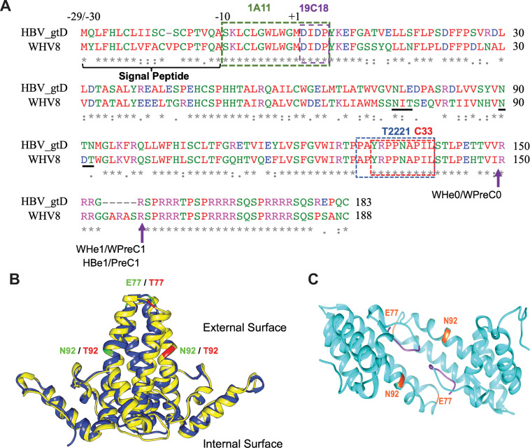
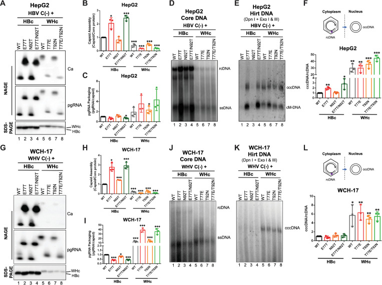
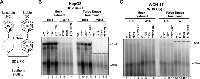
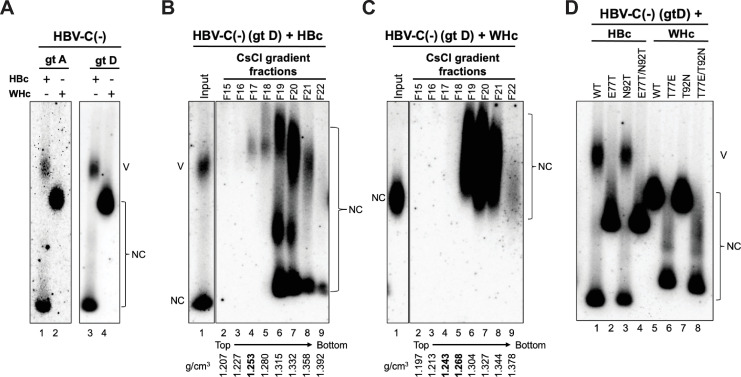
Hepadnaviruses use extensively overlapping genes to expand their coding capacity, especially the precore/core genes encode the precore and core proteins with mostly identical sequences but distinct functions. The precore protein of the woodchuck hepatitis virus (WHV) is N-glycosylated, in contrast to the precore of the human hepatitis B virus (HBV) that lacks N-glycosylation. To explore the roles of the N-linked glycosylation sites in precore and core functions, we substituted T77 and T92 in the WHV precore/core N-glycosylation motifs (75NIT77 and 90NDT92) with the corresponding HBV residues (E77 and N92) to eliminate the sequons. Conversely, these N-glycosylation sequons were introduced into the HBV precore/core gene by E77T and N92T substitutions. We found that N-glycosylation increased the levels of secreted precore gene products from both HBV and WHV. However, the HBV core (HBc) protein carrying the E77T substitution was defective in supporting virion secretion, and during infection, the HBc E77T and N92T substitutions impaired the formation of the covalently closed circular DNA (cccDNA), the critical viral DNA molecule responsible for establishing and maintaining infection. In cross-species complementation assays, both HBc and WHV core (WHc) proteins supported all steps of intracellular replication of the heterologous virus while WHc, with or without the N-glycosylation sequons, failed to interact with HBV envelope proteins for virion secretion. Interestingly, WHc supported more efficiently intracellular cccDNA amplification than HBc in the context of either HBV or WHV. These findings reveal novel determinants of precore secretion and core functions and illustrate strong constraints during viral host adaptation resulting from their compact genome and extensive use of overlapping genes.


Conflict of interest statement

The authors have declared that no competing interests exist.

Figures

Fig 1
Fig 1. WHV, but not HBV, precore/core gene products are N-glycosylated.
(A) Amino acid sequence alignment of HBV (genotype D) and WHV (strain 8) precore/core gene products. The epitopes for mAb 1A11, 19C18, T2221, and C33 are indicated by the dashed boxes. The two putative N-glycosylation sequons 75NIT77 and 90NDT92 in the WHV precore/core gene are underlined. The arrows indicate the putative cleavage sites in the production of WHeAg (WHe0 and WHe1), WHV PreC protein (WPreC0 and WPreC1), and HBeAg and HBV PreC (HBe1 and PreC1) as identified in a previous study [28]. (B) Alignment of the HBc (Protein Data Bank [PDB] accession code 1QGT, blue) and WHc (PDB accession code 6EDJ, yellow) dimer revealed that the two core proteins share highly structural similarity. The T = 4 icosahedral HBV or WHV capsid contains 120 copies of HBc or WHc dimers. Residues E77 and N92 on HBc (in green) and T77 and T92 on WHc (in red) located on the exterior surface of the capsid, which were mutated reciprocally, are highlighted. (C) Structure of HBeAg (PDB accession code 3V6Z). The N-terminal extensions are labeled in purple, and the two residues, E77 and N92, which are located on the protein surface, are highlighted.
Fig 2
Fig 2. Precore N-glycosylation enhanced the secretion of precore gene products.
Immunoblot analysis of (A) WHV and (B) HBV precore gene products in the culture supernatant of Huh7 cells transfected with WHV or HBV precore constructs. The supernatants were concentrated by ultrafiltration and treated with PNGase F or not and resolved by regular SDS-PAGE, followed by sequential immunoblotting with mAb 1A11 and 19C18 on the same membrane. WHV core-transfected cell culture supernatant served as the control for the background bands. WHe1, WHeAg that has the putative cleavage site on the 159th residue at CTD (Fig 1A) as identified previously [28]; mono-gWHe1, mono-glycosylated WHe1; di-gWHe1, doubly-glycosylated WHe1; WHc, WHV core protein; HBe1, HBeAg that has the putative cleavage site on the 154th residue at CTD (Fig 1A); mono-gHBe1, mono-glycosylated HBe1; di-gHBe1, doubly-glycosylated HBe1; PreC1, PreC antigen as previously characterized (Fig 1A); mono-gPreC1, mono-glycosylated PreC1; di-gPreC1, doubly-glycosylated PreC1. *, cross-reactive background bands.
Fig 3
Fig 3. Effects of eliminating N-glycosylation sequons on WHc functions.
The WHV replicon construct expressing the T77E, T92N, or T77E/T92N WHc mutants, or WT WHc was transfected into HpeG2 or WCH-17 cells. (A) The assembled capsids (top) and packaged pgRNA (middle) were detected by the C33 anti-HBc/WHc mAb and anti-sense WHV RNA probe, respectively, following the resolution of cytoplasmic lysates from the transfected HepG2 cells by native agarose gel electrophoresis (NAGE) and transfer to nitrocellulose membrane. Levels of WHc proteins (bottom) were measured by western blot using 19C18 anti-HBc/WHc mAb after SDS-PAGE. Capsid assembly efficiency (B) was determined by normalizing the levels of capsids to those of total WHc protein, and pgRNA packaging efficiency (C) was determined by normalizing the levels of pgRNA to those of capsids, with the efficiencies of WT WHc set to 1.0. (D)-(F) Cytoplasmic lysates from WHV replicon transfected WCH-17 cells were analyzed for capsid assembly and pgRNA packaging, as described for HepG2 cells. (G) WHV core DNA was released from the NCs of cytoplasmic lysate by SDS-proteinase K treatment and detected by Southern blot analysis. WHV PF-DNA was extracted from the transfected cells by the Hirt extraction method. The extracted DNA was treated with Dpn I plus the exonucleases I and III (Exo I & III) to remove all DNA with free 3’ ends. The ssDNA synthesis efficiency (H) was determined by normalizing the levels of ssDNA to those of pgRNA in (A), and the cccDNA formation efficiency (I) was determined by normalizing the levels of cccDNA to those of rcDNA in (G), with the efficiencies of WT WHc set to 1.0. Similarly, core DNA and PF-DNA from WHV replicon transfected WCH-17 cells were analyzed (J)-(L). Data is shown as mean ± SD. Two-tailed unpaired Student’s t test was used to compare the difference of each dataset versus WT WHc (*, p < 0.05; **, p <0.01; ***, p < 0.001). Ca, capsid; pgRNA, pregenomic RNA; WHc, WHV core protein; ssDNA, single-strand DNA; rcDNA, relaxed circular DNA; cccDNA, covalently closed circular DNA; cM-DNA, closed minus strand DNA.
Fig 4
Fig 4. Effects of introducing N-glycosylation sequons on HBc functions.
The HBV replicon construct expressing the E77T, N92T, or E77T/N92T mutant HBc, or WT HBc, or L- that is defective in L-HBs expression was transfected into HepG2 cells. (A) The assembled capsids (top) and packaged pgRNA (middle) were detected by the T2221 anti-HBV precore/core NTD mAb and anti-sense HBV RNA probe, respectively, following the resolution of cytoplasmic lysates by NAGE and transfer to nitrocellulose membrane. Levels of HBc proteins (bottom) were measured by western blot using T2221 mAb after SDS-PAGE. Quantitative results of capsid assembly efficiency (B) and pgRNA packaging efficiency (C) were obtained as described in Fig 3 and are shown. (D) HBV core DNA was released from the NCs of cytoplasmic lysate by SDS-proteinase K treatment and detected by Southern blot analysis. The ssDNA synthesis efficiency (E) was determined by normalizing the levels of ssDNA to those of pgRNA in (A), and the rcDNA synthesis efficiency (F) was determined by normalizing the levels of rcDNA to those of ssDNA in (D), with the efficiencies of WT HBc set to 1.0. (G) HBV PF-DNA was extracted from the transfected cells by the Hirt extraction method. The extracted DNA was treated with Dpn I (left) or Dpn I plus Exo I & III (right) to remove all DNA with free 3’ ends. The PF-rcDNA synthesis efficiency (H) was determined by normalizing the levels of PF-rcDNA to those of rcDNA in (D), and cccDNA formation efficiency (I) was determined by normalizing the levels of cccDNA to those of rcDNA in (D), with the efficiencies of WT HBc set to 1.0. (J) Culture supernatant from HBV replicon transfected HepG2 cells was harvested at day 5 post transfection, concentrated (50X), resolved by agarose gel electrophoresis and transferred to nitrocellulose membrane. HBV DNA, capsids in virions, and envelope proteins in virions and subviral particles were detected sequentially using a 32P-labeled HBV DNA probe, anti-HBV precore/core NTD mAb T2221, and anti-HBs polyclonal antibody, respectively, on the same membrane. Data is shown as mean ± SD. Two-tailed unpaired Student’s t test was used to compare the difference of each dataset versus WT HBc (*, p < 0.05; **, p <0.01; ***, p < 0.001). Ca, capsid; pgRNA, pregenomic RNA; HBc, HBV core protein; ssDNA, single-strand DNA; rcDNA, relaxed circular DNA; cccDNA, covalently closed circular DNA; cM-DNA, closed minus strand DNA; Ca, capsid; V, virion; HBs, HBV surface antigen.
Fig 5
Fig 5. Effects of introducing N-glycosylation sequons on de novo HBV infection.
HBV inocula were prepared from HBV replicon transfected Huh7 cells. (A) Infection of WT, E77T, N92T, and E77T/N92T mutant virus (MOI = 200 GE/cell) in HepG2-huNTCP cells. PF-DNA was extracted from infected cells at 4 dpi by the Hirt extraction method and analyzed by Southern blot analysis, without (lanes 1–4) or with pretreatment by Exo I & III (lanes 5–8). (B) Infection of N92T mutant virus with increased dosage (MOI = 200, 400, 800, or 1600 GE/cell) in HepG2-huNTCP cells. PF-DNA was extracted from infected cells at 4 dpi and analyzed by Southern blot analysis as described above. PF-rcDNA, protein-free rcDNA; cccDNA, covalently closed circular DNA.
Fig 6
Fig 6. Effects of WT and mutant HBc or WHc on supporting the replication of HBV and WHV.
The HBV replicon construct that is defective in HBc expression was co-transfected with WT and mutant HBc or WHc expression constructs in HepG2 cells. (A) The assembled capsids (top) and packaged pgRNA (middle) were detected by the C33 anti-HBc/WHc mAb and anti-sense HBV RNA probe, respectively, following the resolution of cytoplasmic lysates by NAGE and transfer to nitrocellulose membrane. Levels of HBc or WHc proteins (bottom) were measured by western blot using 19C18 anti-HBc/WHc mAb after SDS-PAGE. Quantitative results of capsid assembly efficiency (B) and pgRNA packaging efficiency (C) were obtained as described in Fig 4 and are shown. (D) core DNA was released from the NCs of cytoplasmic lysate by SDS-proteinase K treatment and detected by Southern blot analysis. (E) PF-DNA was extracted by the Hirt extraction method. The extracted DNA was treated with DpnI plus Exo I & III to remove all DNA with free 3’ ends. The cccDNA formation efficiency (F) was determined by normalizing the levels of cccDNA to those of rcDNA, with the efficiency from WT HBc set to 1.0. Similarly, a WHV replicon construct that is defective in WHc expression was co-transfected with WT and mutant HBc or WHc expression constructs in WCH-17 cells. (G) The assembled capsids (top) and packaged pgRNA (middle) were detected by the C33 anti-HBc/WHc mAb and anti-sense WHV RNA probe, respectively. Levels of HBc or WHc proteins (bottom) were measured by western blot using 19C18 anti-HBc/WHc mAb after SDS-PAGE. Capsid assembly efficiency (H) and pgRNA packaging efficiency (I) were determined as described above and are shown. (J) core DNA and (K) PF-DNA from the transfected cells was analyzed by Southern blot analysis. (L) cccDNA formation efficiency of WHV was determined by normalizing the levels of cccDNA to those of rcDNA, with the efficiency from WT HBc set to 1.0. Data is shown as mean ± SD. Two-tailed unpaired Student’s t test was used to compare the difference of each dataset versus WT HBc (*, p < 0.05; **, p <0.01; ***, p < 0.001). Ca, capsid; pgRNA, pregenomic RNA; HBc, HBV core protein; WHc, WHV core protein; ssDNA, single-strand DNA; rcDNA, relaxed circular DNA; cccDNA, covalently closed circular DNA; cM-DNA, closed minus strand DNA.
Fig 7
Fig 7. Effects of HBc or WHc on protecting the rcDNA content inside NCs.
(A) schematic diagram of nuclease treatment of HBV or WHV NCs. Turbo DNase was used to degrade viral DNA inside unstable NCs. Following inactivation of the nuclease, NC-protected DNA was isolated and detected by Southern blot analysis. Core DNA released from the cytoplasmic lysate of WT and mutant HBc or WHc co-transfected with HBV-C(-) from HepG2 cells (B) or with WHV-C(-) from WCH-17 cells (C), with or without prior Turbo DNase digestion, was detected by Southern blot analysis. The rcDNA signals obtained after Turbo DNase treatment is indicated by the dashed, red box for comparison to rcDNA signals present without nuclease treatment. ssDNA, single strand DNA; rcDNA, relaxed circular DNA.
Fig 8
Fig 8. Effects of HBc or WHc on supporting HBV virion secretion.
(A) concentrated culture supernatant of HepG2 cells co-transfected with WT HBc or WHc and HBV-C(-) (genotype A or D) was analyzed by agarose gel electrophoresis. HBV DNA in virions or naked capsids was detected by a HBV DNA probe. Concentrated culture supernatant of HepG2 cells co-transfected with WT HBc or WHc along with HBV-C(-) (genotype D) was fractionated by CsCl density gradient ultracentrifugation. The fractions from WT HBc (B) or WT WHc (C) co-transfected culture supernatant was analyzed by agarose gel electrophoresis. HBV DNA in virions or naked capsids was detected by a HBV DNA probe. The density profile of each fraction is indicated at the bottom. The density of HBV virions (1.250 g/cm3) is highlighted in bold. (D) concentrated cell culture supernatant of HepG2 cells co-transfected with HBV-C(-) (genotype D) and WT or mutant HBc or WHc was analyzed by NAGE and DNA in virions or naked capsids was detected by a HBV DNA probe. NC, nucleocapsid; V, virion.
Fig 9
Fig 9. Summary of the roles of N-glycosylation motifs in precore/core genes on HBV or WHV precore and core functions and their implication in hepadnaviral evolution.
(A) The N-glycosylation on the WHV or HBV precore proteins increased the secretion of e and PreC antigens (eAg and PreC). The E77T mutant that introduced the N-glycosylation sequon in HBc decreased HBV virion secretion and cccDNA formation (i.e., infectivity) during infection (blue box), while HBc-E77T increased cccDNA formation via the intracellular amplification pathway (purple box). The N92T mutant that introduced another N-glycosylation sequon on HBc showed no cccDNA formation during infection (blue box). When a HBV replicon that is defective in HBc expression was complemented with WT or mutant WHc, no HBV virion secretion was detected. However, WHc increased cccDNA formation via the intracellular amplification pathway (purple box) due to the decreased stability of mature NCs. (B) Proposed model of acquisition of N-glycosylation motifs in the WHV precore/core genes but not in HBV precore/core genes during the evolution. See text for details.

Similar articles

Cited by

References

    1. Magnius L, Mason WS, Taylor J, Kann M, Glebe D, Dény P, et al.. ICTV Virus Taxonomy Profile: Hepadnaviridae. Journal of General Virology. 2020:jgv001415. doi: 10.1099/jgv.0.001415 - DOI - PMC - PubMed
    1. Hu J, Seeger C. Hepadnavirus Genome Replication and Persistence. Cold Spring Harb Perspect Med. 2015;5(7):a021386. Epub 2015/07/03. doi: 10.1101/cshperspect.a021386 ; PubMed Central PMCID: PMC4484952. - DOI - PMC - PubMed
    1. WHO. Global hepatitis report 2017: World Health Organization; 2017.
    1. Summers J, Smolec JM, Snyder R. A virus similar to human hepatitis B virus associated with hepatitis and hepatoma in woodchucks. Proceedings of the National Academy of Sciences. 1978;75(9):4533–7. PubMed Central PMCID: PMC336150. doi: 10.1073/pnas.75.9.4533 - DOI - PMC - PubMed
    1. Menne S, Cote PJ. The woodchuck as an animal model for pathogenesis and therapy of chronic hepatitis B virus infection. World J Gastroenterol. 2007;13(1):104–24. Epub 2007/01/09. doi: 10.3748/wjg.v13.i1.104 ; PubMed Central PMCID: PMC4065868. - DOI - PMC - PubMed

Publication types