Review
. 2023 Jan 1;103(1):717-785.
doi: 10.1152/physrev.00044.2021. Epub 2022 Jul 28.

Protease-activated receptors in health and disease

Affiliations
Review

Protease-activated receptors in health and disease

Chloe J Peach et al. Physiol Rev. .

Abstract

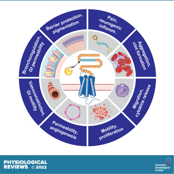
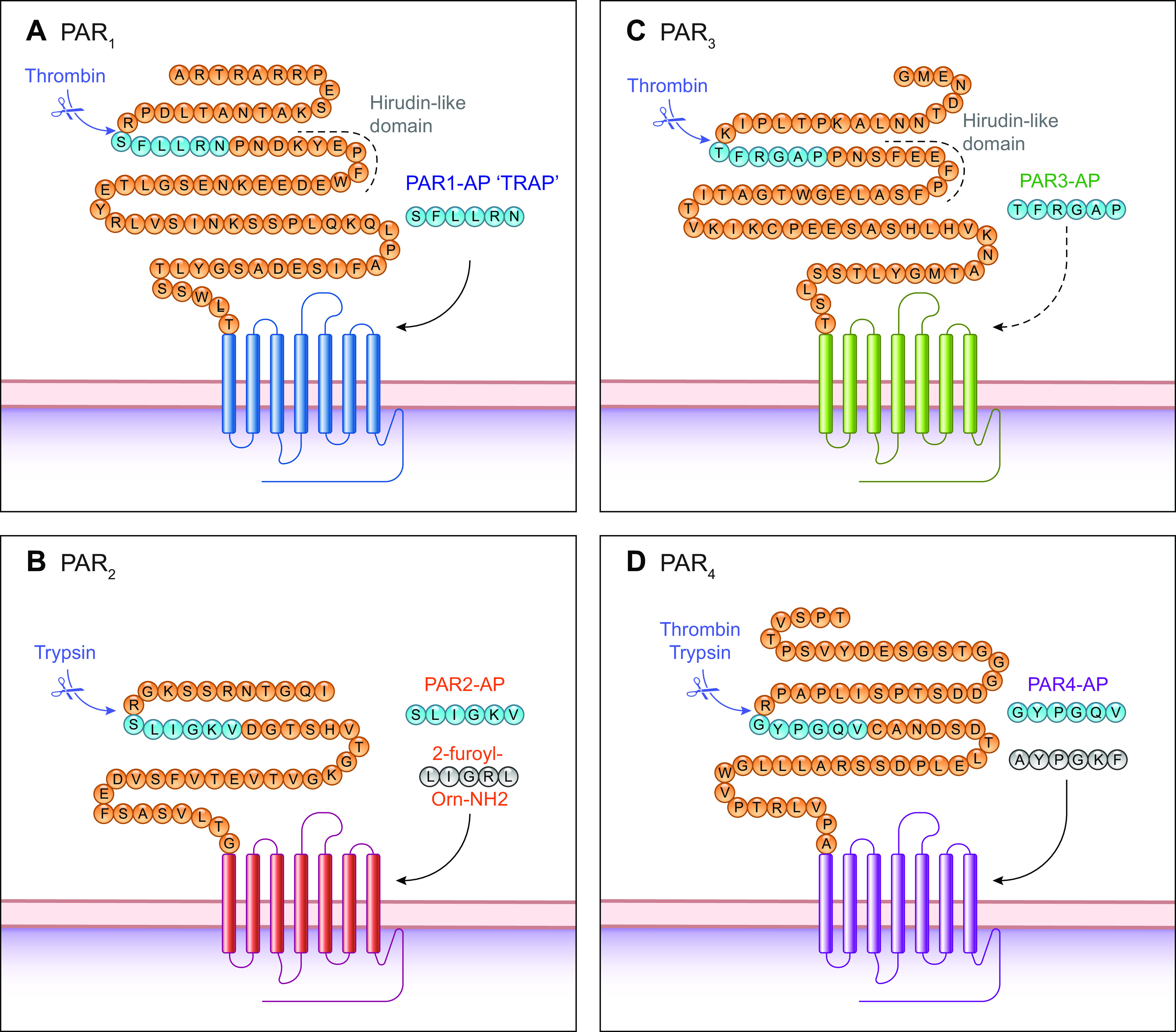
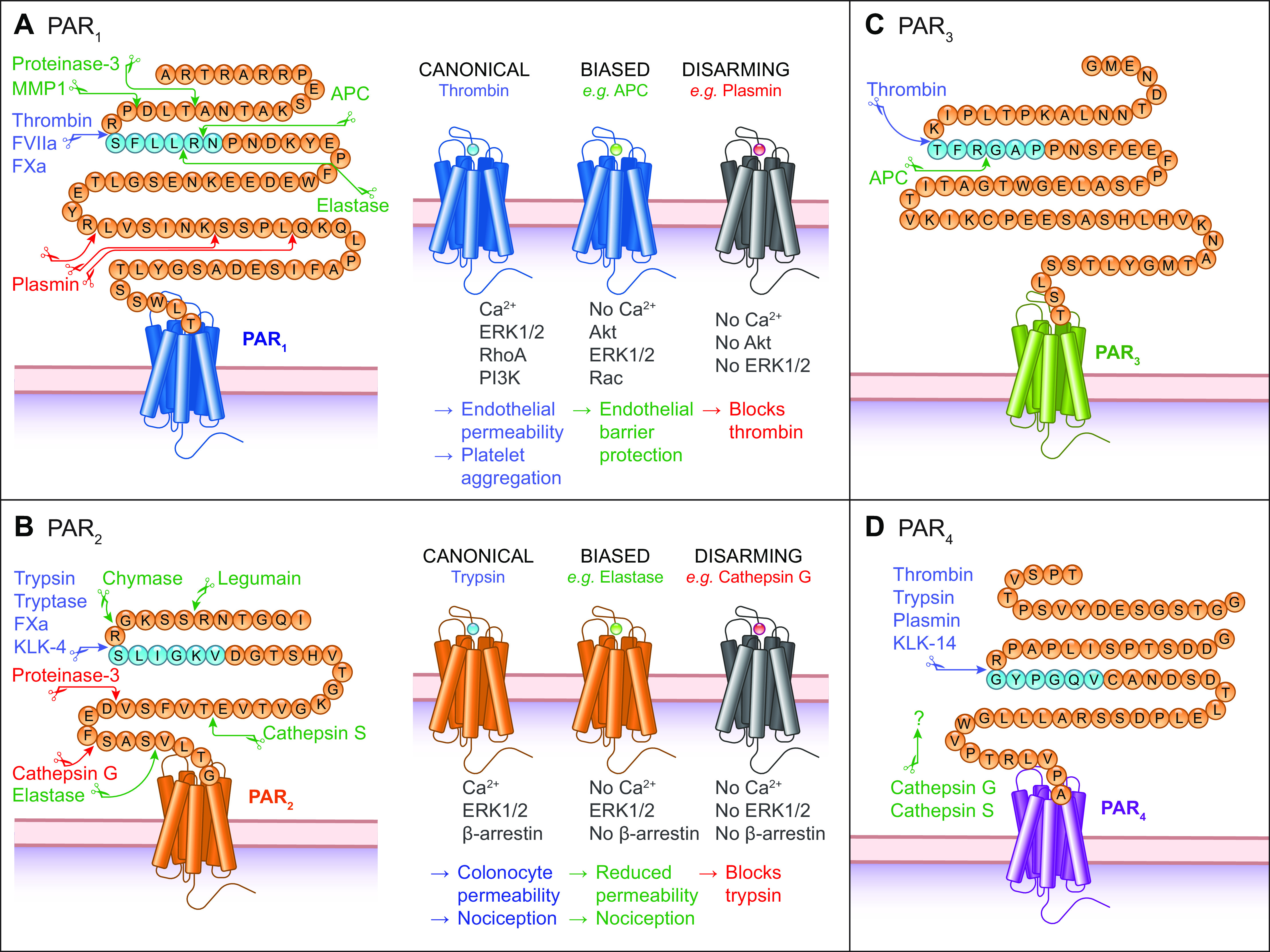
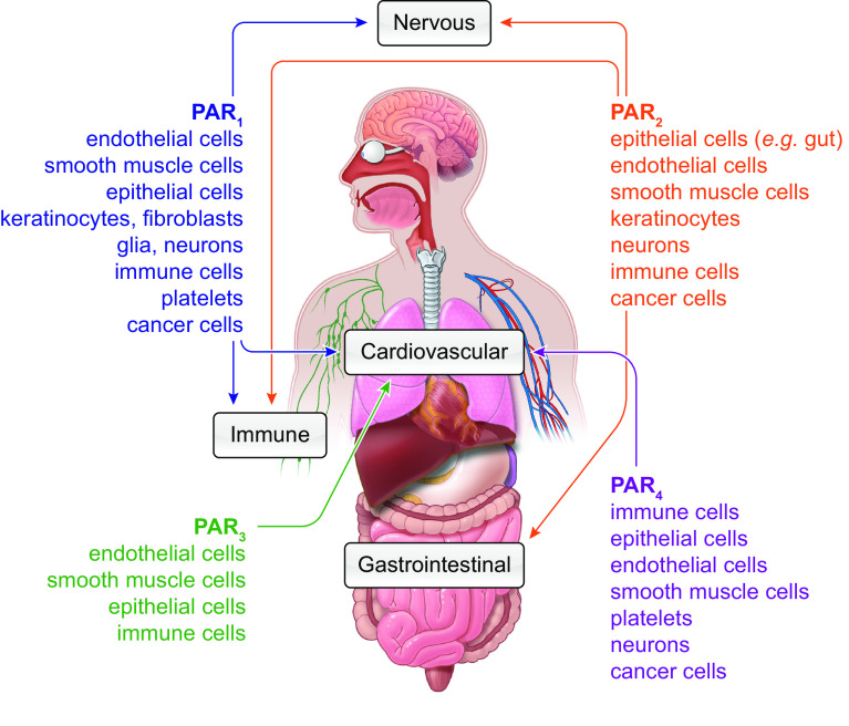
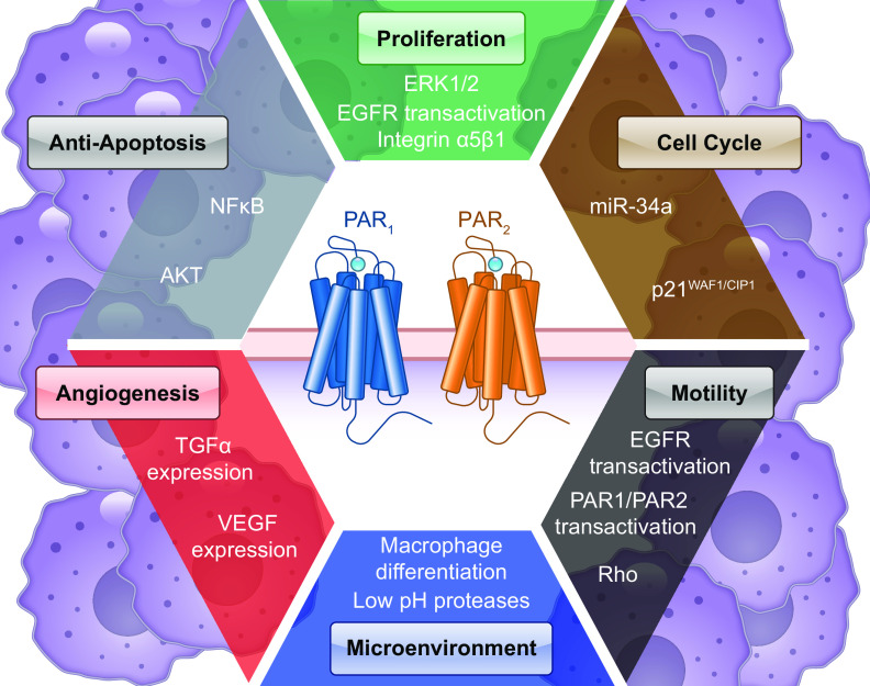
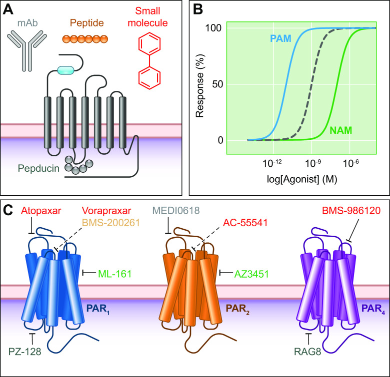
Proteases are signaling molecules that specifically control cellular functions by cleaving protease-activated receptors (PARs). The four known PARs are members of the large family of G protein-coupled receptors. These transmembrane receptors control most physiological and pathological processes and are the target of a large proportion of therapeutic drugs. Signaling proteases include enzymes from the circulation; from immune, inflammatory epithelial, and cancer cells; as well as from commensal and pathogenic bacteria. Advances in our understanding of the structure and function of PARs provide insights into how diverse proteases activate these receptors to regulate physiological and pathological processes in most tissues and organ systems. The realization that proteases and PARs are key mediators of disease, coupled with advances in understanding the atomic level structure of PARs and their mechanisms of signaling in subcellular microdomains, has spurred the development of antagonists, some of which have advanced to the clinic. Herein we review the discovery, structure, and function of this receptor system, highlight the contribution of PARs to homeostatic control, and discuss the potential of PAR antagonists for the treatment of major diseases.

Keywords: antagonists; disease; homeostasis; proteases; receptors.


Conflict of interest statement

N. W. Bunnett is a founding scientist of Endosome Therapeutics Inc. None of the other authors has any conflicts of interest, financial or otherwise, to disclose.

Figures

None
Graphical abstract
FIGURE 1.
FIGURE 1.
Canonical protease-activated receptor (PAR) cleavage sites and activation mechanisms. AD: NH2-terminal domains of PAR1-4 highlighting the canonical cleavage site for thrombin or trypsin in the human receptors. Cleavage can reveal a tethered ligand (blue) from which soluble activating peptides (APs) were derived; PAR3-AP is unable to activate PAR3. APs have been modified to generate selective and potent activating peptides (e.g., for PAR2 and PAR4; gray). TRAP, thrombin receptor-activating peptide.
FIGURE 2.
FIGURE 2.
Cleavage by noncanonical proteases. AD: NH2-terminal domains of protease-activated receptors 1 to 4 (PAR1-4) showing cleavage sites for proteases that activate PARs by canonical (blue) or biased (green) mechanisms or those that disarm the receptor (red). Residues are shown for the human receptors without the predicted signal sequence. APC, activated protein C; MMP, matrix metalloproteases.
FIGURE 3.
FIGURE 3.
Patterns of protease-activated receptors (PAR) expression. Sites of PAR1-4 expression across different organ systems (arrows) and cell types (listed).
FIGURE 4.
FIGURE 4.
Modulation of protease-activated receptors (PAR) signaling. A: heteromers can form between different PAR subtypes, such as PAR1 and PAR2. Tethered ligands from one subtype can transactivate another. B: heteromers can form with other G protein-coupled receptors (GPCRs), such as between the chemokine receptor 4 (CXCR4; shown) or with β-adrenoceptors in cardiac fibroblasts. C: GPCRs transactivate receptor tyrosine kinases (RTKs), such as the epidermal growth factor receptor (EGFR). PAR1-mediated Src activation can phosphorylate (Ph) EGFR (1). PAR1 also activates matrix metalloproteases (MMPs) that liberate membrane-tethered EGFR ligands, such as amphiregulin (2). D: PAR signaling can modulate ion channel signaling, such as PAR2-mediated sensitization of transient receptor potential vanilloid 1 (TRPV1) in sensory neurons. TRPV1 sensitization is mediated by PKC activation downstream of PAR2, causing phosphorylation (Ph) of TRPV1 (Ser residues 502 and 800 in human TRPV1).
FIGURE 5.
FIGURE 5.
Trafficking of protease-activated receptors (PARs). A: PAR2 activation leads to Gα/βγ recruitment, phosphorylation (Ph; red) by G protein-coupled receptor (GPCR)-regulated kinase (GRK), and β-arrestin (βARR) recruitment (1). This triggers clathrin-mediated endocytosis (CME). Internalized PAR2 continues to signal from endosomes (2). Ubiquitination (Ub; blue) of the COOH terminus triggers trafficking to lysosomes for degradation (3). To replace the cleaved receptor, PAR2 signaling induces Gβγ-mediated activation of PKD in the Golgi apparatus, which mobilizes newly synthesized PAR2 to repopulate the plasma membrane (4). B: PAR1 is subject to constitutive CME upon ubiquitination and AP-2 recruitment (1). In contrast to PAR2, it is not subject to βARR-dependent endocytosis. PAR1 can signal from endosomes in a ubiquitin-driven manner with adaptor proteins TABs to drive p38 MAPK signaling (2). Uncleaved PAR1 is returned to the plasma membrane via recycling endosomes. Cleaved PAR1 signals with Gα/βγ. Similarly, PAR1 is then degraded in the lysosome (3).
FIGURE 6.
FIGURE 6.
Role of protease-activated receptors (PARs) in the circulatory system. A: PARs are expressed in various cell types in the vasculature and circulation. B: human platelets expressing PAR1 and PAR4 respond to low- or high-thrombin concentrations to aggregate platelets. C: endothelial PAR1 has distinct signaling outcomes in response to thrombin or activated protein C (APC) bound to endothelial protein C receptor (EPCR). NO, nitric oxide.
FIGURE 7.
FIGURE 7.
Protease-activated receptor (PAR) signaling in the gastrointestinal system. A: colonocytes express PAR1 and PAR2. The apical membrane facing the gut lumen is exposed to proteases, including digestive enzymes and proteases from commensal and pathogenic microorganisms. The basolateral membrane is exposed to proteases released by immune cells, epithelial cells, blood vessels, and other cell types. B: activation of PAR2 at the basolateral region of cells leads to ERK1/2 signaling that causes paracellular permeability via disruption of tight junctions. C: PAR2 activation on sensory neurons, including those that innervate the gut, leads to Ca2+ mobilization. Subsequent PKC activation sensitizes transient receptor potential (TRP) channels, leading to ionic influx and activation of voltage-gated channels to trigger action potentials. This also triggers release of neuropeptides, such as substance P (SP) or calcitonin gene-related peptide (CGRP), which mediate neurogenic inflammation and pain. ZO-1, zona occludens.
FIGURE 8.
FIGURE 8.
Protease-activated receptor (PAR) signaling in pain. A: primary (1º) sensory neurons with cell bodies in dorsal root ganglia (DRG) project fibers to peripheral tissues and the dorsal horn of the spinal cord. Second-order (2º) neurons in the dorsal horn transmit painful signals centrally. B: proteases that are released from immune cells cleave PAR2 expressed on sensory nerve fibers in the periphery. Some proteases (e.g., mast cell tryptase) trigger PAR2 endocytosis and trafficking to signaling endosomes that generate signals leading to sensitization or activation of ion channels. Other proteases (e.g., neutrophil elastase, macrophage cathepsin S, and legumain) activate PAR2 by biased mechanisms that do not evoke endocytosis; PAR2 signals from the plasma membrane to sensitize or activate ion channels. Neurons can release substance P (SP) and calcitonin gene-related peptide (CGRP) locally, causing neurogenic inflammation, and centrally, leading to pain transmission. TRP, transient receptor potential.
FIGURE 9.
FIGURE 9.
Protease-activated receptor (PAR) signaling in cancer. Proteases and PARs contribute to numerous pathways involved in the “hallmarks of cancer” (542). The acidic tumor microenvironment can enhance protease activity. PARs activate downstream signaling pathways and transactivate other receptors that promote cancer. EGFR, epidermal growth factor receptor; TGFα, transforming growth factor-α.
FIGURE 10.
FIGURE 10.
Therapeutic targeting of protease-activated receptors (PARs). A: multiple drug classes have been developed to target PAR signaling, including peptidic and small molecule antagonists and monoclonal antibodies (mAbs). B: receptor signaling can be modified by allosteric modulators that either enhance agonist activity [positive allosteric modulator (PAM)] or decrease signaling [negative allosteric modulator (NAM)]. C: examples of PAR inhibitors, including mAbs (gray), peptides (yellow), small molecules (red), NAMs (green), or pepducins targeting intracellular loops (blue). These can target allosteric sites or the orthosteric binding site of the tethered ligand (dashed).

Similar articles

Cited by

References

    1. Schmidt A. Neue Untersuchungen ueber die Fasserstoffesgerinnung. Pflüger Arch 6: 413–538, 1872. doi:10.1007/BF01612263. - DOI
    1. Van Obberghen-Schilling E, Chambard JC, Lory P, Nargeot J, Pouyssegur J. Functional expression of Ca2(+)-mobilizing alpha-thrombin receptors in mRNA-injected Xenopus oocytes. FEBS Lett 262: 330–334, 1990. doi:10.1016/0014-5793(90)80221-4. - DOI - PubMed
    1. Pipili-Synetos E, Gershengorn MC, Jaffe EA. Expression of functional thrombin receptors in xenopus oocytes injected with human endothelial cell mRNA. Biochem Biophys Res Commun 171: 913–919, 1990. doi:10.1016/0006-291X(90)90770-N. - DOI - PubMed
    1. Vu TK, Hung DT, Wheaton VI, Coughlin SR. Molecular cloning of a functional thrombin receptor reveals a novel proteolytic mechanism of receptor activation. Cell 64: 1057–1068, 1991. doi:10.1016/0092-8674(91)90261-V. - DOI - PubMed
    1. Nystedt S, Emilsson K, Wahlestedt C, Sundelin J. Molecular cloning of a potential proteinase activated receptor. Proc Natl Acad Sci U S A 91: 9208–9212, 1994. doi:10.1073/pnas.91.20.9208. - DOI - PMC - PubMed

Publication types

Substances

LinkOut - more resources