. 2022 Sep;235(6):2365-2377.
doi: 10.1111/nph.18321. Epub 2022 Jul 28.

The innovation of the symbiosome has enhanced the evolutionary stability of nitrogen fixation in legumes

Affiliations

The innovation of the symbiosome has enhanced the evolutionary stability of nitrogen fixation in legumes

Sergio M de Faria et al. New Phytol. 2022 Sep.

Abstract

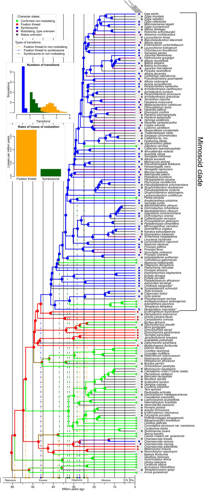
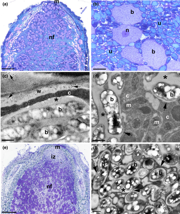
Nitrogen-fixing symbiosis is globally important in ecosystem functioning and agriculture, yet the evolutionary history of nodulation remains the focus of considerable debate. Recent evidence suggesting a single origin of nodulation followed by massive parallel evolutionary losses raises questions about why a few lineages in the N2 -fixing clade retained nodulation and diversified as stable nodulators, while most did not. Within legumes, nodulation is restricted to the two most diverse subfamilies, Papilionoideae and Caesalpinioideae, which show stable retention of nodulation across their core clades. We characterize two nodule anatomy types across 128 species in 56 of the 152 genera of the legume subfamily Caesalpinioideae: fixation thread nodules (FTs), where nitrogen-fixing bacteroids are retained within the apoplast in modified infection threads, and symbiosomes, where rhizobia are symplastically internalized in the host cell cytoplasm within membrane-bound symbiosomes (SYMs). Using a robust phylogenomic tree based on 997 genes from 147 Caesalpinioideae genera, we show that losses of nodulation are more prevalent in lineages with FTs than those with SYMs. We propose that evolution of the symbiosome allows for a more intimate and enduring symbiosis through tighter compartmentalization of their rhizobial microsymbionts, resulting in greater evolutionary stability of nodulation across this species-rich pantropical legume clade.

Keywords: Leguminosae; evolution; fixation threads; nitrogen fixation; nodulation; phylogenomics; symbiosis; symbiosomes.


Figures

Fig. 1
Fig. 1
Evolutionary trajectories of nodulation and nodule type across a time‐calibrated phylogeny of the legume subfamily Caesalpinioideae. Pie charts on nodes show the proportions of the most probable reconstructed character states: nonnodulating, fixation thread (FT‐type nodules), symbiosome (SYM‐type nodules), nodulating but of unknown type, and nodulation status unknown, summarized over 500 simulations. Branch colours denote the nodulation status of the node or tip it subtends and the coloured boxes in front of each taxon name show the character state for that species. Note that in three clades (the Senna + Cassia clade, the Arcoa + Acrocarpus clade and the Peltophorum clade) ‘double’ losses of FT‐type nodules are inferred to have occurred simultaneously in both descendant lineages of that node. For example, the crown node of the Senna + Cassia clade is inferred to be nodulating with FT‐type nodules even though Senna and Cassia are nonnodulating. The dashed orange, blue and dark green vertical lines show the phylogenetic locations and maximum ages of the various character state transitions on the tree. Using the same colours, the histograms show the frequencies of the number of transitions from FT to SYM (blue), from SYM to nonnodulation (green) and from FT to nonnodulation (orange), and the rates of losses of nodulation per million years for SYM to nonnodulation (green) and FT to nonnodulation (orange) across 500 independent character estimations. Note that while the three other character state transitions, from nonnodulating to FT or SYM‐type nodules, and from SYM to FT, were not allowed under our model, and were therefore fixed at zero, an alternative model permitting all character state transitions gave an identical result (Supporting Information Fig. S3). Asterisks after terminal names indicate species which are the focus of detailed nodule anatomical work presented in Figs 2, 3, 4. Ple, Pleistocene; Pli, Pliocene.
Fig. 2
Fig. 2
Nonmimosoid grade caesalpinioid nodules in the genus Erythrophleum contain bacteroids enclosed within fixation threads (FTs). Light (a, b) and transmission electron microscope (c–h) images of sections of nodules from E. ivorense (a–f) and E. suaveolens (g, h). (a) Whole nodule longitudinal profile illustrating the zonation typical of an indeterminate nodule (m, meristem; iz, invasion zone; nf, nitrogen fixing zone). Bar, 250 μm. (b) Higher magnification view of the iz in which newly divided host cells derived from the meristem (m) are being invaded by numerous infection threads (arrows). Bar, 10 μm. (c) Large infection thread containing bacteria (b) invading cells in the iz; the walls of the infection thread are densely immunogold labelled with 10 nm gold particles linked to JIM5 (arrows), a monoclonal antibody which recognizes nonesterified pectin. v, vacuole. Bar, 1 μm. (d) Infection thread in the iz–nf boundary with its cell walls labelled with JIM5 (large arrows) adjacent to an FT (*) with a thinner cell wall that is very sparsely labelled with JIM5 (arrowheads). Bar, 500 nm. (e) Cell in the nf zone packed with FTs (black arrows), including within the nucleus (n) indicated by white arrowheads. Bar, 2 μm. (f) Detail of FTs in the nf zone containing N‐fixing bacteroids (b); the FT walls range from being sparsely labelled with JIM5 (arrowheads) to exhibiting little or no obvious labelling (single gold particles are indicated by arrows). Bar, 500 nm. (g) High‐resolution image of a bacteroid (b) forming within a strand of cytoplasm between vacuoles (v) in an iz cell; the bacteroid is surrounded by a cell wall (w) that is being enveloped in a membrane (arrowheads), stretches of which (arrows) appear to be derived from nearby endoplasmic reticulum/Golgi bodies (er). The intense metabolic activity of this process is suggested by the nearby mitochondria (m) and peroxisomes (p). Bar, 1 μm. (h) Bacteroids (b) in newly formed FTs packed into a new N‐fixing cell in the early nf zone adjacent to the iz; the bacteroids are surrounded by the FT wall (w), which is itself surmounted by a symbiosome membrane (arrowheads). Note the membranes within the cytoplasm that are associated with the FTs (arrows). n, nucleus; m, mitochondrion. Bar, 1 μm.
Fig. 3
Fig. 3
Nodules of caesalpinioids from the Mimosoid clade contain bacteroids enclosed within symbiosomes (SYMs). Light (a, b, e) and transmission electron microscope (c, d, f) images of sections of nodules from Pentaclethra macroloba (a–d) and Chidlowia sanguinea (e, f). (a) Whole P. macroloba nodule longitudinal profile illustrating the zonation typical of an indeterminate nodule (m, meristem; nf, nitrogen fixing zone). Bar, 500 μm. (b) Higher magnification view of the nf zone showing large bacteroid‐containing cells (b) surrounded by smaller and more numerous uninfected cells (u). n, nucleus. Bar, 25 μm. (c) Bacteroids (b) within a symbiosome adjacent to the host cell wall (w) which is immunogold labelled with 10 nm gold particles linked to JIM5 (arrows). The symbiosome peribacteroid space is marked with an asterisk (*); note that there is no cell wall separating the symbiosome from the host cytoplasm (c). Bar, 500 nm. (d) High‐resolution image of bacteroids (b) housed in symbiosomes; the symbiosome membrane separating it from the cytoplasm (c) are marked with arrows, and the peribacteroid space by an asterisk (*). m, mitochondrion. Bar, 500 nm. (e) Whole C. sanguinea nodule longitudinal profile illustrating the zonation typical of an indeterminate nodule (m, meristem; iz, invasion zone; nf, nitrogen fixing zone). Bar, 200 μm. (f) High‐resolution image of bacteroids (b) housed in symbiosomes; the symbiosome membrane separating it from the cytoplasm (c) is marked with arrows, and the peribacteroid space by an asterisk (*). Bar, 500 nm.
Fig. 4
Fig. 4
Fixation threads (FTs) contain cell wall components in addition to unesterified (JIM5) and esterified (JIM7) pectin. Confocal laser scanning microscopy (CLSM) with anti‐rat Alexa Fluor 488 (a–d, i–l) and immunogold transmission electron microscopy with anti‐rat 10 nm gold (e–h, m–p) of Erythrophleum (a–h) and Pentaclethra (i–p) nodules incubated in monoclonal antibodies raised in rat against various plant cell wall components: Lm2 (a, e, i, m), which labels arabinogalactose protein (AGP) glycan; Lm5 (b, f, j, n), which labels the pectic polysaccharide rhamnogalacturonan; and Lm15 (c, g, k, o), which labels the XXXG motif of the nonpectic, noncellullosic polysaccharide xyloglucan. Control sections incubated in buffer alone without a primary antibody are presented in (d, h, l, p). FTs are indicated by arrows in (a–h), and symbiosomes by arrowheads in (i–p). w, host cell wall separating plant cells; b, bacteroid. Bars: (a–d, i–l) 5 μm; (e–h, m–p) 1 μm.

Similar articles

Cited by

References

    1. Ardley J, Sprent J. 2021. Evolution and biogeography of actinorhizal plants and legumes: a comparison. Journal of Ecology 109: 1098–1121.
    1. Batterman SA, Hedin LO, Van Breugel M, Ransijn J, Craven DJ, Hall JS. 2013. Key role of symbiotic dinitrogen fixation in tropical forest secondary succession. Nature 502: 224–227. - PubMed
    1. Brewin NJ. 2004. Plant cell wall remodelling in the rhizobium–legume symbiosis. Critical Reviews in Plant Sciences 23: 293–316.
    1. Bruneau A, Mercure M, Lewis GP, Herendeen PS. 2008. Phylogenetic patterns and diversification in the caesalpinioid legumes. Botany 86: 697–718.
    1. Cardoso D, De Queiroz LP, Toby Pennington R, De Lima HC, Fonty É, Wojciechowski MF, Lavin M. 2012. Revisiting the phylogeny of papilionoid legumes: new insights from comprehensively sampled early‐branching lineages. American Journal of Botany 99: 1991–2013. - PubMed

Publication types

LinkOut - more resources