. 2022 Oct;48(4):435-457.
doi: 10.1037/xan0000332. Epub 2022 Jul 28.

Behavioral studies of spinal conditioning: The spinal cord is smarter than you think it is

Affiliations

Behavioral studies of spinal conditioning: The spinal cord is smarter than you think it is

James W Grau et al. J Exp Psychol Anim Learn Cogn. 2022 Oct.

Abstract

In 1988 Robert Rescorla published an article in the Annual Review of Neuroscience that addressed the circumstances under which learning occurs, some key methodological issues, and what constitutes an example of learning. The article has inspired a generation of neuroscientists, opening the door to a wider range of learning phenomena. After reviewing the historical context for his article, its key points are briefly reviewed. The perspective outlined enabled the study of learning in simpler preparations, such as the spinal cord. The period after 1988 revealed that pain (nociceptive) stimuli can induce a lasting sensitization of spinal cord circuits, laying down a kind of memory mediated by signal pathways analogous to those implicated in brain dependent learning and memory. Evidence suggests that the spinal cord is sensitive to instrumental response-outcome (R-O) relations, that learning can induce a peripheral modification (muscle memory) that helps maintain the learned response, and that learning can promote adaptive plasticity (a form of metaplasticity). Conversely, exposure to uncontrollable stimulation disables the capacity to learn. Spinal cord neurons can also abstract that stimuli occur in a regular (predictable) manner, a capacity that appears linked to a neural oscillator (central pattern generator). Disrupting communication with the brain has been shown to transform how GABA affects neuronal function (an example of ionic plasticity), releasing a brake that enables plasticity. We conclude by presenting a framework for understanding these findings and the implications for the broader study of learning. (PsycInfo Database Record (c) 2022 APA, all rights reserved).


Conflict of interest statement

Author Disclosure Statement : The authors declare no competing financial interests.

Figures

Figure 1.
Figure 1.
(A) Uninjured rats exposed to a few moderately intense (1 mA) shocks to the tail (Intense) exhibit reduced reactivity (antinociception) to a noxious thermal stimulus applied to the tail, relative to a group that was treated the same but did not receive shock (Nothing). Presenting a weak (0.1 mA) shock distractor causes the antinociception to decay more rapidly (Intense→Weak). (B) In rats that have received a high thoracic (T2) transection, a very intense shock (3 mA) to the tail elicits antinociception. Following the intense shock with a weaker (1 mA) shock (“distractor”) causes the antinociception to decay more rapidly. (C) In spinally transected rats, pairing moderate electrical stimulation of the saphenous nerve (the CS) with more intense stimulation of the peroneal (the US) endows the CS with the capacity to elicit a stronger muscle response (the CR), relative to a CS that was presented in an unpaired manner. (D) In spinally transected rats, animals given shock to the tibialis anterior muscle of one hind leg whenever the limb is extended (Master) exhibit a progressive increase in flexion duration relative to animals given the same amount of stimulation independent of leg position (Yoked). Adapted from (Grau, 1987a, 1987b; Grau et al., 2020; Grau et al., 1990).
Figure 2.
Figure 2.
Testing pretrained animals under common conditions. (A) Spinally transected rats were trained with controllable (Master) or uncontrollable (Yoked) stimulation. A third group was set up in the same manner, but received no stimulation (Unshocked). Flexion force and contact electrode depth (response criterion) were then re-equated and animals were tested for 30 min with controllable stimulation applied to the pretrained leg. Prior training with controllable stimulation fostered learning whereas exposure to uncontrollable shock impaired learning. (B) Spinally transected animals received 30 min of training with controllable stimulation and a moderate response criterion (a contact electrode depth of 4 mm). All rats were then tested for 30 min with a higher (8 mm) response criterion. At this higher criterion, previously untrained (Unshocked) rats were unable to learn. Pretained animals were able to learn and this was true independent of whether they were tested on the pretrained (Ipsilateral) or opposite (Contralateral) leg. Adapted from (Grau, 2012).
Figure 3.
Figure 3.
A schematic model illustrating how response contingent (controllable) and non-contingent (uncontrollable) noxious stimulation affect spinal cord function. It is assumed that proprioceptive/sensory cues provide an index of leg position and that the system is prepared to detect the relation between leg position and the onset of a noxious stimulus. Controllable stimulation brings an increase in flexion duration that reduces exposure to noxious stimulation. The early integration of sensory cues related to leg position (the R) and the onset of shock (the O) allows brain systems to directly perceive the underlying R-O relation. With extended training, BDNF is expressed, which has a restorative effect that counters the development of nociceptive sensitization and the consequent learning impairment and allodynia. If no R-O relation is detected after 180 stimuli, a state of over-excitation emerges (coupled to the expression of TNF) that impairs the capacity to learn and enhances mechanical reactivity. This nociceptive sensitization would amplify pain signals relayed to the brain, driving neuropathic pain. Exposure to uncontrollable, but not controllable, stimulation also impairs recovery after a contusion injury of the thoracic spinal cord. Adapted from (Grau, 2014).
Figure 4.
Figure 4.
Schematic illustrating the processes involved in spinal timing and the environmental relations used to explore how the system works. (A) Empirical work suggests that the capacity to time is linked to an internal oscillator that can become entrained to stimuli presented in a regular manner. (B) The oscillator (CPG) can abstract regularity across dermatomes. The abstraction of regularity inhibits the development of the learning impairment induced by uncontrollable stimulation. (C) Stepping and instrumental learning are organized by neurons contained within a portion of the lumbosacral spinal cord between L3 and S2 (Liu et al., 2005). The CPG that drives stepping has been localized to a rostral region (L1–2) (Magnuson et al., 1999). (D) Exposure to variable (VT) shock for 6–30 min (180–900 shocks) induces a learning impairment (Deficit). A brief exposure (180–360 shocks) to shock given in a regular manner (FT) also induces a learning deficit. Continued exposure to FT shock (540 or more shocks) engages a restorative effect that counters the learning impairment. (E) The spinal cord can abstract regularity when stimuli are given across dermatomes (tail and one leg) in a regular (alternating) manner. (F) Increasing the temporal gap between shocks applied to one dermatome, while the gap is reduced for stimuli applied to the other site, yields an irregular (incoherent) pattern of stimulation across dermatomes and results in a learning impairment. When the interval between shocks to each dermatome is shifted by the same amount, the alternating pattern is preserved (coherent) and the restorative effect develops. (G) The spinal cord can also abstract regularity when the site of stimulation is randomly varied over times. (H) The spinal cord can abstract regularity when half the shocks applied to one dermatome are randomly omitted (Unshifted). If the first shock after an omitted one is displaced (Shifted), so that its occurrence does not align with what would be expected given an oscillator, a deficit develops, implying that the internal oscillator was not entrained. Adapted from (Lee et al., 2016).
Figure 5.
Figure 5.
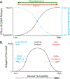
(A) Intracellular Cl is regulated by KCC2, which transports the anion out of the cell. In the uninjured adult CNS, membrane bound KCC2 maintains a low intracellular concentration of Cl. Under these conditions, engaging the GABA-A receptor allows Cl to enter the cell, which has an inhibitory (hyperpolarizing) effect. After SCI, there is a reduction in membrane-bound KCC2 below the injury, recapitulating an earlier developmental state. This leads to a rise in intracellular Cl, which reduces the inward flow of Cl and the inhibitory effect of engaging the GABA-A receptor. At the extreme, engaging this receptor can allow Cl to exit the cell, which would have an excitatory (depolarizing) effect. (B) GABAergic inhibition will normally reduce neural excitability and plasticity in the adult CNS, causing the system to appear immutable (hard wired). The reduction in GABA inhibition (ionic plasticity) can enable learning. A further shift can foster pathological over-excitation. The resultant function is reminiscent of the non-monotonic relation between learning and arousal/stress (Yerkes-Dodson law). Adapted from (Grau & Huang, 2018).
Figure 6.
Figure 6.
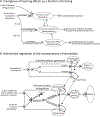
Illustrations of how training affects spinal cord function and the underlying processes. (A) A schematic illustrating the sequential induction of learning and metaplastic processes by alternative forms of stimulation. When noxious intermittent stimulation is given in a response-contingent manner (predicted by cues related to limb position/response criterion), the R-O relation drives an increase in flexion duration. If stimulation is given in the absence of an adaptive behavioral response for six minutes (180 shocks), while the leg is down, a learning impairment is induced that inhibits the capacity to learn. The consequent state of over-excitation enhances mechanical reactivity and blocks learning (the change in response duration normally induced by response-contingent stimulation). Because the induction of this process has a lasting effect (involves a form of plasticity) that interferes with the capacity to learn, it reflects an example of metaplasticity. Because animals given an equivalent amount of controllable stimulation do not develop this effect, it is assumed that processes engaged by a R-O relation inhibit its development. Continued training with controllable stimulation for additional 24 min, or the presentation of regular (FT) shocks (540 or more), induces a restorative effect that counters the adverse consequences of uncontrollable stimulation. Because this restorative effect has a lasting impact on plasticity, it too reflects a form of metaplasticity. Behaviorally, it enables (amplifies) learning, reduces mechanical reactivity, and counters the development of nociceptive sensitization. Adapted from (Baumbauer et al., 2017). (B) The mechanisms thought to underlie spinally-mediated instrumental conditioning, timing, and the consequences of uncontrollable stimulation. It is assumed that the spinal cord contains processes that support sensory-motor integration which can detect the relationship between cues indicative of limb position (and reaching the response criterion) and the onset of noxious stimulation. Our results suggest that the effect of noxious stimulation is initially gated on the basis of leg position; if the leg(s) is flexed, stimulation has no impact on spinal function. If the legs are not flexed, pre-existing circuitry enables the rapid detection of a relationship between the onset of noxious stimulation and sensory cues related to the current leg position (and/or contacting the underlying solution). In this model, the R-O relation is encoded by sensory cues. If there is a clear relation, the stimulation is classified as controllable and the appropriate behavioral response is selected. In addition, with continued training, a process is engaged that facilitates sensory-motor integration. Exposure to regular stimulation can also activate this faciliatory effect through the engagement of a rostral central pattern generator (CPG). If noxious stimulation is given in an uncontrollable/unpredictable manner, it induces a process that impairs sensory-motor integration. Noxious stimulation can also interfere with CPG function and the generation of rhythmic behavior (Bouffard et al., 2014; Caudle et al., 2015). The consequences of training that have a lasting effect are enclosed with dashed ovals. Training with controllable stimulation sensitizes the motor output and, with continued training, induces a modification at the neuromuscular junction that fosters the performance of a flexion response. Exposure to controllable stimulation also has a metaplastic effect that enables the abstraction of R-O relations. Conversely, uncontrollable stimulation has a metaplastic effect that interferes with this process. These metaplastic effects have a general impact that modulates the capacity to detect sensory relations when stimuli are applied to the contralateral limb. Further work is needed to determine whether exposure to irregular stimulation (independent of the R-O relation) interferes with CPG function (indicated with a “?”) or impacts sensory-motor integration. Likewise, we do not know whether controllable stimulation fosters CPG function. Note that in panels A and B, we indicate that uncontrollable stimulation impairs sensory motor integration using a minus sign. While this accurately describes the functional operation of the system, at a neural level uncontrollable stimulation may impair processing by inducing a state of over-excitation (not inhibition; Ferguson et al., 2006). Conversely, the facilitory effects noted in panel B with a plus may reflect a BDNF-dependent reduction in over-excitation after SCI (Cote et al., 2014; Huang et al., 2017; Tashiro et al., 2015).

Similar articles

Cited by

References

    1. Abraham WC (2008). Metaplasticity: tuning synapses and networks for plasticity. Nature Reviews Neuroscience, 9(5), 387–399. doi: 10.1038/nrn2356 - DOI - PubMed
    1. Abraham WC, & Bear MF (1996). Metaplasticity: the plasticity of synaptic plasticity. Trends Neurosci, 19(4), 126–130. - PubMed
    1. Akil H, Watson S, Young E, Lewis ME, Khachaturiam H, & Walker JM (1984). Endogenous opioids: Biology and function. Annual Review of Neuroscience, 7, 223–255. - PubMed
    1. Alkon DL (1983). Learning in a marine snail. Scientific American, 249(1), 70–74. - PubMed
    1. Alluin O, Karimi-Abdolrezaee S, Delivet-Mongrain H, Leblond H, Fehlings MG, & Rossignol S. (2011). Kinematic study of locomotor recovery after spinal cord clip compression injury in rats. Journal of Neurotrauma, 28(9), 1963–1981. doi: 10.1089/neu.2011.1840 - DOI - PubMed

Substances