. 2022 Jul 8;138(3):810-820.
doi: 10.3171/2022.5.JNS22837. Print 2023 Mar 1.

Arousal and salience network connectivity alterations in surgical temporal lobe epilepsy

Affiliations

Arousal and salience network connectivity alterations in surgical temporal lobe epilepsy

Hernán F J González et al. J Neurosurg. .

Abstract

Objective: It is poorly understood why patients with mesial temporal lobe epilepsy (TLE) have cognitive deficits and brain network changes that extend beyond the temporal lobe, including altered extratemporal intrinsic connectivity networks (ICNs). However, subcortical arousal structures project broadly to the neocortex, are affected by TLE, and thus may contribute to these widespread network effects. The authors' objective was to examine functional connectivity (FC) patterns between subcortical arousal structures and neocortical ICNs, possible neurocognitive relationships, and FC changes after epilepsy surgery.

Methods: The authors obtained resting-state functional magnetic resonance imaging (fMRI) in 50 adults with TLE and 50 controls. They compared nondirected FC (correlation) and directed FC (Granger causality laterality index) within the salience network, default mode network, and central executive network, as well as between subcortical arousal structures; these 3 ICNs were also compared between patients and controls. They also used an fMRI-based vigilance index to relate alertness to arousal center FC. Finally, fMRI was repeated in 29 patients > 12 months after temporal lobe resection.

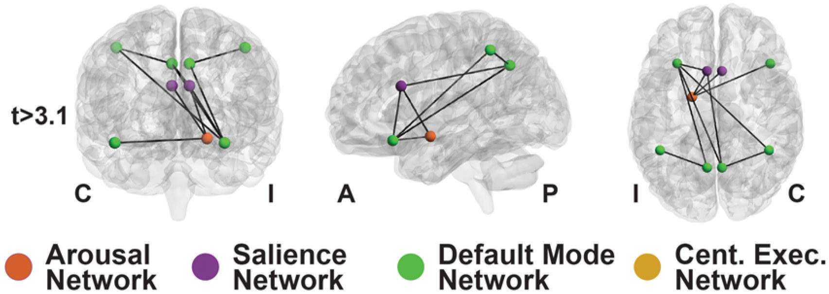
Results: Nondirected FC within the salience (p = 0.042) and default mode (p = 0.0008) networks, but not the central executive network (p = 0.79), was decreased in patients in comparison with controls (t-tests, corrected). Nondirected FC between the salience network and subcortical arousal structures (nucleus basalis of Meynert, thalamic centromedian nucleus, and brainstem pedunculopontine nucleus) was reduced in patients in comparison with controls (p = 0.0028-0.015, t-tests, corrected), and some of these connectivity abnormalities were associated with lower processing speed index, verbal comprehension, and full-scale IQ. Interestingly, directed connectivity measures suggested a loss of top-down influence from the salience network to the arousal nuclei in patients. After resection, certain FC patterns between the arousal nuclei and salience network moved toward control values in the patients, suggesting that some postoperative recovery may be possible. Although an fMRI-based vigilance measure suggested that patients exhibited reduced alertness over time, FC abnormalities between the salience network and arousal structures were not influenced by the alertness levels during the scans.

Conclusions: FC abnormalities between subcortical arousal structures and ICNs, such as the salience network, may be related to certain neurocognitive deficits in TLE patients. Although TLE patients demonstrated vigilance abnormalities, baseline FC perturbations between the arousal and salience networks are unlikely to be driven solely by alertness level, and some may improve after surgery. Examination of the arousal network and ICN disturbances may improve our understanding of the downstream clinical effects of TLE.

Keywords: arousal structures; epilepsy surgery; fMRI; focal epilepsy; functional connectivity; networks; neuroimaging; temporal lobe epilepsy.


Conflict of interest statement

Disclosures

The authors report no conflict of interest concerning the materials or methods used in this study or the findings specified in this paper.

Figures

FIG. 1.
FIG. 1.
The relationships between subcortical arousal networks and ICNs in the triple network model. The arousal network (burnt orange) contains subcortical structures (brainstem ascending reticular activating system nuclei, intralaminar thalamic nuclei, and NBM) that project to subcortical and cortical networks to modulate arousal levels.,, The triple network model consists of the SN, DMN, and CEN. The SN (royal purple) is composed of the insula and anterior cingulate cortex. The DMN (lime green) includes the posterior cingulate cortex, medial prefrontal cortex, precuneus, and angular gyrus/lateral parietal cortices. The CEN (mustard yellow) is a frontoparietal network containing the dorsolateral prefrontal cortex and lateral posterior parietal cortex. The DMN and CEN are 2 typically anticorrelated (indicated by the dashed blue arrow) networks with antithetical functions. The DMN is primarily active when a person is in quiet repose and deactivated by attention-demanding tasks., The CEN is active during goal-directed actions., The SN is responsible for detecting stimuli and engaging networks for task-positive functions, helping to switch activations between the DMN and CEN.,,, Net. = network.
FIG. 2.
FIG. 2.
Patients with TLE exhibited decreased nondirected connectivity within ICNs. The y-axis (A–C) shows mean nondirected functional connectivity (determined with Fisher z-transformed Pearson correlation analysis) between all components of each network. Mean connectivity values within the SN (A) and within the DMN (B) were decreased in patients with TLE compared with controls. We detected no differences in CEN connectivity between patients and controls (C). *p < 0.05 and ***p < 0.001 according to the paired t-test with BHC. The red lines indicate median values. Each value in each matrix shows the p value (t-test, uncorrected) for connectivity between network region pairs in patients versus controls (D–F). We included 50 patients with TLE and 50 healthy controls. AC = anterior cingulate; Caud = caudal; Cing = cingulate; Co = contralateral; Exec. = executive; FC = functional connectivity; Fr = frontal; Inf = inferior; Ip = ipsilateral; Lat = lateral; Med = medial; Net. = network; Oper = opercularis; Orb = orbital; Post = posterior; Rost = rostral; Sup = superior.
FIG. 3.
FIG. 3.
In patients with TLE, subcortical arousal structures exhibited decreased nondirected connectivity with the SN. The y-axis of each panel (A–C) shows nondirected functional connectivity (determined with Fisher z-transformed Pearson correlation analysis) between the SN and 3 arousal structures. In patients with TLE, the PPN (A), CM intralaminar thalamic nuclei ipsilateral to the seizure onset zone (B), and NBM ipsilateral to the seizure onset zone (C) each demonstrated decreased connectivity with the SN. *p < 0.05 and **p < 0.01 according to the paired t-test with BHC. The red lines indicate the median values. We included 50 patients with TLE and 50 healthy controls. Nuc. = nucleus.
FIG. 4.
FIG. 4.
A key network with decreased connectivity was identified in TLE patients. Network-based statistic revealed that a key central network of nodes and edges was decreased in TLE patients in comparison with controls, which included the ipsilateral NBM, SN, and DMN. This component network was tested at t-statistic > 3.1, and the network contained 9 nodes and 10 edges. Network-based statistic yielded p = 0.001 with family-wise error correction. We included 50 TLE patients and 50 healthy controls. A = anterior; C = contralateral to epileptogenic temporal lobe; Cent. = central; Exec. = executive; I = ipsilateral to epileptogenic temporal lobe; P = posterior.
FIG. 5.
FIG. 5.
Directed connectivity influence between the arousal structures and SN was lost in patients with TLE. The y-axis of each panel shows directed functional connectivity as measured with GCLI between the arousal structures and SN (A) or between the PPN and SN (B). Positive GCLI values indicate greater directed connectivity influence from arousal structures to cortical networks. Negative GCLI values indicate greater influence from cortical networks to arousal structures. In healthy controls, we found that the SN influenced the arousal structures in general (A), with a strong relationship seen in the PPN (B). However, in patients, this directionality of influence was lost, with GCLI values approaching 0. **p < 0.01 according to the paired t-test with BHC. The red lines indicate median values. We included 50 patients with TLE and 50 healthy controls.
FIG. 6.
FIG. 6.
After surgery, abnormal functional connectivity between the arousal structures and SN recovered in patients with TLE. The y-axis shows nondirected functional connectivity (Fisher z-transformed Pearson correlation analysis) between the SN and arousal structures (A) or NBM ipsilateral to the epileptogenic side of the brain and SN (B). We found that the nondirected connectivity of both the average arousal structures and ipsilateral NBM with SN did not differ between patients and healthy controls after surgery. ANOVA with honest significant difference criterion for multiple corrections was performed. *p < 0.05 and **p < 0.01. The red lines indicate median values. We included 29 patients with TLE and ≥ 1-year postoperative follow-up and 29 healthy controls.

Similar articles

Cited by

References

    1. Englot DJ, Konrad PE, Morgan VL. Regional and global connectivity disturbances in focal epilepsy, related neurocognitive sequelae, and potential mechanistic underpinnings. Epilepsia. 2016; 57(10): 1546–1557. - PMC - PubMed
    1. Bell B, Lin JJ, Seidenberg M, Hermann B. The neurobiology of cognitive disorders in temporal lobe epilepsy. Nat Rev Neurol. 2011; 7(3): 154–164. - PMC - PubMed
    1. Mueller SG, Bateman LM, Nei M, Goldman AM, Laxer KD. Brainstem atrophy in focal epilepsy destabilizes brainstem-brain interactions: preliminary findings. Neuroimage Clin. 2019; 23: 101888. - PMC - PubMed
    1. Serafini A, Kuate C, Gelisse P, et al. Sleep before and after temporal lobe epilepsy surgery. Seizure. 2012; 21(4): 260–265. - PubMed
    1. Englot DJ, Morgan VL, Chang C. Impaired vigilance networks in temporal lobe epilepsy: mechanisms and clinical implications. Epilepsia. 2020; 61(2): 189–202. - PMC - PubMed

Publication types

LinkOut - more resources