. 2022 Sep:201:104713.
doi: 10.1016/j.beproc.2022.104713. Epub 2022 Jul 25.

Age-related changes in the organization of spontaneously occurring behaviors

Affiliations

Age-related changes in the organization of spontaneously occurring behaviors

J R Osterlund Oltmanns et al. Behav Processes. 2022 Sep.

Abstract

Age-related changes in spatial and temporal processing have been documented across a range of species. Rodent studies typically investigate differences in performance between adult and senescent animals; however, progressive loss of neurons in the hippocampus and cortex has been observed to occur as early as after adolescence. Therefore, the current study evaluated the effects of age in three- and ten-month-old female rats on the organization of movement in open field and food protection behaviors, two tasks that have previously dissociated hippocampal and cortical pathology. Age-related differences were observed in general measures of locomotion, spatial orientation, and attentional processing. The results of the current study are consistent with age-related changes in the processing of spatial information and motivation that occur earlier in life than previously anticipated. These observations establish a foundation for future studies evaluating interventions that influence these age-related differences in performance.

Keywords: Exploration; Food protection; Movement kinematics; Open field; Rats; Senescence.


Figures

Figure 1:
Figure 1:
Topographic profiles of progressions (black) and stops (red) are plotted for a representative three-month-old (A) and ten-month-old (B) rat for a 10-minute sample. Moment-to-moment speeds are plotted for a representative three-month-old (C) and ten-month-old (D) rat. Three-month-old rats traveled a significantly shorter distance (E) and exhibited slower average peak speeds (F), but there were no group differences in stop time (G) compared to ten-month-old rats across samples. *p < 0.050
Figure 2:
Figure 2:
Position and duration (as represented by the diameter of circles) of stops are plotted for a representative three-month-old rat across the four, five-minute samples (A). Within (color matches sample) and between sample (white line) average heading (direction of line) and concentration (length of line) are plotted for the same representative rat (B). No group differences were observed for between sample stop concentration (C), nor percent of stops on the periphery of the open field (D).
Figure 3:
Figure 3:
A sequence of four progressions is plotted for a representative three-month-old (A) rat. The white circles indicate the starting point, the black circles represent the location of the peak speed during the progression, and the red circles represent the stop locations where changes in heading occurred. Degree of change in heading between each stop is indicated near the red circle. Three-month-old rats made smaller changes in heading compared to ten-month-old rats (B). Histograms plot change in heading counts for the three-month (C) and ten-month (D) groups in five-degree steps. Line plots represent each animal’s kernel density estimation curve across all possible change in heading values. The group average change in heading is plotted for small, medium, and large angle classes. *p < 0.050
Figure 4:
Figure 4:
A representative long (red), medium (green), and short (blue) progression speed is plotted for a representative three-month-old rat (A). The path circuity observed for a respective long (red), medium (green), and short (blue) is plotted with the Euclidean path marked in white (B). Both groups decreased peak speeds as progression class shortened with three-month-old rats maintaining significantly lower peak speeds compared to ten-month-old rats (C). Additionally, both groups on average had relatively non-circuitous progressions; however, differences emerged dependent on length class of the progression, and three-month-old rats trended toward less path circuity in long and medium progressions compared to ten-month-old rats (D). *p < 0.050
Figure 5:
Figure 5:
Average eat time (s) is plotted with (A) and without (B) a conspecific by age. The total number of food protection behaviors are plotted by age (C) with the ten-month-old rats exhibiting significantly more behaviors than the three-month-old rats. The total number of thefts is plotted by age (D). *p < 0.050
Figure 6:
Figure 6:
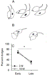
A representative dodge (A) is displayed with the dodger (unmarked belly) transferring the pellet from their forelimbs to their mouth while executing a full body turn, resulting in their forelimbs touching the floor. Additionally, a representative brace (B) is depicted with the conspecific approaching the dodger not resulting in a transfer of the pellet, but rather a pivot. The percent of doges is plotted across early and late timepoints in the trial, grouped by age (C). All rats displayed a decrease in percent dodging, and subsequent increase in percent of bracing, from early to late timepoints.
Figure 7:
Figure 7:
Representative nose distances (yellow lines) from dodger and conspecific are displayed for long (A) and short (B) distances. The average nose distance (cm) is plotted by age group with the ten-month-old rats initiating food protection behaviors when the conspecifics were at a shorter distance. *p < 0.050

Similar articles

Cited by

References

    1. Anderson RI, Bush PC, & Spear LP (2013). Environmental manipulations alter age differences in attribution of incentive salience to reward-paired cues. Behavioural brain research, 257, 83–89. - PMC - PubMed
    1. Anson E, & Jeka J (2016). Perspectives on aging vestibular function. Frontiers in neurology, 6, 269. - PMC - PubMed
    1. Ayyildiz M, Kozan R, Agar E, & Kaplan S (2008). Sexual dimorphism in the medial vestibular nucleus of adult rats: stereological study. Anatomical science international, 83(3), 131–139. - PubMed
    1. Bagarinao E, Watanabe H, Maesawa S, Mori D, Hara K, Kawabata K, Yoneyama N, Ohdake R, Imai K, & Masuda M (2019). Reorganization of brain networks and its association with general cognitive performance over the adult lifespan. Scientific reports, 9(1), 1–15. - PMC - PubMed
    1. Banovetz MT, Lake RI, Blackwell AA, Oltmanns JRO, Schaeffer EA, Yoder RM, & Wallace DG (2021). Effects of acquired vestibular pathology on the organization of mouse exploratory behavior. Experimental Brain Research, 1–15. - PubMed

LinkOut - more resources