. 2022 Oct;17(10):1957-1968.
doi: 10.1007/s11548-022-02716-x. Epub 2022 Jul 29.

Reconstruction of dental roots for implant planning purposes: a feasibility study

Affiliations

Reconstruction of dental roots for implant planning purposes: a feasibility study

Leonard Simon Brandenburg et al. Int J Comput Assist Radiol Surg. 2022 Oct.

Abstract

Purpose: Modern virtual implant planning is a time-consuming procedure, requiring a careful assessment of prosthetic and anatomical factors within a three-dimensional dataset. In order to facilitate the planning process and provide additional information, this study examines a statistical shape model (SSM) to compute the course of dental roots based on a surface scan.

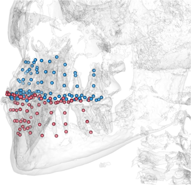
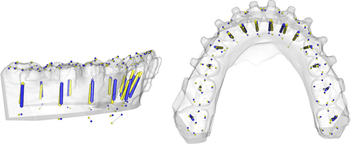
Material and methods: Plaster models of orthognathic patients were scanned and superimposed with three-dimensional data of a cone-beam computer tomography (CBCT). Based on the open-source software "R", including the packages Morpho, mesheR, Rvcg and RvtkStatismo, an SSM was generated to estimate the tooth axes. The accuracy of the calculated tooth axes was determined using a leave-one-out cross-validation. The deviation of tooth axis prediction in terms of angle or horizontal shift is described with mean and standard deviation. The planning dataset of an implant surgery patient was additionally analyzed using the SSM.

Results: 71 datasets were included in this study. The mean angle between the estimated tooth-axis and the actual tooth-axis was 7.5 ± 4.3° in the upper jaw and 6.7 ± 3.8° in the lower jaw. The horizontal deviation between the tooth axis and estimated axis was 1.3 ± 0.8 mm close to the cementoenamel junction, and 0.7 ± 0.5 mm in the apical third of the root. Results for models with one missing tooth did not differ significantly. In the clinical dataset, the SSM could give a reasonable aid for implant positioning.

Conclusions: With the presented SSM, the approximate course of dental roots can be predicted based on a surface scan. There was no difference in predicting the tooth axis of existent or missing teeth. In clinical context, the estimation of tooth axes of missing teeth could serve as a reference for implant positioning. However, a higher number of training data must be achieved to obtain increasing accuracy.

Keywords: Anatomical reconstruction; Implant planning; Statistical shape model; Virtual planning.


Conflict of interest statement

The authors declare that they have no conflict of interest.

Figures

Fig. 1
Fig. 1
Superimposition of plaster cast and segmented CBCT scan. The deviation of the teeth surfaces after registration was throughout below 0.05 mm
Fig. 2
Fig. 2
Procedure for placing landmarks along the roots of multi-rooted teeth. The first landmark was set at the level of the CEJL. Three further landmarks were placed with 2 mm distance along the apical direction. A fifth landmark was set at the mid-point of the apical end of all roots
Fig. 3
Fig. 3
By augmentation of the CBCT-scan with the corresponding plaster cast model, all structures of the teeth could be captured with landmarks. Thus, every individual received 270 landmarks to encode the morphology of the tooth crowns and teeth
Fig. 4
Fig. 4
Measurements performed for evaluation of the calculated tooth axis (red) in comparison with the actual tooth axis (black). One section each at the level of the CEJL and the apical landmark LM-4 were measured, as well as the angular deviation between the two axes
Fig. 5
Fig. 5
Example for the reconstruction of a fully toothed upper arch. Predicted axes (blue) and actual axes (yellow) are shown in one dataset
Fig. 6
Fig. 6
Example for the reconstruction of a fully toothed lower arch. Predicted axes (blue) and actual axes (yellow) are shown in one dataset
Fig. 7
Fig. 7
Using CoDiagnostix, a superimposition of the CBCT-scan with the axes reconstructed by the SSM and a dental wax up (both in yellow) was performed. The axes calculated by the SSM were depicted as yellow cylinders. Therefore, in this horizontal cross section, they appear as a dot. The yellow dots mostly match with the hypodense area of the root canals of the teeth (if existent)
Fig. 8
Fig. 8
Paramedian cut of the CBCT-scan superimposed with a dental wax-up and the SSM-based tooth axis reconstruction. The predicted tooth axis of tooth 23 (yellow) is located at a similar position as the actual root canal of the tooth 23. The comparison of calculated tooth axis with dental roots of existent teeth enables to presume the validity of the SSM-based prediction of missing roots
Fig. 9
Fig. 9
Paramedian cut of the CBCT-scan superimposed with a dental wax-up and the SSM-based tooth axis reconstruction (both in yellow). The SSM-based prediction of the tooth axis of tooth 12 appears to be close to parallel to the planned implantation axis
Fig. 10
Fig. 10
Paramedian cut of the CBCT-scan superimposed with a dental wax-up and the SSM-based tooth axis reconstruction (both in yellow). The SSM-based prediction of the tooth axis of tooth 11 deviates from the planned implantation axis in oro-vestibular direction. Due to vestibular bone loss, the implantation axis was adjusted to guarantee sufficient bone thickness in all dimensions

References

    1. Testori T, Weinstein T, Scutellà F, Wang H-L. Zucchelli G (2018) Implant placement in the esthetic area: criteria for positioning single and multiple implants. Periodontol. 2000;77:176–196. doi: 10.1111/prd.12211. - DOI - PubMed
    1. Scutellà F, Weinstein T, Lazzara R, Testori T. Buccolingual implant position and vertical abutment finish line geometry: two strictly related factors that may influence the implant esthetic outcome. Implant Dent. 2015;24:343–348. doi: 10.1097/ID.0000000000000235. - DOI - PubMed
    1. Morton D, Phasuk K, Polido WD, Lin W-S. Consideration for contemporary implant surgery. Dent Clin North Am. 2019;63:309–329. doi: 10.1016/j.cden.2018.11.010. - DOI - PubMed
    1. Scherer MD. Presurgical implant-site assessment and restoratively driven digital planning. Dent Clin North Am. 2014;58:561–595. doi: 10.1016/j.cden.2014.04.002. - DOI - PubMed
    1. Talluri S, Vaddamanu SK, Apparaju V, Vyas R, Ahuja S, Kanji MA. Evaluating cortico-cancellous ratio using virtual implant planning and its relation with immediate and long-term stability of a dental implant- A CBCT-assisted prospective observational clinical study. Niger J Clin Pract. 2019;22:982. doi: 10.4103/njcp.njcp_22_19. - DOI - PubMed

Substances

LinkOut - more resources