

Angle Kappa agreement between Scheimpflug tomography, combined placido Scheimpflug and combined slit scanning placido systems
- PMID: 35902424
- PMCID: PMC9971106
- DOI: 10.1007/s10792-022-02433-z
Angle Kappa agreement between Scheimpflug tomography, combined placido Scheimpflug and combined slit scanning placido systems
Abstract
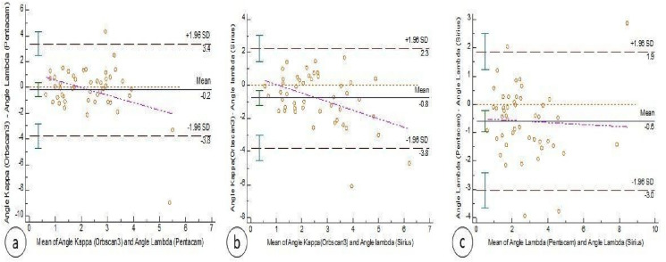
Purpose: To compare the measured or calculated angle Kappa using Oculus pentacam HR, Sirius and Orbscan III devices.
Patients and methods: A prospective randomized cohort study, conducted on 47 eyes of 47 healthy orthotropic individuals, with an age range of 18-50 years and a corrected Snellen's distance visual acuity (CDVA) of 0.8 decimal or better. Angle Kappa is assessed directly using Orbscan® III software version 1.8.165.1. (Bausch and Lomb Rochester, New York, United States), while Pentacam® HR 1.21r.65 (Oculus Optikgeräte GmbH, Wetzlar, Germany) and Sirius device (CSO, version 3.2.1.60, Costruzione Strumenti Oftalmici, Florence, Italy) were used to calculate angle kappa indirectly.
Results: Least mean difference of estimated angle Kappa was between Orbscan and Pentacam devices (- 0.18° ± 1.8), and it was statistically insignificant (p value = 0.1294). Differences between both Orbscan and Sirius, and Pentacam and Sirius were statistically significant (p value = 0.0004 and < 0.0001 consecutively). Bland Altman analysis showed a 95% confidence interval between Orbscan III and Pentacam of - 3.76 to 3.4 and between Orbscan III and Sirius of - 3.79 to 2.26.
Conclusion: Pentacam parameters can be used as a reliable method to calculate angle kappa indirectly, without usage of any additional measurements from other machine. Sirius device parameters could also be used, but with less accurate results. A simple modification to those devices' software to calculate it, and incorporate it in the printout is possible, and highly recommended.
Keywords: Kappa angle; Orbscan III; Pentacam; Sirius device.
© 2022. The Author(s).
Conflict of interest statement
The authors have no relevant financial or non-financial interests to disclose.
Figures
References
-
- Kermani O, Schmeidt K, Oberheide U, Gerten G. (2005) Hyperopic laser in situ keratomileusis with 5.5-, 6.5-, and 7.0-mm optical zones. J Refract Surg 21(1):52–8. - PubMed
Publication types
MeSH terms
LinkOut - more resources
Full Text Sources
