

Somatic GJA4 gain-of-function mutation in orbital cavernous venous malformations
- PMID: 35902510
- PMCID: PMC9908695
- DOI: 10.1007/s10456-022-09846-5
Somatic GJA4 gain-of-function mutation in orbital cavernous venous malformations
Abstract
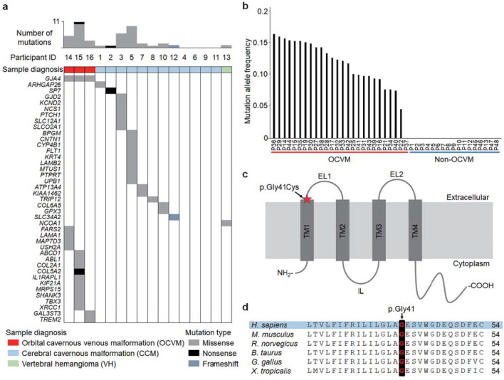
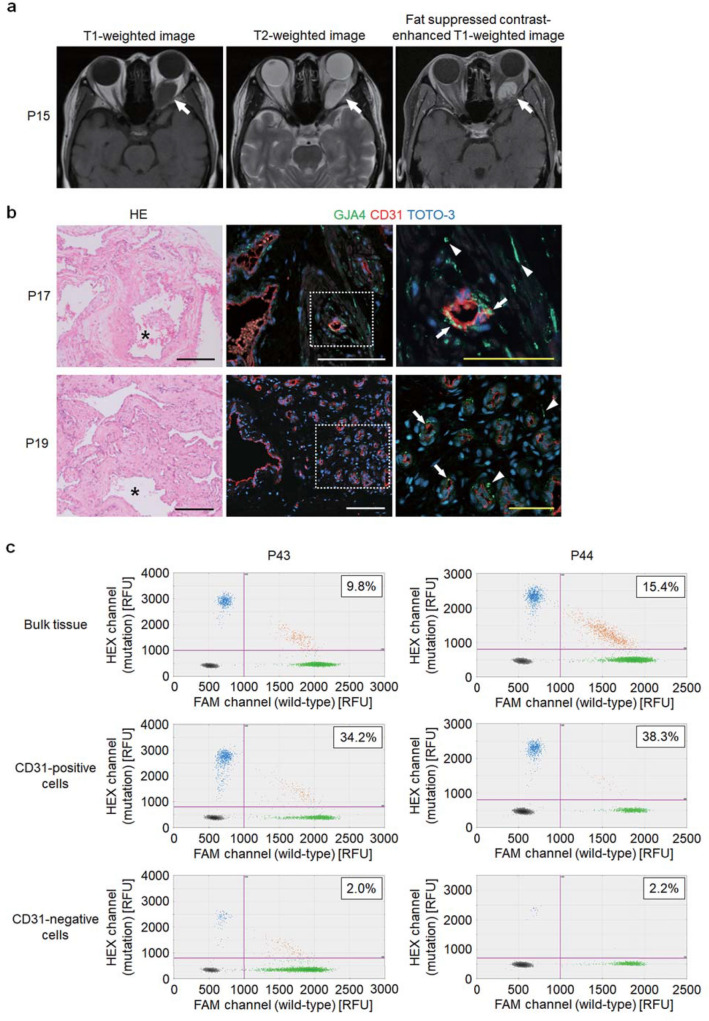
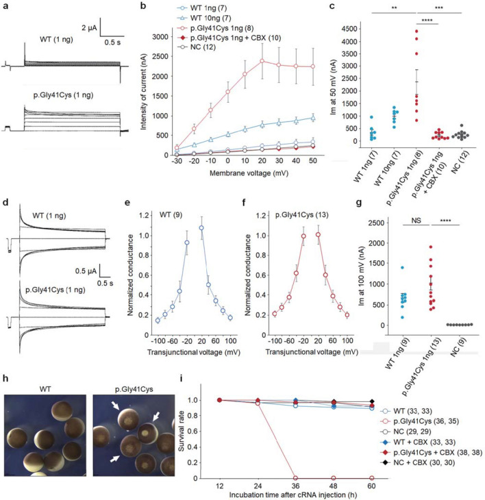
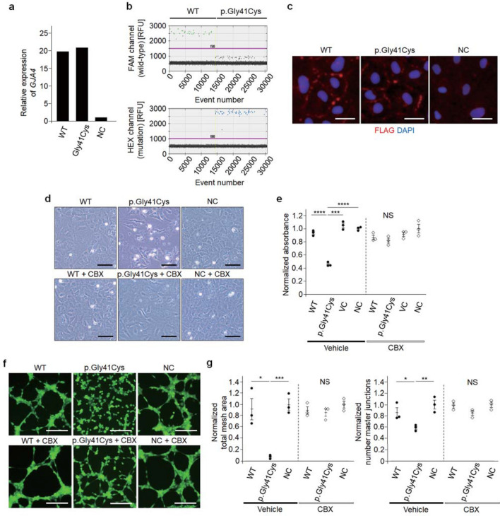
Orbital cavernous venous malformation (OCVM) is a sporadic vascular anomaly of uncertain etiology characterized by abnormally dilated vascular channels. Here, we identify a somatic missense mutation, c.121G > T (p.Gly41Cys) in GJA4, which encodes a transmembrane protein that is a component of gap junctions and hemichannels in the vascular system, in OCVM tissues from 25/26 (96.2%) individuals with OCVM. GJA4 expression was detected in OCVM tissue including endothelial cells and the stroma, through immunohistochemistry. Within OCVM tissue, the mutation allele frequency was higher in endothelial cell-enriched fractions obtained using magnetic-activated cell sorting. Whole-cell voltage clamp analysis in Xenopus oocytes revealed that GJA4 c.121G > T (p.Gly41Cys) is a gain-of-function mutation that leads to the formation of a hyperactive hemichannel. Overexpression of the mutant protein in human umbilical vein endothelial cells led to a loss of cellular integrity, which was rescued by carbenoxolone, a non-specific gap junction/hemichannel inhibitor. Our data suggest that GJA4 c.121G > T (p.Gly41Cys) is a potential driver gene mutation for OCVM. We propose that hyperactive hemichannel plays a role in the development of this vascular phenotype.
Keywords: Connexin; Endothelial cell; Gap junction protein; Orbital disease; Vascular malformations; Whole-cell voltage clamp.
© 2022. The Author(s).
Conflict of interest statement
The authors have no competing interests to declare that are relevant to the content of this article.
Figures
Similar articles
-
Cutaneous and hepatic vascular lesions due to a recurrent somatic GJA4 mutation reveal a pathway for vascular malformation.HGG Adv. 2021 Apr 8;2(2):100028. doi: 10.1016/j.xhgg.2021.100028. Epub 2021 Mar 1. HGG Adv. 2021. PMID: 33912852 Free PMC article.
-
Orbital angioleiomyoma with GJA4 mutation mimicking cavernous venous malformation: a case report and comprehensive review.Orbit. 2025 Jul 18:1-7. doi: 10.1080/01676830.2025.2515594. Online ahead of print. Orbit. 2025. PMID: 40679935
-
Somatic GJA4 mutation in intracranial extra-axial cavernous hemangiomas.Stroke Vasc Neurol. 2023 Dec 29;8(6):453-462. doi: 10.1136/svn-2022-002227. Stroke Vasc Neurol. 2023. PMID: 37072338 Free PMC article.
-
A clinicopathological reappraisal of orbital vascular malformations and distinctive GJA4 mutation in cavernous venous malformations.Hum Pathol. 2022 Dec;130:79-87. doi: 10.1016/j.humpath.2022.10.002. Epub 2022 Oct 6. Hum Pathol. 2022. PMID: 36209871 Review.
-
Emerging issues of connexin channels: biophysics fills the gap.Q Rev Biophys. 2001 Aug;34(3):325-472. doi: 10.1017/s0033583501003705. Q Rev Biophys. 2001. PMID: 11838236 Review.
Cited by
-
Pathological angiogenesis: mechanisms and therapeutic strategies.Angiogenesis. 2023 Aug;26(3):313-347. doi: 10.1007/s10456-023-09876-7. Epub 2023 Apr 15. Angiogenesis. 2023. PMID: 37060495 Free PMC article. Review.
-
Mesenchymal non-meningothelial tumors of the central nervous system: a literature review and diagnostic update of novelties and emerging entities.Acta Neuropathol Commun. 2023 Feb 3;11(1):22. doi: 10.1186/s40478-023-01522-z. Acta Neuropathol Commun. 2023. PMID: 36737790 Free PMC article. Review.
-
Epidemiology and Aetiology of Cerebral Cavernous Malformations.Acta Neurochir Suppl. 2025;136:143-149. doi: 10.1007/978-3-031-89844-0_18. Acta Neurochir Suppl. 2025. PMID: 40632265 Review.
-
Increased Hemichannel Activity Displayed by a Connexin43 Mutation Causing a Familial Connexinopathy Exhibiting Hypotrichosis with Follicular Keratosis and Hyperostosis.Int J Mol Sci. 2023 Jan 22;24(3):2222. doi: 10.3390/ijms24032222. Int J Mol Sci. 2023. PMID: 36768546 Free PMC article.
-
Biological identity of orbital cavernous venous malformations.Arq Bras Oftalmol. 2024 Apr 22;87(2):e2023. doi: 10.5935/0004-2749.2023-0338. eCollection 2024. Arq Bras Oftalmol. 2024. PMID: 38655941 Free PMC article. Review.
References
-
- Rootman DB, Heran MK, Rootman J, White VA, Luemsamran P, Yucel YH. Cavernous venous malformations of the orbit (so-called cavernous haemangioma): a comprehensive evaluation of their clinical, imaging and histologic nature. Br J Ophthalmol. 2014;98(7):880–888. doi: 10.1136/bjophthalmol-2013-304460. - DOI - PubMed
Publication types
MeSH terms
LinkOut - more resources
Full Text Sources
Molecular Biology Databases
Miscellaneous
