. 2022 Jul 28;13(7):658.
doi: 10.1038/s41419-022-05118-8.

PAK6 promotes homologous-recombination to enhance chemoresistance to oxaliplatin through ATR/CHK1 signaling in gastric cancer

Affiliations

PAK6 promotes homologous-recombination to enhance chemoresistance to oxaliplatin through ATR/CHK1 signaling in gastric cancer

Weicai Huang et al. Cell Death Dis. .

Abstract

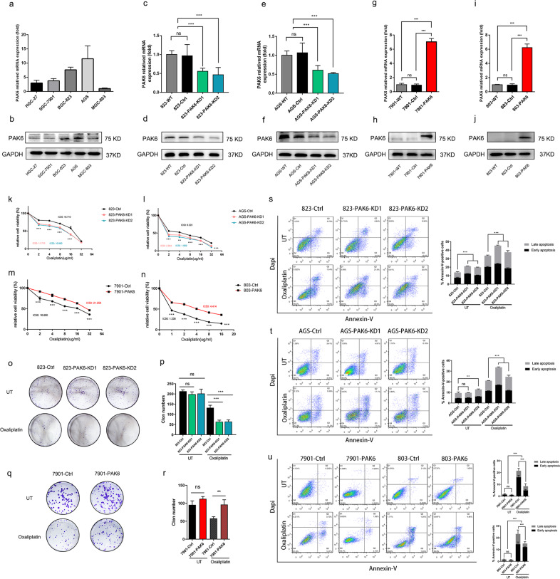
Chemoresistance remains the primary challenge of clinical treatment of gastric cancer (GC), making the biomarkers of chemoresistance crucial for treatment decision. Our previous study has reported that p21-actived kinase 6 (PAK6) is a prognostic factor for selecting which patients with GC are resistant to 5-fluorouracil/oxaliplatin chemotherapy. However, the mechanistic role of PAK6 in chemosensitivity remains unknown. The present study identified PAK6 as an important modulator of the DNA damage response (DDR) and chemosensitivity in GC. Analysis of specimens from patients revealed significant associations between the expression of PAK6 and poorer stages, deeper invasion, more lymph node metastases, higher recurrence rates, and resistance to oxaliplatin. Cells exhibited chemosensitivity to oxaliplatin after knockdown of PAK6, but showed more resistant to oxaliplatin when overexpressing PAK6. Functionally, PAK6 mediates cancer chemoresistance by enhancing homologous recombination (HR) to facilitate the DNA double-strand break repair. Mechanistically, PAK6 moves into nucleus to promote the activation of ATR, thereby further activating downstream repair protein CHK1 and recruiting RAD51 from cytoplasm to the DNA damaged site to repair the broken DNA in GC. Activation of ATR is the necessary step for PAK6 mediated HR repair to protect GC cells from oxaliplatin-induced apoptosis, and ATR inhibitor (AZD6738) could block the PAK6-mediated HR repair, thereby reversing the resistance to oxaliplatin and even promoting the sensitivity to oxaliplatin regardless of high expression of PAK6. In conclusion, these findings indicate a novel regulatory mechanism of PAK6 in modulating the DDR and chemoresistance in GC and provide a reversal suggestion in clinical decision.


Conflict of interest statement

The authors declare no competing interests.

Figures

Fig. 1
Fig. 1. PAK6 is associated with poor prognosis and the therapeutic efficacy of oxaliplatin in GC.
a Kaplan–Meier survival curves of GC patients with PAK6 expression in the KM-Plotter database (www.kmplot.com). b Subgroup survival analysis according to the PAK6 expression with GC patients treated with surgery only in the KM-Plotter database. c Subgroup survival analysis of PAK6 expression with GC patients treated with chemotherapy in the KM-Plotter database. d Representative graphs of PAK6 expression (high expression and low expression) in the tumor tissues. e Quantification of the immunohistochemical staining intensity of the PAK6 expression in GC patients. f Quantification of immunohistochemical staining intensity of the PAK6 expression in different TNM stages. g Percentage of GC patients with high PAK6 expression in the groups classified by TNM stage, tumor invasion, lymph node metastasis, and recurrence. h Kaplan–Meier survival curves of disease-free survival of the GC patients according to the PAK6 expression. i Subgroup survival analysis of disease-free survival of GC patients treated with surgery only according to the PAK6 expression. j Subgroup survival analysis of disease-free survival of GC patients treated with chemotherapy of oxaliplatin/5 FU after surgery according to the PAK6 expression. k Subgroup survival analysis of disease-free survival of GC patients treated with chemotherapy of capecitabine after surgery according to the PAK6 expression. l Kaplan–Meier survival curves of overall survival of the GC patients according to the PAK6 expression. m Subgroup survival analysis of overall survival of GC patients treated with surgery only according to the PAK6 expression. n Subgroup survival analysis of overall survival of GC patients treated with chemotherapy of oxaliplatin/5 FU after surgery according to the PAK6 expression. o Subgroup survival analysis of overall survival of GC patients treated with chemotherapy of capecitabine after surgery according to the PAK6 expression. *: P < 0.05, **: P < 0.01, ns: no statistical difference.
Fig. 2
Fig. 2. PAK6 increases the chemoresistance to oxaliplatin in GC cells.
a, b The expression of PAK6 in HGC-27, SGC-7901, BGC-823, AGS, and MGC-803 GC cell lines. cf Knock down of the expression of PAK6 in 823 (c, d) and AGS (e, f) GC cells. gj Overexpressing the expression of PAK6 in 7901 (g, h) and 803 (i, j) GC cells. kn Dose-response curves of control cells or PAK6 knocked down cells in 823 (k) and AGS (l) GC cells after treated with oxaliplatin for 24 h. The IC50 of the oxaliplatin in the 823 cell line are 18.701(823-Ctrl cells), 11.710 (823-PAK6-KD1 cells), and 10.960 (823-AK6-KD2 cells) μg/ml. The IC50 of the oxaliplatin in the AGS cell line are 6.233 (AGS-Ctrl cells), 2.904 (AGS-PAK6-KD1 cells), and 1.969 (AGS-PAK6-KD2 cells) μg/ml. Dose-response curves of control cells or PAK6 overexpression cells in 7901 (m) and 803 (n) GC cells after treated with oxaliplatin for 24 h. The IC50 of the oxaliplatin in the cells are 10.890 (7901-Ctrl cells), 21.250 (7901-PAK6 cells), 1.236 (803-Ctrl cells), and 4.414 (803-PAK6 cells) μg/ml. or Colony formation ability of PAK6 knocked down cells in 823 GC cell line (o, p) and PAK6 overexpression cells in 7901 (q, r) GC cell line treated with or without oxaliplatin (0.3 μg/ml in 823 cells or 0.5 μg/ml in 7901 cells). s, t Apoptosis analysis of control cells and PAK6 knocked down cells in 823 and AGS GC cells treated with or without oxaliplatin. u Apoptosis analysis of control cells and PAK6 overexpression cells in 7901 and 803 GC cells treated with or without oxaliplatin. Data presented as mean ± SD of three independent replicates. *: P < 0.05, **: P < 0.01, ***: P < 0.001, ns: no statistical difference.
Fig. 3
Fig. 3. PAK6 reduces DNA damage induced by oxaliplatin in GC cells.
ad Go analysis of the gene pathways differentially expressed between PAK6-high expression and PAK6-low expression gastric cancer samples in TCGA database. Four representative GSEA-enrichment plots were shown. e Left: PAK6 staining (red), γH2A.X staining (green), and DAPI staining (blue) in the control cells and PAK6 overexpression cells in 803 GC cell line; Right: Quantification of mean γH2A.X foci per cell. f Left: PAK6 staining (red), γH2A.X staining (green), and DAPI staining (blue) in the control cells and PAK6 overexpression cells in 803 GC cell line after treated with oxaliplatin for 12 h; Right: Quantification of mean γH2A.X foci per cell. g Left: PAK6 staining (red), γH2A.X staining (green), and DAPI staining (blue) in the control cells and PAK6 overexpression cells in 803 GC cell line after recovery from oxaliplatin treating (12 h) for 12 h; Right: Quantification of mean γH2A.X foci per cell. At least 30 cells per group were included for the counting and quantification. h, i Left: Representative images of neutral comet assays of control cells, PAK6 overexpression cells, and PAK6 knocked down cells in the absence and presence of oxaliplatin; Right: Quantification of the percentages of DNA tail moments. At least 30 cells per group were included for the counting and quantification. UT: untreated. *: P < 0.05, ***: P < 0.001, ****: P < 0.0001, ns: no statistical difference.
Fig. 4
Fig. 4. PAK6 regulates the efficiency of HR.
a Schematic illustration of HR in HEK 293T-HR-EGFP reporter cells. b PAK6 overexpression leads to increase HR efficiency. HEK 293T-HR-EGFP cells were cotransfected with either empty controls or PAK6 and Myc-I-SceI plasmids. The expression of EGFP, PAK6, and Myc-I-SceI was detected by WB analysis. The percentage of EGFP positive cells were examined by flow cytometry. cf Real-time PCR analysis of the mRNA level of the key molecules involved in HR pathway in PAK6 knocked down and overexpression cells in the absence (c, e) and presence (d, f) of oxaliplatin. Data presented as mean ± SD of three independent replicates. g, h WB analysis of HR related key genes, PAK6, γH2A.X, cleaved caspase 3, and GAPDH in PAK6 knocked down cells of 823 and AGS GC cell lines (g) and PAK6 overexpression cells of 7901 and 803 GC cell lines (h) in the absence and presence of oxaliplatin. i, j Left: Representative images of PAK6 staining (red), RAD51 staining (green), and DAPI staining (blue) of control cells and PAK6 overexpression cells in 7901 (i) and 803 (j) GC cell lines in the absence and presence of oxaliplatin. Right: Quantification of mean γH2A.X foci per cell. At least 30 cells per group were included for the counting and quantification. *: P < 0.05, ***: P < 0.001, ns: no statistical difference.
Fig. 5
Fig. 5. ATR/CHK1 signaling activation is required for PAK6 mediated HR repair.
a, b WB analysis of HR related key genes, PAK6, γH2A.X, cleaved caspase 3, and GAPDH in control cells and PAK6 overexpression cells in 7901 (a) and 803 (b) GC cell lines. c, d WB analysis of PAK6, RAD51, GAPDH, and lamin B1 protein in nucleus in control cells and PAK6 overexpression cells in 7901 (c) and 803 (d) GC cell lines. Cells were untreated, subjected to oxaliplatin, subjected to combination of oxaliplatin and ATR inhibitor AZD6738, or subjected to combination of oxaliplatin and ATM inhibitor AZD0156 for 24 h. e Left: Representative images of γH2A.X staining (red), RAD51 staining (green), and DAPI staining (blue) of control cells and PAK6 overexpression cells in 803 GC cell lines in the presence of oxaliplatin. Right: Representative images of γH2A.X staining (red), RAD51 staining (green), and DAPI staining (blue) of control cells and PAK6 overexpression cells in 803 GC cell lines in the presence of combination of oxaliplatin and ATR inhibitor AZD6738. f Quantification of mean γH2A.X and RAD51 foci per cell. At least 30 cells per group were included for the counting and quantification. g Immunoprecipitation assay with anti-PAK6 antibody from control cells and PAK6 overexpression cells in 7901 GC cell line. Cells were untreated or subjected to oxaliplatin for 24 h. And protein was lysed from cytoplasm and nucleus respectively. h, i Relative protein band intensity of p-ATR (h) and p-CHK1 (i) protein normalized against of PAK6. UT: untreated. ATRi: ATR inhibitor AZD6738. ATMi: ATM inhibitor AZD0156. ***: P < 0.001.
Fig. 6
Fig. 6. ATR inhibitor AZD6738 combinates with oxaliplatin could re-sensitize PAK6 overexpression GC cells to oxaliplatin.
a, b Dose-response curves of control cells or PAK6 overexpression cells in 7901 (a) and 803 (b) GC cell line after treated with oxaliplatin or combination of ATR inhibitor AZD6738 and oxaliplatin for 24 h. Data presented as mean ± SD of three independent replicates. c Left: Representative images of neutral comet assays of control cells and PAK6 overexpression cells in 7901 GC cell lines in the presence of oxaliplatin or combination of ATR inhibitor AZD6738 and oxaliplatin; Right: Quantification of the percentages of DNA tail moments. At least 30 cells per group were included for the counting and quantification. d Apoptosis analysis of control cells and PAK6 overexpression cells in 7901 and 803 GC cell lines in the presence of oxaliplatin or combination of ATR inhibitor AZD6738 and oxaliplatin. e The percentages of apoptotic cells were displayed by the bar chart. Data presented as mean ± SD of three independent replicates. ATRi: ATR inhibitor AZD6738. OX: oxaliplatin. *P < 0.05, **P < 0.01, ***P < 0.001, ns: no statistical difference.
Fig. 7
Fig. 7. ATR inhibitor AZD6738 enhances antitumor efficacy of oxaliplatin in PAK6 overexpression cell line xenograft.
a Tumor picture for each group was displayed. b Tumor growth of indicated 803 xenografts in each group. c Animal weight for indicated treatments. d HE staining and IHC staining of PAK6, RAD51, γH2A.X, and cleaved caspase 3 for the tumor isolated from the indicated treatments. Data presented as mean ± SD of three independent replicates. OX: oxaliplatin; ATRi: ATR inhibitor AZD6738; *: P < 0.05, ns: no statistical difference.
Fig. 8
Fig. 8. A model for PAK6 induced chemoresistance to oxaliplatin in GC.
In PAK6 high expression GC cells, when DSB is initiated by the treatment of oxaliplatin, PAK6 could behave as an upper DDR factor to move into nucleus from cytoplasm to promote the phosphorylation of ATR. And then the activated p-ATR could further phosphorylate CHK1 to arrest the cell cycle in G2/M phase, and promote the recruitment of RAD51 to the nucleus to perform HR repair, thereby promoting the chemoresistance to oxaliplatin, ultimately leading to cells survival. However, failure to maintain the appropriate phosphorylation of ATR after conducting AZD6738 based on oxaliplatin treatment results in deficiency of recruitment of RAD51 to the nucleus to perform HR repair, ultimately leading to cell apoptosis. Combination of AZD6738 and oxaliplatin could overcome the chemoresistance to oxaliplatin induced by PAK6.

Similar articles

Cited by

References

    1. Smyth EC, Nilsson M, Grabsch HI, van Grieken NCT, Lordick F. Gastric cancer. Lancet. 2020;396:635–48. doi: 10.1016/S0140-6736(20)31288-5. - DOI - PubMed
    1. Sung H, Ferlay J, Siegel RL, Laversanne M, Soerjomataram I, Jemal A, et al. Global Cancer Statistics 2020: GLOBOCAN Estimates of Incidence and Mortality Worldwide for 36 Cancers in 185 Countries. CA Cancer J Clin. 2021;71:209–49. doi: 10.3322/caac.21660. - DOI - PubMed
    1. Thrift AP, El-Serag HB. Burden of gastric cancer. Clin Gastroenterol Hepatol. 2020;18:534–42. doi: 10.1016/j.cgh.2019.07.045. - DOI - PMC - PubMed
    1. Al-Batran S-E, Hartmann JT, Probst S, Schmalenberg H, Hollerbach S, Hofheinz R, et al. Phase III trial in metastatic gastroesophageal adenocarcinoma with fluorouracil, leucovorin plus either oxaliplatin or cisplatin: a study of the Arbeitsgemeinschaft Internistische Onkologie. J Clin Oncol. 2008;26:1435–42. doi: 10.1200/JCO.2007.13.9378. - DOI - PubMed
    1. Raymond E, Chaney SG, Taamma A, Cvitkovic E. Oxaliplatin: a review of preclinical and clinical studies. Ann Oncol. 1998;9:1053–71. doi: 10.1023/A:1008213732429. - DOI - PubMed

Publication types

MeSH terms