. 2022 Jul 28;13(1):4374.
doi: 10.1038/s41467-022-31442-w.

PACAP-PAC1R modulates fear extinction via the ventromedial hypothalamus

Affiliations

PACAP-PAC1R modulates fear extinction via the ventromedial hypothalamus

E R Velasco et al. Nat Commun. .

Abstract

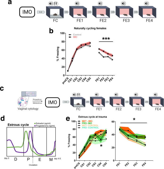
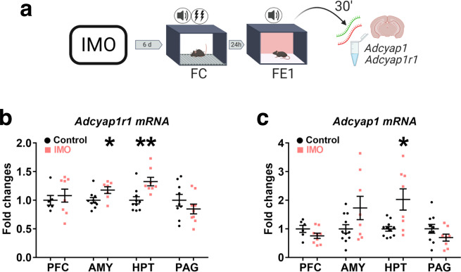
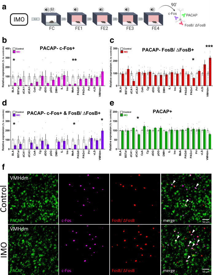
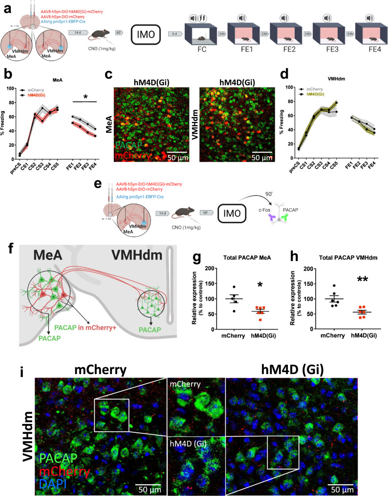
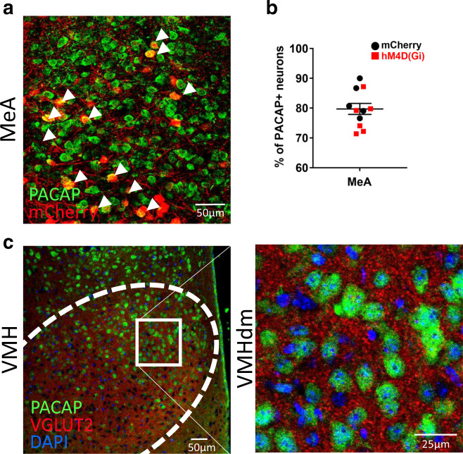
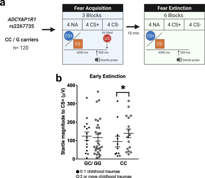
Exposure to traumatic stress can lead to fear dysregulation, which has been associated with posttraumatic stress disorder (PTSD). Previous work showed that a polymorphism in the PACAP-PAC1R (pituitary adenylate cyclase-activating polypeptide) system is associated with PTSD risk in women, and PACAP (ADCYAP1)-PAC1R (ADCYAP1R1) are highly expressed in the hypothalamus. Here, we show that female mice subjected to acute stress immobilization (IMO) have fear extinction impairments related to Adcyap1 and Adcyap1r1 mRNA upregulation in the hypothalamus, PACAP-c-Fos downregulation in the Medial Amygdala (MeA), and PACAP-FosB/ΔFosB upregulation in the Ventromedial Hypothalamus dorsomedial part (VMHdm). DREADD-mediated inhibition of MeA neurons projecting to the VMHdm during IMO rescues both PACAP upregulation in VMHdm and the fear extinction impairment. We also found that women with the risk genotype of ADCYAP1R1 rs2267735 polymorphism have impaired fear extinction.


Conflict of interest statement

The authors declare no competing interests.

Figures

Fig. 1
Fig. 1. Long-lasting alterations of behavior after trauma and its association with the estrous cycle phase.
a Schematic representation of the behavioral protocol. b Fear learning and fear extinction in naturally cycling females undergoing IMO or compensatory handling (control: n = 31, IMO: n = 32) (P = 0.001). c Methods used to monitor the estrous cycle before the behavioral procedures. d Estrous cycle and related hormonal levels in female C57BL/6 mice. e Fear learning, and fear extinction in proestrus or metestrus females (n = 7 per group) (P = 0.029). Data are expressed as mean ± SEM. *P ≤ 0.05, ***P ≤ 0.001. In b, e, main effect stress, main effect estrous cycle, main effect FE session or CS, and FE session* stress or CS*stress, estrous cycle*stress, FE session * estrous cycle, FE session * stress * estrous cycle interactions were analyzed using repeated-measures ANOVA, Bonferroni corrections were made for multiple comparisons. Asterisks above a line indicate significant main effect stress in repeated-measures ANOVA. CS conditioned stimulus, D diestrus, E estrus, FC fear conditioning, FE fear extinction session, IMO immobilization stress, M metestrus, Met metestrus, P proestrus, Pro proestrus. Source data are provided as a Source Data file.
Fig. 2
Fig. 2. Adcyap1-Adcyap1r1 regulation during early FE (FE1).
a Representation of the methods used to evaluate Adcyap1, Adcyap1r1 regulation after FE1. b Adcyap1r1 mRNA regulation 30 min after FE1 in prefrontal cortex (PFC) (control: n = 6, IMO: n = 8), amygdala (AMY) (control: n = 10, IMO: n = 7) (P = 0.030), hypothalamus (HPT) (control: n = 10, IMO: n = 8) (P = 0.002), and periaqueductal gray (PAG) (control: n = 9, IMO: n = 9). c Adcyap1 in PFC (control: n = 6, IMO: n = 9), AMY (control: n = 11, IMO: n = 8), HPT (control: n = 11, IMO: n = 8) (P = 0.020), and PAG (control: n = 10, IMO: n = 8). Data are means ± SEM. *P ≤ 0.05, **P ≤ 0.01. Two-tailed t tests were conducted. IMO immobilization stress, FC fear conditioning, FE fear extinction. Source data are provided as a Source Data file.
Fig. 3
Fig. 3. Immobilization stress induces changes in PACAP+ neuronal activation after a fear extinction task.
a Schematic representation of the behavioral and immunohistochemical methods. b Quantification of cellular colocalization of PACAP-c-Fos+ (BLA and MeA n = 5 per group) (BLA: P = 0.023, MeA: P = 0.005). c Quantification of cellular colocalization of PACAP-FosB/ΔFosB+ (PAGvl n = 6 per group, VMHdm control: n = 4, IMO: n = 6) (PAGvl: P = 0.037, VMHdm: P = 0.001). d Quantification of double colocalization of PACAP-c-Fos+ and FosB/ΔFosB+ (BLA and MeA n = 5 per group; VMH control: n = 4, IMO: n = 6) (BLA: P = 0.015, MeA: P = 0.040, VMHdm: P = 0.030). e Quantification of PACAP+ cells (dCA3 n = 4 per group) (dCA3: P = 0.028). f Representative confocal images of PACAP, c-Fos, FosB/ΔFosB colocalization in the VMHdm. White arrowheads signal triple colocalization. Scale bar 50 μm. Results are presented as relative expression compared to controls (in %) and extracted from cells per mm2 normalized to DAPI. Data are presented as means ± SEM. *P ≤ 0.05, **P ≤ 0.01, ***P ≤ 0.001. Two-tailed t tests or Mann–Whitney’s U tests were used. BLA basolateral amygdala, BSTal bed nucleus of the stria terminalis anterolateral part, dCA1 dorsal CA1, dCA3 dorsal CA3, CeA central amygdala, Cg cingulate cortex, dDG dorsal dentate gyrus, pDg posterior dentate gyrus, DMH dorsomedial hypothalamus, FC fear conditioning, FE fear extinction, IL infralimbic cortex, IMO immobilization stress, Ins insular cortex, MeA medial amygdala, PAGdl dorsolateral periaqueductal gray, PAGvl ventrolateral periaqueductal gray, Prl prelimbic cortex, vLS ventral lateral septum, VMHdm ventromedial hypothalamus dorsomedial nucleus. Source data are provided as a Source Data file.
Fig. 4
Fig. 4. Effect of the chemogenetic inhibition of the medial amygdala-ventromedial hypothalamus circuit during immobilization stress.
a Schematic representation of the methods and timeline of experiments. b Fear learning and fear extinction in mice with a temporal inhibition of MeA to VMHdm projections (hM4D(Gi)) during IMO vs controls (mCherry) (mCherry: n = 10, hM4D(Gi): n = 8) (P = 0.036). c, left panel: representative confocal images showing PACAP+ (green) neurons and mCherry+ (red) cell bodies in the MeA in animals receiving hM4D(Gi) in MeA; right panel: cell bodies in the VMHdm in animals receiving hM4D(Gi) in VMHdm. Scale bar 50 μm. d Fear learning, and fear extinction in mice with a temporal inhibition of VMHdm to MeA projections (hM4D(Gi)) during IMO vs controls (mCherry) (mCherry: n = 6, hM4D(Gi): n = 8). e, f Methods used to assess PACAP expression shortly after IMO in animals with inhibited MeA to VMHdm circuitry (hM4D(Gi) vs controls (mCherry)). g, h Effect of a temporal inhibition of MeA to VMHdm projections on PACAP expression shortly after IMO in the MeA (mCherry: n = 5, hM4D(Gi): n = 6) (P = 0.015) and VMHdm (mCherry: n = 6, hM4D(Gi): n = 6) (P = 0.005). Results are presented as relative expression compared to controls (in %) and extracted from PACAP levels normalized to DAPI (n = 5–6 per group). i Representative confocal images displaying differences in the expression of PACAP in the VMHdm of animals receiving a temporal inhibition of MeA to VMHdm projections during IMO (hM4D(Gi)) left panel vs controls (mCherry) right panel. Respective magnifications are shown in the middle part upper panel (mCherry) and lower panel (hM4D(Gi)). Scale bar: 50 μm. Data are means ± SEM. *P ≤ 0.05, **P ≤ 0.01. In b, d, main effect treatment, main effect FE session, and FE session * treatment interaction were analyzed using a repeated-measures ANOVA. Asterisks above a line indicate significant main effect treatment in repeated-measures ANOVA. In g, h, two-tailed t tests were used. CNO clozapine N-oxide, FC fear conditioning, FE fear extinction, IMO immobilization stress, MeA medial amygdala, VMHdm ventromedial hypothalamus dorsomedial nucleus. Source data are provided as a Source Data file.
Fig. 5
Fig. 5. Sites of delivery of viral vectors in MeA and VMHdm.
a Representative image showing the colocalization of mCherry+ neurons with PACAP+ immunolabeling in the MeA. White arrowheads signal colocalization. b Quantification of the proportion of mCherry+ cells with PACAP+ immunolabeling of animals receiving AAV5rg-pmSyn-EBFP-Cre in the VMHdm and either AAV-hM4Di-DREADD or AAV-DIO-mCherry in the MeA (mCherry: n = 5, hM4D(Gi): n = 6). c Representative image showing a prominent VGLUT2 expression in the VMHdm where PACAP+ neurons are enriched. Data are expressed as mean ± SEM. Scale bars a, 50 μm; c left panel scale bar 50 μm, right panel 25 μm. MeA medial amygdala, VMHdm ventromedial hypothalamus dorsomedial nucleus. Source data are provided as a Source Data file.
Fig. 6
Fig. 6. Post-traumatic symptoms and menstrual cycle phases.
a Menstrual cycle and related hormonal levels in women. b Post-traumatic symptom scores of women at 3 weeks post trauma analyzed by menstrual cycle phase at trauma (EF: n = 38, LF: n = 29, LUT: n = 47) c, or PTSD diagnosis within the first year of follow-up (PTSD: n = 16, No PTSD: n = 69) (P = 0.009). Data are expressed as mean ± SEM. **P ≤ 0.01. In b, the main effect menstrual cycle was analyzed in a one-way ANOVA. In c, post-traumatic symptoms were analyzed using a two-tailed t test. EF early follicular, LF late follicular, LUT luteal phase, PTSD post-traumatic stress disorder. Source data are provided as a Source Data file.
Fig. 7
Fig. 7. ADCYAP1R1 rs2267735 genotype effects during early FE in highly traumatized individuals.
a Methods used for the assessment of the influence of rs2267735 genotype over FE (n = 71, 49). b Fear-potentiated startle magnitude of cycling women (≤40 years old) with CC or GC/GG genotypes during early FE and its relation to childhood trauma exposure (n = 71) (P = 0.030). Analyses for >40 years old (n = 49) are shown in Supplementary Fig. 9c. Data are means ± SEM. *P ≤ 0.05. Fear-potentiated startle data in b were analyzed with an ANCOVA with extinction as repeated measures variable, age, genotype, and childhood trauma as between-subjects variables. Covariates included GWAS principal components, total lifetime trauma, baseline startle. None of the CS+ presentations was paired with an US during the FE phase. CS+ reinforced conditioned stimulus, CS- non-reinforced conditioned stimulus, NA noise probe alone, US unconditioned stimulus. Source data are provided as a Source Data file.

References

    1. WHO. Global health risks: mortality and burden of disease attributable to selected major risks. Bull. World Health Organ. 2009;87:646–646. doi: 10.2471/BLT.09.070565. - DOI - PMC - PubMed
    1. American Psychiatric Association. Diagnostic and Statistical Manual of Mental Disorders (American Psychiatric Association, 2013).
    1. Jovanovic T, et al. Impaired fear inhibition is a biomarker of PTSD but not depression. Depress Anxiety. 2010;27:244–251. doi: 10.1002/da.20663. - DOI - PMC - PubMed
    1. Bryant, R. A. Posttraumatic stress disorder. in The Science of Cognitive Behavioral Therapy (eds Hofmann, S. & Asmundson, G.) 319–336 (Elsevier, 2017).
    1. Tortella-Feliu M, et al. Risk factors for posttraumatic stress disorder: an umbrella review of systematic reviews and meta-analyses. Neurosci. Biobehav. Rev. 2019;107:154–165. doi: 10.1016/j.neubiorev.2019.09.013. - DOI - PubMed

Publication types

MeSH terms

Substances