Review
. 2022 Jul 28;23(1):35.
doi: 10.1186/s12860-022-00433-6.

Chromatin structure in cancer

Affiliations
Review

Chromatin structure in cancer

Meng Wang et al. BMC Mol Cell Biol. .

Abstract

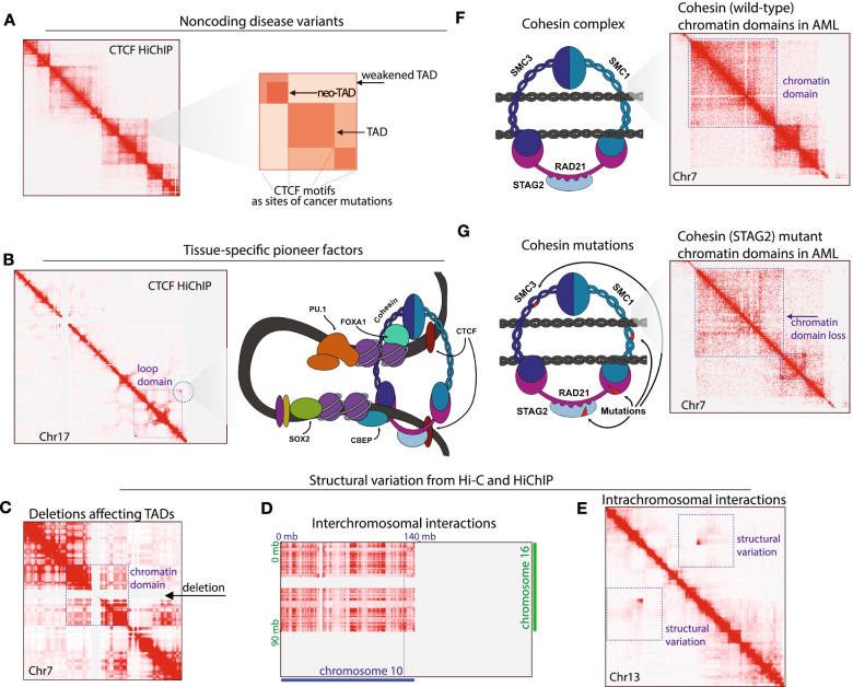
In the past decade, we have seen the emergence of sequence-based methods to understand chromosome organization. With the confluence of in situ approaches to capture information on looping, topological domains, and larger chromatin compartments, understanding chromatin-driven disease is becoming feasible. Excitingly, recent advances in single molecule imaging with capacity to reconstruct "bulk-cell" features of chromosome conformation have revealed cell-to-cell chromatin structural variation. The fundamental question motivating our analysis of the literature is, can altered chromatin structure drive tumorigenesis? As our community learns more about rare disease, including low mutational frequency cancers, understanding "chromatin-driven" pathology will illuminate the regulatory structures of the genome. We describe recent insights into altered genome architecture in human cancer, highlighting multiple pathways toward disruptions of chromatin structure, including structural variation, noncoding mutations, metabolism, and de novo mutations to architectural regulators themselves. Our analysis of the literature reveals that deregulation of genome structure is characteristic in distinct classes of chromatin-driven tumors. As we begin to integrate the findings from single cell imaging studies and chromatin structural sequencing, we will be able to understand the diversity of cells within a common diagnosis, and begin to define structure-function relationships of the misfolded genome.

Keywords: Cancer epigenetics; Chromatin imaging; Chromatin structure; Genome sequencing; Sarcoma; Structural variation.


Conflict of interest statement

There are no competing interests to declare.

Figures

Fig. 1
Fig. 1
Major chromatin structural attributes of cancer. A Disease variants are associated with chromatin interactions. We illustrate non-coding mutations affecting CTCF binding sites in the context of weakened TADs, neo-TADs, and TAD boundaries. CTCF HiChIP data visualized (from [85]) with annotations for how these structural elements may be altered in tumors. B Tissue-specific pioneer transcription factors at loop anchors. We illustrate chromatin loop domains, visualized (from [85]) with CTCF HiChIP, and annotations for how transcription factors would occupy the termini of loop domains. C Structural variants can alter chromatin domains. We illustrate how deletions alter the visualization of TADs and chromatin domains. Data visualization is from IMR90 cells [6]. D Illustration of interchromosomal rearrangements revealed in in Hi-C experiments, with interactions spanning chromosome 10 and chromosome 16 from GM12878 cells, visualized from available Hi-C data [6]. E Intrachromosomal structural variation with rearrangements occurring within a chromosome, viewed from HiChIP experiments in AML cells, focused on chromosome 13 [85]. F Mammalian cohesin complexes are illustrated with the major subunits SMC1, SMC3, RAD21, STAG2 shown (left). Chromatin domains are shown in AML cells with wild-type cohesion complexes (right; chromosome 7), visualized from available data [85]. G Cohesin mutations affect chromatin interactions. Cohesin loss can occur through alterations in the major subunits, as shown (left). The result of Cohesin mutations on chromatin interactions is the substantial loss of TAD-level interactions, as visualized from available data on AML cells with STAG2 loss (right; chromosome 7, [85])

References

    1. Lander ES, Linton LM, Birren B, Nusbaum C, Zody MC, Baldwin J, Devon K, Dewar K, Doyle M, FitzHugh W, et al. Initial sequencing and analysis of the human genome. Nature. 2001;409(6822):860–921. doi: 10.1038/35057062. - DOI - PubMed
    1. Consortium EP. Snyder MP, Gingeras TR, Moore JE, Weng Z, Gerstein MB, Ren B, Hardison RC, Stamatoyannopoulos JA, Graveley BR, et al. Perspectives on ENCODE. Nature. 2020;583(7818):693–698. doi: 10.1038/s41586-020-2449-8. - DOI - PMC - PubMed
    1. Simonis M, Klous P, Splinter E, Moshkin Y, Willemsen R, de Wit E, van Steensel B, de Laat W. Nuclear organization of active and inactive chromatin domains uncovered by chromosome conformation capture-on-chip (4C) Nat Genet. 2006;38(11):1348–1354. doi: 10.1038/ng1896. - DOI - PubMed
    1. Nora EP, Lajoie BR, Schulz EG, Giorgetti L, Okamoto I, Servant N, Piolot T, van Berkum NL, Meisig J, Sedat J, et al. Spatial partitioning of the regulatory landscape of the X-inactivation centre. Nature. 2012;485(7398):381–385. doi: 10.1038/nature11049. - DOI - PMC - PubMed
    1. Lieberman-Aiden E, van Berkum NL, Williams L, Imakaev M, Ragoczy T, Telling A, Amit I, Lajoie BR, Sabo PJ, Dorschner MO, et al. Comprehensive mapping of long-range interactions reveals folding principles of the human genome. Science. 2009;326(5950):289–293. doi: 10.1126/science.1181369. - DOI - PMC - PubMed