. 2022 Jul 28;11(1):37.
doi: 10.1186/s40035-022-00311-3.

Olfactory swab sampling optimization for α-synuclein aggregate detection in patients with Parkinson's disease

Affiliations

Olfactory swab sampling optimization for α-synuclein aggregate detection in patients with Parkinson's disease

Matilde Bongianni et al. Transl Neurodegener. .

Erratum in

Abstract

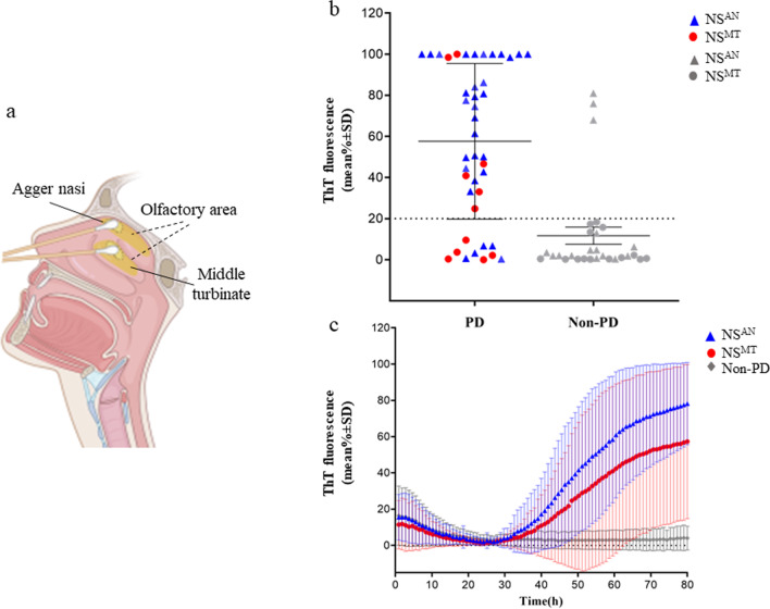
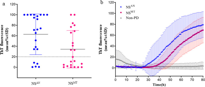
Background: In patients with Parkinson's disease (PD), real-time quaking-induced conversion (RT-QuIC) detection of pathological α-synuclein (α-syn) in olfactory mucosa (OM) is not as accurate as in other α-synucleinopathies. It is unknown whether these variable results might be related to a different distribution of pathological α-syn in OM. Thus, we investigated whether nasal swab (NS) performed in areas with a different coverage by olfactory neuroepithelium, such as agger nasi (AN) and middle turbinate (MT), might affect the detection of pathological α-syn.

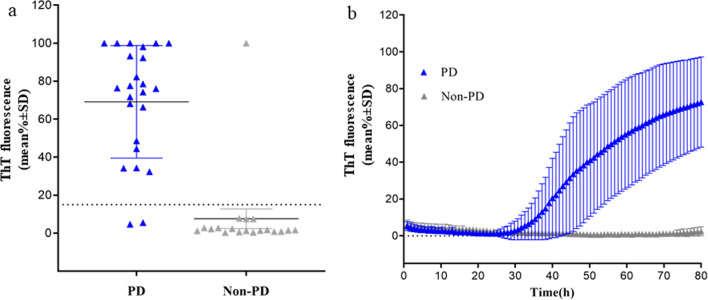
Methods: NS was performed in 66 patients with PD and 29 non-PD between September 2018 and April 2021. In 43 patients, cerebrospinal fluid (CSF) was also obtained and all samples were analyzed by RT-QuIC for α-syn.

Results: In the first round, 72 OM samples were collected by NS, from AN (NSAN) or from MT (NSMT), and 35 resulted positive for α-syn RT-QuIC, including 27/32 (84%) from AN, 5/11 (45%) from MT, and 3/29 (10%) belonging to the non-PD patients. Furthermore, 23 additional PD patients underwent NS at both AN and MT, and RT-QuIC revealed α-syn positive in 18/23 (78%) NSAN samples and in 10/23 (44%) NSMT samples. Immunocytochemistry of NS preparations showed a higher representation of olfactory neural cells in NSAN compared to NSMT. We also observed α-syn and phospho-α-syn deposits in NS from PD patients but not in controls. Finally, RT-QuIC was positive in 22/24 CSF samples from PD patients (92%) and in 1/19 non-PD.

Conclusion: In PD patients, RT-QuIC sensitivity is significantly increased (from 45% to 84%) when NS is performed at AN, indicating that α-syn aggregates are preferentially detected in olfactory areas with higher concentration of olfactory neurons. Although RT-QuIC analysis of CSF showed a higher diagnostic accuracy compared to NS, due to the non-invasiveness, NS might be considered as an ancillary procedure for PD diagnosis.

Keywords: Alpha-synuclein; Cerebrospinal fluid; Olfactory mucosa; Parkinson disease; Real-time quaking-induced conversion assay.


Conflict of interest statement

All authors report no competing interests.

Figures

Fig. 1
Fig. 1
RT-QuIC detection of α-syn seeding activity in the nasal swabs (NSs) of patients with PD and non-PD. a Illustration of olfactory mucosa (OM) swab sampling from agger nasi (AN) (NSAN) and middle turbinate (MT) (NSMT) (created with BioRender.com). b Average thioflavin T (ThT) fluorescence from four replicate readings obtained in NS for each subject with PD (n = 43) and non-PD (n = 29) at 80 h. NSAN are shown as blue triangles (n = 46) while red dots denote NSMT (n = 26). c Traces represent the relative average percentage of ThT fluorescence readings from four replicate reactions (normalized as described in the Methods section) from PD and non-PD samples. The means (thick lines) with standard deviations (thin lines) are shown as a function of RT-QuIC reaction time
Fig. 2
Fig. 2
RT-QuIC detection of α-synuclein seeding activity in the nasal swabs (NS) of PD patients at agger nasi (AN) and middle turbinate (MT). a The final average relative ThT fluorescence from four replicate readings obtained from NS of each individual case (n = 23) at the level of AN and through the MT at 80 h. Bars show the average ± SD for all the cases in each group. The dashed line shows the fluorescence threshold for a positive result. b Traces represent the relative average percentage of ThT fluorescence readings of positive samples from AN (blue trace) and the MT (magenta trace), and non-PD (grey trace) as negative controls. The means (thick lines) with SDs (thin lines) of those averages are shown as a function of RT-QuIC reaction time
Fig. 3
Fig. 3
Immunocytochemical analyses of nasal swabs (NS) of controls and PD patients. a, b OM cell preparations from NS of AN (a) and MT (b) of a PD patient and immunostained with anti-β-tubulin III antibody (green) (scale bars 25 µm, magnification 20×). Thin and elongated β-III tubulin-positive cells with a neuronal-shape morphology are dominant in AN (inset, scale bar 25 µm) compared to MT (inset, scale bar 25 µm). c, d Immunostaining with anti-α-syn (red) and anti-phospho-α-syn (green) antibodies of NS from the PD patient showed α-syn and phospho-α-syn deposits in neuronal-shaped and ciliated cells. In contrast, NS from a non-PD patient (e) and normal control (f) showed a weak positivity (scale bar 25 µm, magnification 40×). Nuclei were stained with DAPI (blue)
Fig. 4
Fig. 4
RT-QuIC detection of α-syn seeding activity in CSF samples of patients with PD and non-PD. a The average of relative ThT fluorescence from four replicate readings obtained from CSF of each individual cases with PD (n = 24) and non-PD (n = 19) at 80 h. Bars show the average ± SD for each group. The dashed line shows the fluorescence threshold for a positive result. b Traces represent the average percentage of ThT fluorescence readings from positive 22/24 CSF samples from patients with PD and 1/19 non-PD . The means (thick lines) with SD (thin lines) of those averages are shown as a function of RT-QuIC reaction time

References

    1. Poewe W, Seppi K, Tanner CM, Halliday GM, Brundin P, Volkmann J, et al. Parkinson disease. Nat Rev Dis Prim. 2017;3:1–21. - PubMed
    1. Postuma RB, Berg D, Stern M, Poewe W, Olanow CW, Oertel W, et al. MDS clinical diagnostic criteria for Parkinson’s disease. Mov Disord. 2015;30(12):1591–1601. doi: 10.1002/mds.26424. - DOI - PubMed
    1. Emre M, Aarsland D, Brown R, Burn DJ, Duyckaerts C, Mizuno Y, et al. Clinical diagnostic criteria for dementia associated with Parkinson’s disease. Mov Disord. 2007;22(12):1689–1707. doi: 10.1002/mds.21507. - DOI - PubMed
    1. Braak H, Del Tredici K, Rüb U, De Vos RAI, Jansen Steur ENH, Braak E. Staging of brain pathology related to sporadic Parkinson’s disease. Neurobiol Aging. 2003;24(2):197–211. doi: 10.1016/S0197-4580(02)00065-9. - DOI - PubMed
    1. Fairfoul G, McGuire LI, Pal S, Ironside JW, Neumann J, Christie S, et al. Alpha-synuclein RT-QuIC in the CSF of patients with alpha-synucleinopathies. Ann Clin Transl Neurol. 2016;3(10):812–818. doi: 10.1002/acn3.338. - DOI - PMC - PubMed

Publication types

Substances