Review
. 2022 Jul 12:3:931331.
doi: 10.3389/fragi.2022.931331. eCollection 2022.

Pathobiology of the Klotho Antiaging Protein and Therapeutic Considerations

Affiliations
Review

Pathobiology of the Klotho Antiaging Protein and Therapeutic Considerations

Gérald J Prud'homme et al. Front Aging. .

Abstract

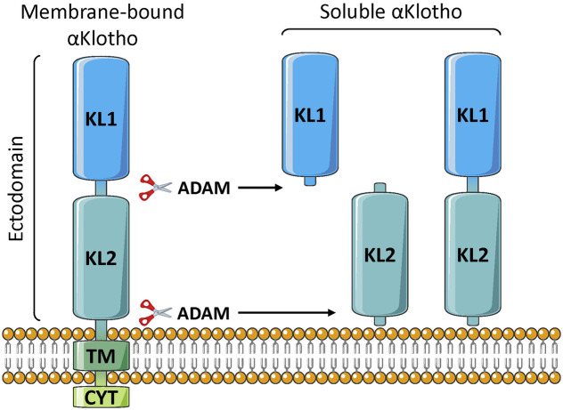
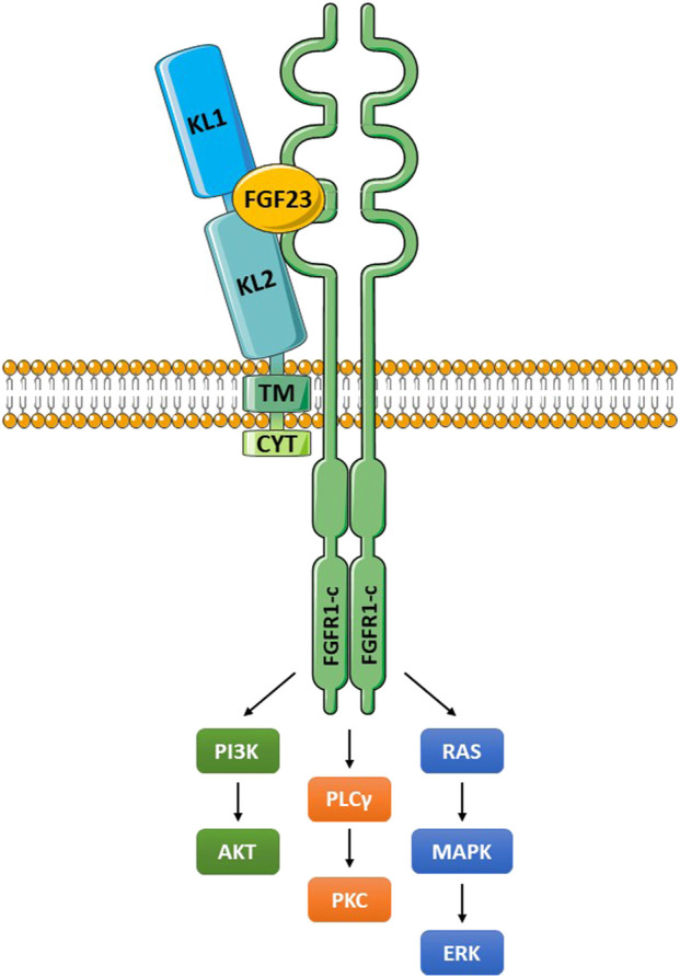
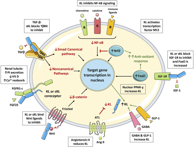
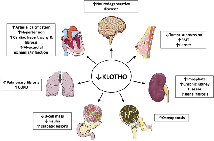
The α-Klotho protein (henceforth denoted Klotho) has antiaging properties, as first observed in mice homozygous for a hypomorphic Klotho gene (kl/kl). These mice have a shortened lifespan, stunted growth, renal disease, hyperphosphatemia, hypercalcemia, vascular calcification, cardiac hypertrophy, hypertension, pulmonary disease, cognitive impairment, multi-organ atrophy and fibrosis. Overexpression of Klotho has opposite effects, extending lifespan. In humans, Klotho levels decline with age, chronic kidney disease, diabetes, Alzheimer's disease and other conditions. Low Klotho levels correlate with an increase in the death rate from all causes. Klotho acts either as an obligate coreceptor for fibroblast growth factor 23 (FGF23), or as a soluble pleiotropic endocrine hormone (s-Klotho). It is mainly produced in the kidneys, but also in the brain, pancreas and other tissues. On renal tubular-cell membranes, it associates with FGF receptors to bind FGF23. Produced in bones, FGF23 regulates renal excretion of phosphate (phosphaturic effect) and vitamin D metabolism. Lack of Klotho or FGF23 results in hyperphosphatemia and hypervitaminosis D. With age, human renal function often deteriorates, lowering Klotho levels. This appears to promote age-related pathology. Remarkably, Klotho inhibits four pathways that have been linked to aging in various ways: Transforming growth factor β (TGF-β), insulin-like growth factor 1 (IGF-1), Wnt and NF-κB. These can induce cellular senescence, apoptosis, inflammation, immune dysfunction, fibrosis and neoplasia. Furthermore, Klotho increases cell-protective antioxidant enzymes through Nrf2 and FoxO. In accord, preclinical Klotho therapy ameliorated renal, cardiovascular, diabetes-related and neurodegenerative diseases, as well as cancer. s-Klotho protein injection was effective, but requires further investigation. Several drugs enhance circulating Klotho levels, and some cross the blood-brain barrier to potentially act in the brain. In clinical trials, increased Klotho was noted with renin-angiotensin system inhibitors (losartan, valsartan), a statin (fluvastatin), mTOR inhibitors (rapamycin, everolimus), vitamin D and pentoxifylline. In preclinical work, antidiabetic drugs (metformin, GLP-1-based, GABA, PPAR-γ agonists) also enhanced Klotho. Several traditional medicines and/or nutraceuticals increased Klotho in rodents, including astaxanthin, curcumin, ginseng, ligustilide and resveratrol. Notably, exercise and sport activity increased Klotho. This review addresses molecular, physiological and therapeutic aspects of Klotho.

Keywords: FGF23; IGF-1; NF-KappaB; TGF-beta; Wnt; aging; hyperphosphatemia; klotho.


Conflict of interest statement

Author QW was employed by the company Shanghai Yinuo Pharmaceutical Co., Ltd. QW holds GLP-1 related patents. The remaining authors declare that the research was conducted in the absence of any commercial or financial relationships that could be construed as a potential conflict of interest.

Figures

FIGURE 1
FIGURE 1
Klotho structure. The membrane-bound form is single-pass, and consists of two extracellular domains (KL1 and KL2), a transmembrane segment (TM), and a short non-signaling cytoplasmic tail (CYT). The soluble form is generated by proteolytic cleavage, usually by either ADAM10 or ADAM17 enzymes, to release the large soluble form (s-Klotho). This is the major form found in the circulation. It can be further cleaved to generate independent KL1 and KL2 fragments, but these are either minor forms or undetectable in the plasma.
FIGURE 2
FIGURE 2
The Klotho/FGFR/FGF23 signaling complex. Klotho interacts with a FGFR (frequently FGFR1c) through an extension of its KL2 domain. FGF23 fits into a groove formed by components of KL1, KL2 and the FGFR. The membrane-bound and soluble Klotho forms (KL1/KL2 domains) can both bind to FGFR1c and function as coreceptors (Chen G, et al., 2018). These molecular interactions create a high affinity binding site for FGF23. The activated FGFR signals through multiple pathways as illustrated, and outlined in the text. Abbreviations: ERK, extracellular signal-regulated kinase; FGF23, fibroblast growth factor 23; FGFR1-c, FGF receptor 1c; MAPK, mitogen-activated protein kinase; PI3K, phosphoinositide 3-kinase; PKC, protein kinase C; PLCγ, phospholipase Cγ.
FIGURE 3
FIGURE 3
The multiple functions of Klotho. FGF23 binds to Klotho/FGFR1c receptor in the renal tubule to increase excretion of phosphate (phosphaturic effect), regulate vitamin D metabolism, and increase calcium reabsorption. Klotho binds to the TGF-β receptor (TβRII component) to block TGF-β action. Klotho inhibits activation of the inflammatory NF-κB pathway by preventing nuclear translocation of the active form. Klotho increases signaling in the Nrf2 pathway; inducing multiple antioxidant enzymes and inhibiting NF-κB. Klotho blocks IGF-1 receptor signaling; this increases activation of FoxO and antioxidant responses. Klotho also blocks activation of the Wnt pathway by binding to soluble Wnt ligands. Klotho expression is increased by PPAR-γ activation, and also by GLP-1 and GABA stimulation; but is suppressed by angiotensin II activation of the AT1 receptor. Abbreviations: Ang II, angiotensin II; Ca2+, calcium ion; FoxO, forkhead boxprotein O; GABA, γ-aminobutyric acid; GLP-1, glucagon-like peptide 1; IGF-1, insulin-like growth factor 1; IGFR, IGF-1 receptor; KL, membrane-bound αKlotho; NF-κB, nuclear factor κB; Nrf2, nuclear factor-erythroid 2-related factor 2; Pi, inorganic phosphate; PPAR-γ, peroxisome proliferator-activated receptors γ; R, receptor; sKL, soluble αKlotho; TGF-β, transforming growth factor β; TβRII, TGF-β receptor type II; Vit D, vitamin D.
FIGURE 4
FIGURE 4
Klotho deficiency associates with multiple age-related diseases. As outlined in the captions, depressed Klotho levels are linked to hyperphosphatemia, chronic kidney diseases, multiple cardiovascular conditions, neurodegenerative diseases, several types of cancer, pulmonary fibrosis, COPD, bone disease and diabetes (reduced β-cell mass in the pancreas). References are listed in Table 1. Abbreviations: COPD, chronic obstructive pulmonary disease; EMT, epithelial-mesenchymal transition.

References

    1. Aashaq S., Batool A., Mir S. A., Beigh M. A., Andrabi K. I., Shah Z. A. (2022). TGF‐β Signaling: A Recap of SMAD‐independent and SMAD‐dependent Pathways. J. Cell. Physiology 237 (1), 59–85. 10.1002/jcp.30529 - DOI - PubMed
    1. Abolghasemi M., Yousefi T., Maniati M., Qujeq D. (2019). The Interplay of Klotho with Signaling Pathway and microRNAs in Cancers. J Cell. Biochem. 120 (9), 14306–14317. 10.1002/jcb.29022 - DOI - PubMed
    1. Abramovitz L., Rubinek T., Ligumsky H., Bose S., Barshack I., Avivi C., et al. (2011). KL1 Internal Repeat Mediates Klotho Tumor Suppressor Activities and Inhibits bFGF and IGF-I Signaling in Pancreatic Cancer. Clin. Cancer Res. 17 (13), 4254–4266. 10.1158/1078-0432.CCR-10-2749 - DOI - PubMed
    1. Adeli S., Zahmatkesh M., Tavoosidana G., Karimian M., Hassanzadeh G. (2017). Simvastatin Enhances the Hippocampal Klotho in a Rat Model of Streptozotocin-Induced Cognitive Decline. Prog. Neuro-Psychopharmacology Biol. Psychiatry 72, 87–94. 10.1016/j.pnpbp.2016.09.009 - DOI - PubMed
    1. Ali M. F., Venkatarayappa S. K. B., Benny M., Rojas C., Yousefi K., Shehadeh L. A., et al. (2020). Effects of Klotho Supplementation on Hyperoxia-Induced Renal Injury in a Rodent Model of Postnatal Nephrogenesis. Pediatr. Res. 88, 565–570. 10.1038/s41390-020-0803-z - DOI - PMC - PubMed