. 2022 Jul 12:9:905468.
doi: 10.3389/fmolb.2022.905468. eCollection 2022.

Insulin-Like Growth Factor Binding Protein (IGFBP-6) as a Novel Regulator of Inflammatory Response in Cystic Fibrosis Airway Cells

Affiliations

Insulin-Like Growth Factor Binding Protein (IGFBP-6) as a Novel Regulator of Inflammatory Response in Cystic Fibrosis Airway Cells

Onofrio Laselva et al. Front Mol Biosci. .

Abstract

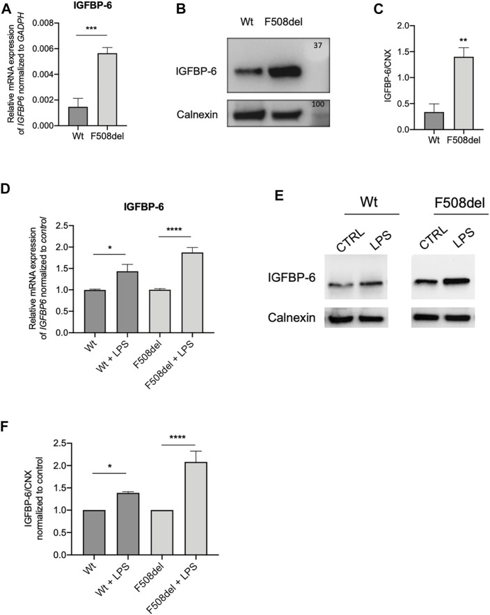
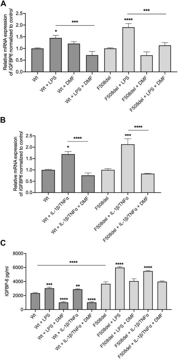
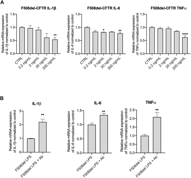
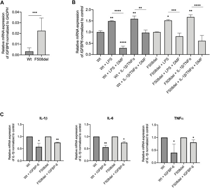
Cystic Fibrosis (CF) patients are prone to contracting bacterial lung infections with opportunistic pathogens, especially Pseudomonas aeruginosa. Prolonged P. aeruginosa infections have been linked to chronic inflammation in the CF lung, whose hallmarks are increased levels of cytokines (i.e., TNF-α, IL-1β, IL-6) and neutrophil attraction by chemokines, like IL-8. Recently, insulin-like growth factor binding protein 6 (IGFBP-6) has been shown to play a putative role in the immune system and was found at higher levels in the sera and synovial tissue of rheumatoid arthritis patients. Moreover, it has been demonstrated that IGFBP-6 has chemoattractant properties towards cells of the innate (neutrophils, monocytes) and adaptive (T cells) immunity. However, it is not known whether IGFBP-6 expression is dysregulated in airway epithelial cells under infection/inflammatory conditions. Therefore, we first measured the basal IGFBP-6 mRNA and protein levels in bronchial epithelial cells lines (Wt and F508del-CFTR CFBE), finding they both are upregulated in F508del-CFTR CFBE cells. Interestingly, LPS and IL-1β+TNFα treatments increased the IGFBP-6 mRNA level, that was reduced after treatment with an anti-inflammatory (Dimethyl Fumarate) in CFBE cell line and in patient-derived nasal epithelial cultures. Lastly, we demonstrated that IGFBP-6 reduced the level of pro-inflammatory cytokines in both CFBE and primary nasal epithelial cells, without affecting rescued CFTR expression and function. The addition of a neutralizing antibody to IGFBP-6 increased pro-inflammatory cytokines expression under challenge with LPS. Together, these data suggest that IGFBP-6 may play a direct role in the CF-associated inflammation.

Keywords: IGFBP-6; TRIKAFTA; airway epithelial cells; cystic fibrosis; cytokine; dimethyl fumarate.


Conflict of interest statement

The authors declare that the research was conducted in the absence of any commercial or financial relationships that could be construed as a potential conflict of interest.

Figures

FIGURE 1
FIGURE 1
IGFBP-6 shows higher expression in F508del-CFTR CFBE cells and is further increased after LPS treatment. (A) Total RNA was extracted and qRT-PCR was performed in order to quantify IGFBP-6 mRNA normalized to GADPH as housekeeping gene. Data represent the mean ± SEM (n = 3). Statistical significance tested using paired two-tailed t-test. (B) Immunoblots of basal expression of IGFBP-6 in Wt and F508del-CFTR CFBE cells. (C) Data represent the mean ± SEM of the ratio IGFBP-6/Calnexin (n = 3). Statistical significance tested using paired two-tailed t-test. (D,E) Cells were treated with 1 μg/ml LPS for 4 h. Total RNA was extracted and qRT-PCR was performed in order to quantify IGFBP-6 mRNAs normalized first to GADPH as housekeeping gene and then to control untreated cells. Data represent the mean ± SEM (n = 4). Statistical significance tested using two-way ANOVA with Tukey’s multiple comparisons test (F) Immunoblots of IGFBP-6 following treatment with 1 μg/ml LPS for 24 h. (G) Data represent the mean ± SEM of the ratio IGFBP-6/Calnexin normalized to Calnexin control (n = 3–4). *p < 0.05; **p < 0.01; ****p < 0.0001.
FIGURE 2
FIGURE 2
IGFBP-6 is reduced by Dimethyl fumarate. (A) Cells were treated with 0.1% DMSO, 1 μg/ml LPS ± 50 µM DMF or (B) PBS, 30 ng/ml IL-1β + 30 ng/ml TNFα ± 50 µM DMF for 4 h. Total RNA was extracted and qRT-PCR was performed in order to quantify IGFBP mRNA normalized to GADPH as housekeeping gene and then to control untreated cells. Data represent the mean ± SEM (n = 4). (C) IGFBP-6 secretion after stimulation with 0.1% DMSO, 1 μg/ml LPS, 30 ng/ml IL-1β + 30 ng/ml TNFα ± 50 µM DMF for 24 h. Data represent the mean ± SEM (n = 3). *p < 0.05; **p < 0.01; ***p < 0.001; ****p < 0.0001. Statistical significance tested using two-way ANOVA with Tukey’s multiple comparisons test to the control (Wt or F508del) for each cell line.
FIGURE 3
FIGURE 3
Anti-IGFBP-6 antibodies increased pro-inflammatory cytokines expression. (A) F508del-CFTR CFBE cells were treated with PBS or 0.2–200 ng/ml IGFBP-6 for 4 h. Statistical significance tested using two-way ANOVA with Tukey’s multiple comparisons test. (B) Cells were treated with 1 μg/ml LPS ± an antibody directed against IGFBP-6 (1 μg/ml) for 4 h. Total RNA was extracted and qRT-PCR was performed in order to quantify IL-1β, IL-6 and TNF-α mRNAs normalized to GADPH as housekeeping gene and then to control untreated cells. Data represent the mean ± SEM (n = 3). Statistical significance tested using paired two-tailed t-test.
FIGURE 4
FIGURE 4
IGFBP-6 shows higher expression in F508del/F508del nasal epithelial cells from three CF donors and reduces pro-inflammatory cytokines expression. (A) Total RNA was extracted from 2 patients homozygous for F508del mutation and 2 non-CF donors. qRT-PCR was performed in order to quantify IGFBP-6 mRNA normalized to GADPH as housekeeping gene. Data represent the mean ± SEM (n = 4). Statistical significance tested using paired two-tailed t-test (B) Cells were treated with 0.1% DMSO, 1 μg/ml LPS, 30 ng/ml IL-1β + 30 ng/ml TNFα ± 50 µM DMF for 4 h. Total RNA was extracted and qRT-PCR was performed in order to quantify IGFBP mRNA normalized to control untreated cells. Data represent the mean ± SEM (n = 4). (C) Cells were treated with 200 ng/ml IGFBP-6 for 4 h. Total RNA was extracted and qRT-PCR was performed in order to quantify IL-1β, IL-6 and TNFα mRNAs normalized to control untreated cells. Data represent the mean ± SEM (n = 4). *p < 0.05; **p < 0.01; ***p < 0.001; ****p < 0.0001. Statistical significance tested using two-way ANOVA with Tukey’s multiple comparisons test to the control for (Wt or F508del) each cell line.

Similar articles

Cited by

References

    1. Abbattiscianni A. C., Favia M., Mancini M. T., Cardone R. A., Guerra L., Monterisi S., et al. (2016). Correctors of Mutant CFTR Enhance Subcortical cAMP-PKA Signaling through Modulating Ezrin Phosphorylation and Cytoskeleton Organization. J. Cell Sci. 129, 1128–1140. 10.1242/jcs.177907 - DOI - PubMed
    1. Alunno A., Bistoni O., Manetti M., Cafaro G., Valentini V., Bartoloni E., et al. (2017). Insulin-Like Growth Factor Binding Protein 6 in Rheumatoid Arthritis: A Possible Novel Chemotactic Factor? Front. Immunol. 8, 554. 10.3389/fimmu.2017.00554 - DOI - PMC - PubMed
    1. Bach L. A. (2016). Current Ideas on the Biology of IGFBP-6: More Than an IGF-II Inhibitor? Growth Hormone IGF Res. 30-31, 81–86. 10.1016/j.ghir.2016.09.004 - DOI - PubMed
    1. Broxmeyer H. E., Sherry B., Cooper S., Lu L., Maze R., Beckmann M. P., et al. (1993). Comparative Analysis of the Human Macrophage Inflammatory Protein Family of Cytokines (Chemokines) on Proliferation of Human Myeloid Progenitor Cells. Interacting Effects Involving Suppression, Synergistic Suppression, and Blocking of Suppression. J. Immunol. 150, 3448–3458. - PubMed
    1. Cacalano G., Lee J., Kikly K., Ryan A. M., Pitts-Meek S., Hultgren B., et al. (1994). Neutrophil and B Cell Expansion in Mice that Lack the Murine IL-8 Receptor Homolog. Science 265, 682–684. 10.1126/science.8036519 - DOI - PubMed