Review
. 2022 Jul 12:10:925951.
doi: 10.3389/fped.2022.925951. eCollection 2022.

Cerebral Oxygenation and Metabolism After Hypoxia-Ischemia

Affiliations
Review

Cerebral Oxygenation and Metabolism After Hypoxia-Ischemia

Simerdeep K Dhillon et al. Front Pediatr. .

Abstract

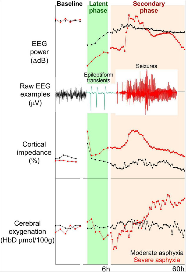
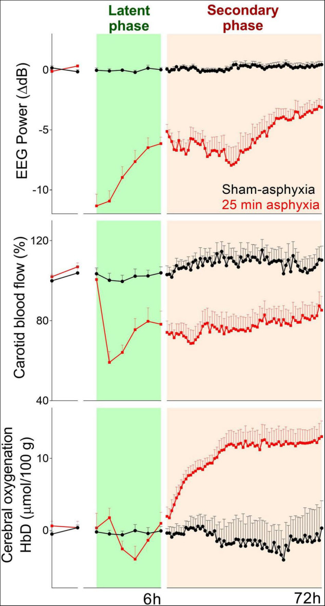
Perinatal hypoxia-ischemia (HI) is still a significant contributor to mortality and adverse neurodevelopmental outcomes in term and preterm infants. HI brain injury evolves over hours to days, and involves complex interactions between the endogenous protective and pathological processes. Understanding the timing of evolution of injury is vital to guide treatment. Post-HI recovery is associated with a typical neurophysiological profile, with stereotypic changes in cerebral perfusion and oxygenation. After the initial recovery, there is a delayed, prolonged reduction in cerebral perfusion related to metabolic suppression, followed by secondary deterioration with hyperperfusion and increased cerebral oxygenation, associated with altered neurovascular coupling and impaired cerebral autoregulation. These changes in cerebral perfusion are associated with the stages of evolution of injury and injury severity. Further, iatrogenic factors can also affect cerebral oxygenation during the early period of deranged metabolism, and improving clinical management may improve neuroprotection. We will review recent evidence that changes in cerebral oxygenation and metabolism after HI may be useful biomarkers of prognosis.

Keywords: biomarkers; cerebral blood flow; fetal sheep; hypoxia-ischemia brain; monitoring; neonatal encephalopathy.


Conflict of interest statement

The authors declare that the research was conducted in the absence of any commercial or financial relationships that could be construed as a potential conflict of interest.

Figures

FIGURE 1
FIGURE 1
Examples of changes in EEG power (top panel), cortical impedance (middle panel), and cerebral oxygenation (difference in oxygenated and deoxygenated hemoglobin, bottom panel) during baseline and 60 h post-HI recovery in near-term (0.85 gestation) fetal sheep subjected to 15 min (n = 2) or 18 min (n = 2) complete umbilical cord occlusion. Fifteen and eighteen minutes of asphyxia reflect moderate and severe HI, respectively. In the moderate group, total EEG power remained suppressed, but epileptiform transient activity on a suppressed background was seen during the latent phase. High amplitude, stereotypic seizures developed during the secondary phase. EEG power in the severe group increased with the onset of status epilepticus and subsequently fell below the moderate group. A secondary rise in impedance (delayed cell swelling) was only seen in the severe group. Cerebral oxygenation remained stable in the moderate group, but the severe group had early reduction and subsequent increase in cerebral oxygenation during the secondary phase.
FIGURE 2
FIGURE 2
Changes in EEG power (top panel), carotid blood flow (index of cerebral perfusion) (middle panel), and cerebral oxygenation (bottom panel) during baseline and 72 h of post-HI recovery in preterm (0.7 gestation) fetal sheep subjected to sham occlusion (n = 4) or 25 min of asphyxia (n = 8). Hypoperfusion during the latent phase was associated with a transient decrease in cerebral oxygenation. Despite the sustained reduction in cerebral blood flow, there was an increase in cerebral oxygenation during the secondary phase.

References

    1. Perin J, Mulick A, Yeung D, Villavicencio F, Lopez G, Strong KL, et al. Global, regional, and national causes of under-5 mortality in 2000-19: an updated systematic analysis with implications for the sustainable development goals. Lancet Child Adolesc Health. (2022) 6:106–15. 10.1016/s2352-4642(21)00311-4 - DOI - PMC - PubMed
    1. Campbell H, Eddama O, Azzopardi D, Edwards AD, Strohm B, Rivero-Arias O. Hypothermia for perinatal asphyxia: trial-based quality of life at 6-7 years. Arch Dis Child. (2018) 103:654–9. 10.1136/archdischild-2017-313733 - DOI - PMC - PubMed
    1. Yates N, Gunn AJ, Bennet L, Dhillon SK, Davidson JO. Preventing brain injury in the preterm infant-current controversies and potential therapies. Int J Mol Sci. (2021) 22:1671. 10.3390/ijms22041671 - DOI - PMC - PubMed
    1. Osredkar D, Verdenik I, Gergeli AT, Gersak K, Lucovnik M. Apgar score and risk of cerebral palsy in preterm infants: a population-based cohort study. Neuropediatrics. (2021) 52:310–5. 10.1055/s-0041-1729181 - DOI - PubMed
    1. Sarda SP, Sarri G, Siffel C. Global prevalence of long-term neurodevelopmental impairment following extremely preterm birth: a systematic literature review. J Int Med Res. (2021) 49:3000605211028026. 10.1177/03000605211028026 - DOI - PMC - PubMed

LinkOut - more resources