Review
. 2022 Jun 6:9:884247.
doi: 10.3389/fsurg.2022.884247. eCollection 2022.

Minimally-Invasive Assisted Robotic Spine Surgery (MARSS)

Affiliations
Review

Minimally-Invasive Assisted Robotic Spine Surgery (MARSS)

Ramiro A Pérez de la Torre et al. Front Surg. .

Abstract

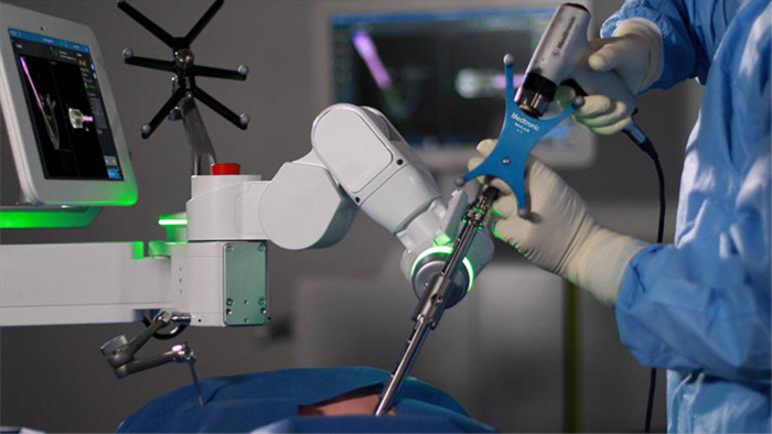
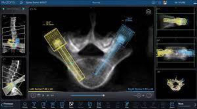
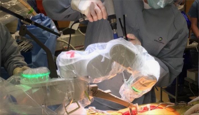
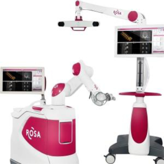
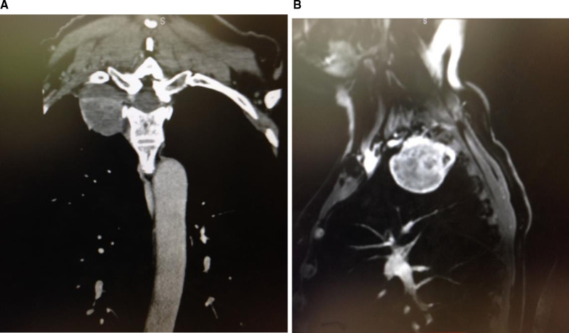
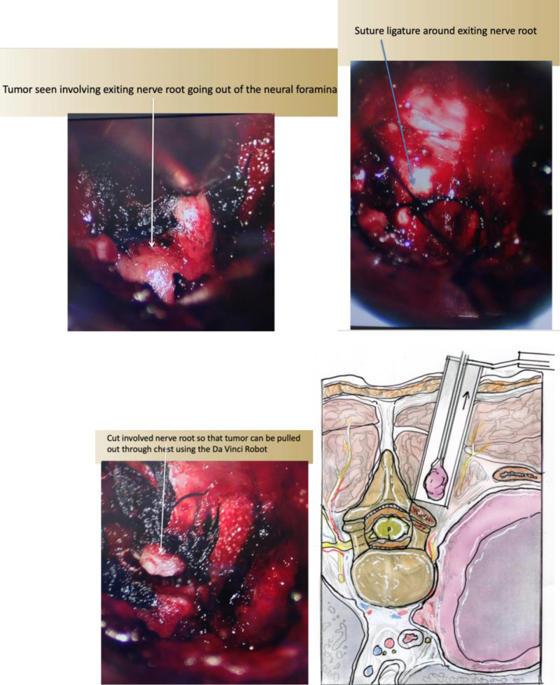
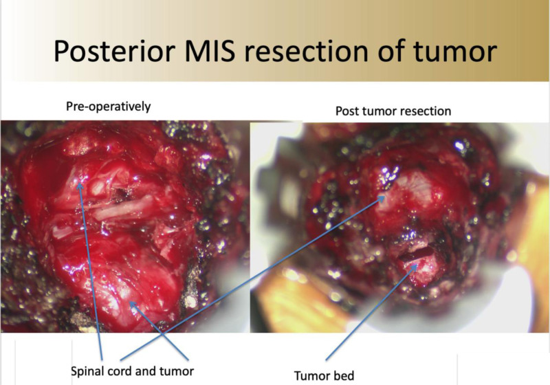
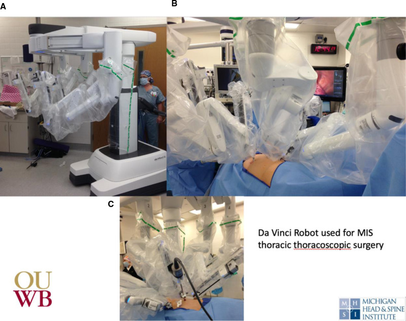
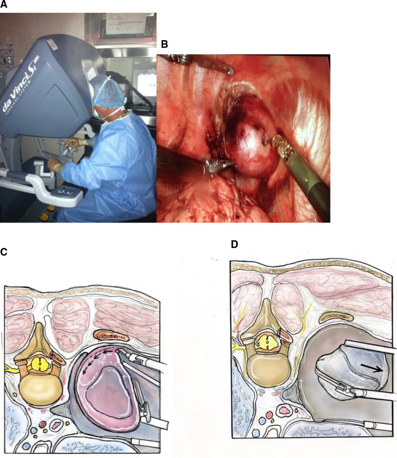
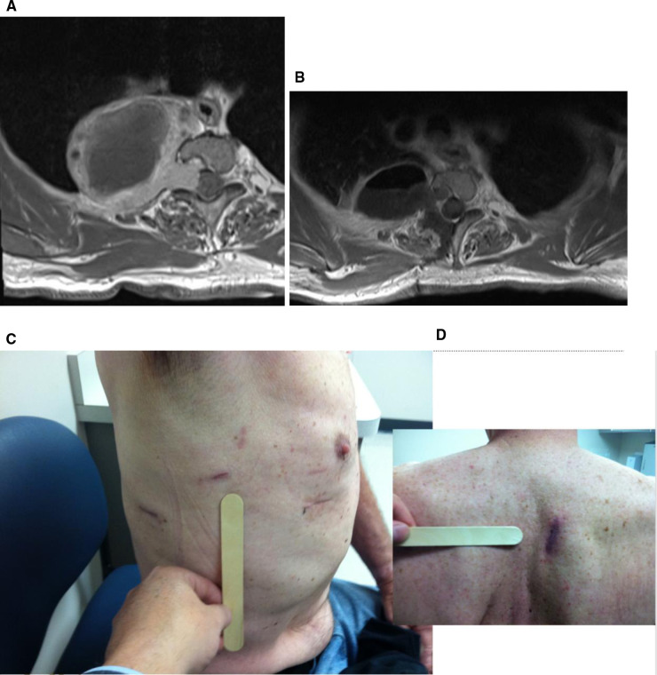
Minimally-Invasive robotic spine surgery (MARSS) has expanded the surgeons armamentarium to treat a variety of spinal disorders. In the last decade, robotic developments in spine surgery have improved the safety, accuracy and efficacy of instrumentation placement. Additionally, robotic instruments have been applied to remove tumors in difficult locations while maintaining minimally invasive access. Gross movements by the surgeon are translated into fine, precise movements by the robot. This is exemplified in this chapter with the use of the da Vinci robot to remove apical thoracic tumors. In this chapter, we will review the development, technological advancements, and cases that have been conducted using MARSS to treat spine pathology in a minimally invasive fashion.

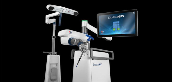
Keywords: da Vinci; mazor X; minimally invasive spine surgery (MISS); minimally-invasive robotic spine surgery (MARSS); robotic; stereotactic transformation.


Figures

Figure 1
Figure 1
Photo of Mazor X (Mazor Stealth technologies, Medtronic). A commercial system designed to extend the working options, including imaging processing and robot-based interaction.
Figure 2
Figure 2
Software interaction with Mazor X, including pedicle screw selection along with optimization of construct definition. Proper pedicle screw diameter and length can be selected to conform to the patient's individual anatomy based on pre-operative imaging using the work-station.
Figure 3
Figure 3
Mazor X in working position during drilling of the pedicle for pedicle screw application.
Figure 4
Figure 4
Excelcius GPS equipment. A robotic arm-based technology along with multiple intuitive functions for working environment.
Figure 5
Figure 5
ROSA technologies (Zimmer Spine Inc.).
Figure 6
Figure 6
Pre-operative coronal and sagittal thoracic MRI images showing high apical chest tumor. Thin slice CT images can be very helpful in identifying the neural foramen of tumor origin. This is critical in safely detaching the tumor from the spinal canal before final removal through the chest cavity.
Figure 7
Figure 7
(A) Intra-operative photo showing (A) tumor extending outside the neural foramen, (B) silk suture ligature around the nerve giving rise to the tumor, (C) ligation of the nerve leading to the tumor. (D) Illustration of removal of intra-spinal canal portion of the tumor via a posterior approach through a tubular retractor.
Figure 8
Figure 8
Intra-operative photos after minimally invasive posterior approach (A) tumor and intra-canal neural foramen tumor exposed and (B) after resection of the tumor with residual tumor bed.
Figure 9
Figure 9
Intra-operative photos of (A) da Vinci robot, (B) used for anterior thoracoscopic approach the spine to (C) removal apical thoracic tumor.
Figure 10
Figure 10
Intra-operative photo of (A) surgeon using the da Vinci robot to remove (B) apical thoracic tumor. Illustrations showing (C) resection of apical thoracic tumor from the chest wall and (D) removal of the tumor. (Illustrations from: An Anatomical Approach to Minimally Invasive Spine Surgery, Editors; M. Perez-Cruet, R. Fessler, M. Wang, Thieme Publishing Inc. NY, 2019).
Figure 11
Figure 11
(A) Pre- and (B) post-operative axial MRI showing gross total tumor resection. Post-operative (C) anterior and (D) posterior thoracic incision after use of the da Vinci robot to remove apical thoracic tumor.

References

    1. Carsten S, Ringel F, Stoffel M, Reinke A, Behr M, Meyer B. Robotic technology in spine surgery: current applications and future developments. Acta Neurochir Suppl. (2011) 109:241–5. 10.1007/978-3-211-99651-5_38 - DOI - PubMed
    1. Booher G, Vardiman A. Navigated robotic assisted thoracic pedicle screw placement for metastatic renal cell carcinoma. Neurosurg Focus. (2018) 45(VideoSuppl1):V5. 10.3171/2018.7.FocusVid.18121 - DOI - PubMed
    1. Tian W, Liu YJ, Liu B, He D, Wu JY, Han XG, et al. Guideline for thoracolumbar pedicle screw placement assisted by orthopaedic surgical robot. Orthop Surg. (2019) 11(2):153–9. 10.1111/os.12453 - DOI - PMC - PubMed
    1. Zhang Q, Han XG, Xu YF, Fan MX, Zhao JW, Liu YJ, et al. Robotic navigation during spine surgery. Expert Rev Med Devices. (2020) 17(1):27–32. 10.1080/17434440.2020.1699405 - DOI - PubMed
    1. Li QH, Zamorano L, Pandya A, Perez R, Gong J, Diaz F. The application accuracy of the NeuroMate robot–a quantitative comparison with frameless and frame-based surgical localization systems. Comput Aided Surg. (2002) 7(2):90–8. 10.1002/igs.10035 - DOI - PubMed

LinkOut - more resources