. 2022 Jul;71(1):22-28.
doi: 10.3164/jcbn.22-9. Epub 2022 May 13.

Copper in the tumor microenvironment and tumor metastasis

Affiliations

Copper in the tumor microenvironment and tumor metastasis

Tetsuro Kamiya. J Clin Biochem Nutr. 2022 Jul.

Abstract

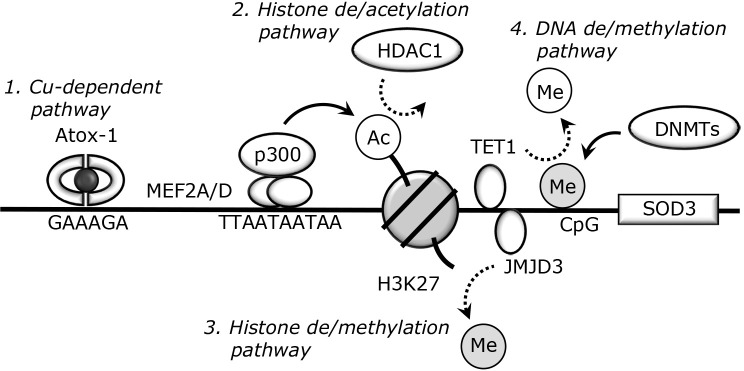
Copper (Cu), an essential micronutrient, plays an essential role in several physiological processes, including cell proliferation and angiogenesis; however, its dysregulation induces oxidative stress and inflammatory responses. Significant Cu accumulation is observed in several tumor tissues. The bioavailability of intracellular Cu is tightly controlled by Cu transporters, including Cu transporter 1 (CTR1) and Cu-transporting P-type ATPase α and β (ATP7A and ATP7B), and Cu chaperones, including Cu chaperone for superoxide dismutase 1 (CCS) and antioxidant-1 (Atox-1). In several tumor tissues, these abnormalities that induce intra-cellular Cu accumulation are involved in tumor progression. In addition, functional disturbance in Cu-containing secretory enzymes, such as superoxide dismutase 3 (SOD3), and lysyl oxidase enzymes (LOX and LOXL1-4) with abnormal Cu dynamics plays a key role in tumor metastasis. For example, the loss of SOD3 in tumor tissues induces oxidative stress, which promotes neovascularization and epithelial-to-mesenchymal transition (EMT). LOX promotes collagen crosslinking, which functions in the metastatic niche formation. Accordingly, restricted Cu regulation may be a novel strategy for the inhibition of tumor metastasis. However, it is unclear how these Cu disturbances occur in tumor tissues and the exact molecular mechanisms underlying Cu secretory enzymes. In this review article, I discuss the role of Cu transporters, Cu chaperones, and Cu-containing secretory enzymes in tumor progression to better understand the role of Cu homeostasis in tumor tissues.

Keywords: copper; copper chaperone; copper transporters; copper-containing secretory enzymes; tumor metastasis.


Conflict of interest statement

No potential conflicts of interest were disclosed.

Figures

Fig. 1.
Fig. 1.
Proposed models of intracellular Cu-trafficking. STEAPs participate in extracellular Cu (II) reduction, and then reduced Cu (I) is taken up into the cells through membrane-bound CTR1. CTR1 delivers Cu (I) to Cu chaperone CCS, COX17, and Atox-1. These chaperones transfer Cu (I) to SOD1, CCO, and ATP7A, respectively. Atox-1 also translocates into the nucleus, binds to Atox-1 response element (GAAAGA), and functions as a Cu-dependent transcription factor for cyclin D1, NADPH p47phox, and SOD3. ATP7A delivers Cu (I) to Cu-containing secretory enzymes, including SOD3, and functions in the Cu egress to maintain the level of intracellular Cu (I).
Fig. 2.
Fig. 2.
Cu-dependent and epigenetic SOD3 regulation. 1. Cu-dependent pathway. The Cu chaperone Atox-1 is expressed in the nucleus under several conditions and binds to the proximal promoter region of SOD3. The transcription activity of Atox-1 has been confirmed in several cells such as smooth muscle cells and monocytes/macrophages. 2. Histone de/acetylation pathway. The de/acetylation within the SOD3 promoter is involved in its regulation. Our previous studies suggested that MEF2 proteins bind to MEF2 response motif (TTAATAATAA) and function as scaffold proteins that interact with histone acetyltransferase p300. On the other hand, HDAC1-mediated histone deacetylation plays a key role in SOD3 silencing. The lined or dotted arrows indicate the addition or removal of histone or DNA marks, respectively. 3. Histone de/methylation pathway. Trimethylated-H3K27 is well recognized as the marker of gene reduction. We demonstrated that JMJD3, a histone demethylase, reduces the level of H3K27me3 within the SOD3 promoter region and is involved in its induction. 4. DNA de/methylation pathway. DNMTs transfer methyl residues to cytosines within CpG sequences and reduce target gene expression. TET1, a DNA demethylase, plays a key role in the removal of methyl residues from the SOD3 promoter region, which induces its expression.
Fig. 3.
Fig. 3.
The functional role of LOXs in tumor progression. 1. Collagen crosslinking. LOXs catalyze the conversion of lysine residue to aldehyde allysine, and spontaneous reaction with other lysine and allysine residues creates crosslinks in collagen. 2. EMT induction. During the crosslinking of collagen, LOXs produce H2O2 and NH3 as byproducts. The generated H2O2 activates several signal pathways, including FAK/Src signaling, which facilitates EMT processes. 3. Exosomal secretion. Recent studies revealed that LOXs are extracellularly secreted through exosomes. Exosomal secretion of LOXs generates H2O2 in recipient cells and functions in the acquisition of metastatic properties. 4. Gene regulation. There is accumulating evidence supporting the involvement of LOXs in the deamination of lysine residues in trimethylated H3K4, a transcription marker. At present, LOXs are considered to catalyze deamination in H3K4 within the CDH1 promoter and reduce its expression.

Similar articles

Cited by

References

    1. Harris ED. Copper transport: an overview. Proc Soc Exp Biol Med 1991; 196: 130–140. - PubMed
    1. Bull PC, Cox DW. Wilson disease and Menkes disease: new handles on heavy-metal transport. Trends Genet 1994; 10: 246–252. - PubMed
    1. Rodríguez JP, Rios S, Gonzalez M. Modulation of the proliferation and differentiation of human mesenchymal stem cells by copper. J Cell Biochem 2002; 85: 92–100. - PubMed
    1. Oe S, Miyagawa K, Honma Y, Harada M. Copper induces hepatocyte injury due to the endoplasmic reticulum stress in cultured cells and patients with Wilson disease. Exp Cell Res 2016; 347: 192–200. - PubMed
    1. Singh A, Kukreti R, Saso L, Kukreti S. Oxidative stress: a key modulator in neurodegenerative diseases. Molecules 2019; 24: 1583. - PMC - PubMed