

Cardiac electrical instability in Erdheim-Chester disease: a case report
- PMID: 35903613
- PMCID: PMC9318901
- DOI: 10.1093/omcr/omac071
Cardiac electrical instability in Erdheim-Chester disease: a case report
Abstract
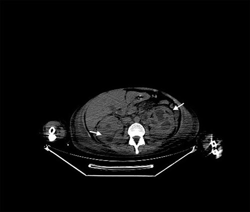
Erdheim-Chester disease (ECD) is a rare multisystemic disorder of non-Langerhans histiocytic cells with a pleomorphic clinical presentation. It affects bones, skin, central nervous system, pituitary gland, ocular tissue, kidneys and perirenal tissue and lungs. Cardiac involvement presents usually with pericardial effusion and right atrial masses, but rarely with conduction system infiltration and subsequent arrhythmic events. Following the discovery of mutations of activating signaling kinase proteins (BRAF, MEK, ALK), the therapeutic landscape has changed to a more precise targeted treatment. Currently vemurafenib is approved for patient with end-organ dysfunction and BRAF-V600E mutation and the prognosis has dramatically improved. Here we present a case of ECD with electrical instability as main clinically relevant manifestation of cardiac involvement.
© The Author(s) 2022. Published by Oxford University Press. All rights reserved. For Permissions, please email: journals.permissions@oup.com.
Figures


References
-
- Goyal G, Heaney ML, Collin M, Cohen-Aubart F, Vaglio A, Durham Bet al. . Erdheim-Chester disease: consensus recommendation for evaluation, diagnosis, and treatment in the molecular era. Blood 2020;135:1929–45. - PubMed
-
- Campochiaro C, Tomelleri A, Cavalli G, Berti A, Dagna L. Erdheim-Chester disease. Eur J Intern Med 2015;26:223–9. - PubMed
-
- Ghotra AS, Thompson K, Lopez-Mattei J, Bawa D, Hernandez R, Banchs Jet al. . Cardiovascular manifestation of Erdheim-Chester disease. Echocardiography 2019;36:229–36. - PubMed
