Case Reports
. 2022 Jul 12:16:877479.
doi: 10.3389/fnins.2022.877479. eCollection 2022.

Diabetic Striatopathy Complicated With Acute Ischemic Stroke: A Case Report

Affiliations
Case Reports

Diabetic Striatopathy Complicated With Acute Ischemic Stroke: A Case Report

Xiao Huang et al. Front Neurosci. .

Abstract

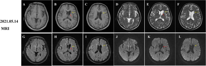
Diabetic striatopathy (DS) is a rare complication secondary to hyperglycemia, featured by the choreiform movements and reversible striatal abnormalities on neuroimaging. Several studies have described the clinical characteristics of DS, however, the simultaneous occurrence of DS and acute ischemic stroke (AIS) in the striatum has not been reported. Herein, we report a 68-year-old man with uncontrolled type 2 diabetes who experienced the progressive involuntary movement of the right upper and lower limbs for 10 days. We initially considered this patient as an AIS with hemorrhage in the left basal ganglia and adjacent area because his brain magnetic resonance imaging (MRI) showed hyperintensity on fluid-attenuated inversion recovery (FLAIR) and diffusion-weighted imaging (DWI) images, as well as slight T1-hyperintensity around T1-hypointensity. However, his symptoms worsen persistently, which was inconsistent with neuroimaging findings. Further computed tomography (CT) scan revealed an extensive hyper-density and focal low-density in the left striatum, suggesting the diagnosis of DS and AIS. His symptoms were in complete remission after 2 months of glucose control. However, striatal hyperintensity on T1 images was significantly increased compared to the initial images, which disappeared 18 months later. Additionally, DWI hyperintensity on infarction lesions disappeared, while softening lesions and gliosis were observed on the follow-up MRI images. Therefore, we finally diagnosed the patient as DS complicated with AIS. This report highlights that DS and AIS could occur simultaneously in the striatum after hyperglycemia, which is easily misdiagnosed as AIS with hemorrhage and requires clinicians to pay more attention to avoid misdiagnosis and delayed treatment.

Keywords: acute ischemic stroke; chorea; diabetic striatopathy; hyperglycemia; involuntary movement.


Conflict of interest statement

The authors declare that the research was conducted in the absence of any commercial or financial relationships that could be construed as a potential conflict of interest.

Figures

Figure 1
Figure 1
Computed tomography (CT) and MRI of brain at admission. Diabetic striatopathy (DS)-associated striatal abnormalities were marked by red arrows, while infarction focuses were marked by yellow arrows. Typical imaging manifestations of DS were found in the left striatum with T1 hyperintensity [(A,B), coronal images; (P,Q), sagittal images] on MRI and hyper-density on CT images (S,T). The hypointensity on T2 (D,E), fluid-attenuation inversion recovery (FLAIR) (G,H), and apparent diffusion coefficient (ADC) (M,N) images, as well as equal signal intensity on DWI images (J,K) were also observed in DS-associated striatal lesions. Subacute cerebral infarctions showed T1 hypointensity [(B,C) coronal images; (Q) sagittal image], T2 hyperintensity (E,F), FLAIR hyperintensity (H,I), DWI hyperintensity (K,L), and slight ADC hyperintensity (N,O) on MRI images, as well as hypo-density on CT images (T,U). MRA images disclosed obvious stenosis of the left middle cerebral artery [(R), green arrow]. There were several artifacts on ADC images [(J,K), blue arrows].
Figure 2
Figure 2
Follow-up magnetic resonance imaging (MRI) of brain 2 months after onset. Diabetic striatopathy (DS)-associated striatal abnormalities were marked by red arrows, while infarction focuses were marked by yellow arrows. DS-associated T1 hyperintensity on the left striatum was obviously extended [(A), coronal image; (P,Q) (R): sagittal images). DS-associated T2 and FLAIR hypointensity changed to slight hyperintensity (D,G). The left striatum showed an equal signal intensity on DWI images (J). DS-associated initial ADC hypointensity changed to equal signal intensity (M). DWI hyperintensity on infarction lesions disappeared (K,L) and ADC hyperintensity increased (N,O). Abnormal T1 hypointensity (B,C), T2 hyperintensity (E,F), and FLAIR hypointensity on the infarction core (H,I), as well as FLAIR hyperintensity around the infarction core (H,I) suggested the formation of softening lesions and secondary gliosis.
Figure 3
Figure 3
Follow-up magnetic resonance imaging (MRI) of brain 18 months after onset. Diabetic striatopathy (DS)-associated striatal hyperintensity on T1 and T2 and fluid-attenuation inversion recovery (FLAIR) images were disappeared (A,D,G,J), but atrophy of the left caudate nucleus with resultant dilatation of frontal horn of left lateral ventricle was noted [(B,E,H,K) red arrows]. Remote cerebral infarctions were observed on T1, T2, FLAIR and diffusion weighted imaging (DWI) images [(B,C,E,F,H,I,K,L), yellow arrows].

Similar articles

Cited by

References

    1. Abe Y., Yamamoto T., Soeda T., Kumagai T., Tanno Y., Kubo J., et al. . (2009). Diabetic striatal disease: clinical presentation, neuroimaging, and pathology. Intern. Med. 48, 1135–1141. 10.2169/internalmedicine.48.1996 - DOI - PubMed
    1. Ahlskog J. E., Nishino H., Evidente V. G., Tulloch J. W., Forbes G. S., Caviness J. N., et al. . (2001). Persistent chorea triggered by hyperglycemic crisis in diabetics. Mov. Disord. 16, 890–898. 10.1002/mds.1171 - DOI - PubMed
    1. Altafullah I., Pascual-Leone A., Duvall K., Anderson D. C., Taylor S. (1990). Putaminal hemorrhage accompanied by hemichorea-hemiballism. Stroke. 21, 1093–1094. 10.1161/01.STR.21.7.1093 - DOI - PubMed
    1. Carrion D. M., Carrion A. F. (2013). Non-ketotic hyperglycaemia hemichorea-hemiballismus and acute ischaemic stroke. BMJ Case Rep. 2013, bcr2012008359. 10.1136/bcr-2012-008359 - DOI - PMC - PubMed
    1. Chang K. H., Tsou J. C., Chen S. T., Ro L. S., Lyu R. K., Chang H. S., et al. . (2010). Temporal features of magnetic resonance imaging and spectroscopy in non-ketotic hyperglycemic chorea-ballism patients. Eur. J. Neurol. 17, 589–593. 10.1111/j.1468-1331.2009.02867.x - DOI - PubMed

Publication types

LinkOut - more resources