. 2022 Dec;70(12):2276-2289.
doi: 10.1002/glia.24251. Epub 2022 Jul 29.

Piezo channels contribute to the regulation of myelination in Schwann cells

Affiliations

Piezo channels contribute to the regulation of myelination in Schwann cells

Jenica Acheta et al. Glia. 2022 Dec.

Abstract

Peripheral nerves and Schwann cells have to sustain constant mechanical constraints, caused by developmental growth as well as stretches associated with movements of the limbs and mechanical compressions from daily activities. In Schwann cells, signaling molecules sensitive to stiffness or stretch of the extracellular matrix, such as YAP/TAZ, have been shown to be critical for Schwann cell development and peripheral nerve regeneration. YAP/TAZ have also been suggested to contribute to tumorigenesis, neuropathic pain, and inherited disorders. Yet, the role of mechanosensitive ion channels in myelinating Schwann cells is vastly unexplored. Here we comprehensively assessed the expression of mechanosensitive ion channels in Schwann cells and identified that PIEZO1 and PIEZO2 are among the most abundant mechanosensitive ion channels expressed by Schwann cells. Using classic genetic ablation studies, we show that PIEZO1 is a transient inhibitor of radial and longitudinal myelination in Schwann cells. Contrastingly, we show that PIEZO2 may be required for myelin formation, as the absence of PIEZO2 in Schwann cells delays myelin formation. We found an epistatic relationship between PIEZO1 and PIEZO2, at both the morphological and molecular levels. Finally, we show that PIEZO1 channels affect the regulation of YAP/TAZ activation in Schwann cells. Overall, we present here the first demonstration that PIEZO1 and PIEZO2 contribute to mechanosensation in Schwann cells as well myelin development in the peripheral nervous system.

Keywords: Fam38a; Fam38b; PIEZO1; PIEZO2; Schwann cell; myelin.


Conflict of interest statement

CONFLICT OF INTEREST

The authors declare no competing financial interests.

Figures

FIGURE 1
FIGURE 1
Polymerase chain reaction (PCR) and real time quantitative PCR (RTq-PCR) analysis of TRP and Piezo channels in sciatic nerves at 30 days of age and in primary Schwann cells. (a, b) Twenty-five TRP channels (1 TRPA, 7 TRPC, 8 TRPM, 3 PKD/TRPP, and 6 TRPV) and 2 PIEZO channels were examined with conventional PCR. Twenty TRP and 2 PIEZO channels were detected in sciatic nerves. Seven TRP and 2 PIEZO channels were detected in primary Schwann cells. Gapdh was used as a control. Genes written in red are expressed in sciatic nerves (a) or Schwann cells (b). M, DNA length marker (100 bp ladder). s, sciatic nerve. o, optic nerve. sc, Schwann cell. (c), 13 TRP channels (1 TRPC (yellow), 4 TRPM (orange), 2 PKD (green), and 1 TRPV (blue)) and 2 PIEZO (purple) channels were examined with RTq-PCR. Data represent fold differences relative to a control gene Rps20. N = 3 experiments. Data are represented in means ± SD.
FIGURE 2
FIGURE 2
Immunofluorescence staining and quantification of the nuclear/cytoplasmic ratio of TAZ in Schwann cell cultures. (a) Schwann cells were treated with GsMTx-4 (100 μM) for 48 h. (b) Schwann cells were starved for 12 h, then treated with Yoda1 (5 μM) for 30 or 60 min. Cultures were stained for TAZ (yellow), and DAPI (cyan). N = 4–10 coverslips from three distinct experiments. Data are represented in means ± SD. Scale bars 10 μm. All images were acquired at the same magnification. Two-tailed unpaired Student’s t test with Bonferroni correction. *p value <.05, **p value <.01, ***p value <.001
FIGURE 3
FIGURE 3
Immunofluorescence staining and quantification of the MBP segments in Schwann cell and dorsal root ganglia neuron cocultures. Cocultures were treated with ascorbic acid and Yoda1 (5 μM) for 7 days. Cocultures were stained for MBP (yellow), Neurofilament H (magenta), and DAPI (cyan). N = 10 coverslips from three distinct experiments. Data are represented in means ± SD. Scale bars 50 μm. All images were acquired at the same magnification. Two-tailed unpaired Student’s t test. ***p value <.001, ****p value <.0001
FIGURE 4
FIGURE 4
Generation and characterization of PiezocKO mice. (a) Western blot analysis shows that PIEZO1 and PIEZO2 protein levels are expressed in sciatic nerves. Western blot analysis was performed on C57BL/6 sciatic nerves at 6, 15, 30, and 60 days of age. *, PIEZO2 band. Also, PIEZO1 and PIEZO2 are expressed by primary rat Schwann cells (d). Calnexin was used as a protein loading control. (b) Mpz-Cre directed recombination in Piezo1cKO and Piezo2cKO mutant mice. Mpz activates Cre recombinase expression in Schwann cells from E13.5. Upon Cre expression, exons between LoxP sites are excised, that is, 20–23 of the Piezo1 alleles and/or 43–45 of the Piezo2 alleles. (c) Western blot analysis shows that PIEZO1 and PIEZO2 protein levels are decreased in sciatic nerves of Piezo1/2cKO mice at 15 days of age. (d) Western blot analysis shows that PIEZO1 and PIEZO2 protein levels are not increased in Piezo2cKO and Piezo1cKO mice, respectively, at 15 days of age. Calnexin was used as a protein loading control
FIGURE 5
FIGURE 5
Myelination in Piezo1cKO mice. Semithin analysis of control and Piezo1cKO sciatic nerves at P6, P15, and P30. The number of myelinated fibers and the thickness of myelin (g ratio) were measured. N = 3–4 animals per genotype. Data are represented in means ± SEM. Scale bars 10 μm. All images were acquired at the same magnification. Two-tailed unpaired Student’s t test. *p value <.05
FIGURE 6
FIGURE 6
Myelination in Piezo2cKO mice. Semithin analysis of control and Piezo2cKO sciatic nerves at P6, P15, and P30. The number of myelinated fibers and the thickness of myelin (g ratio) were measured. N = 4 animals per genotype. Data are represented in means ± SEM. Scale bars 10 μm. All images were acquired at the same magnification. Two-tailed unpaired Student’s t test. *p value <.05
FIGURE 7
FIGURE 7
Myelination in Piezo2cKO mice. Semithin analysis of control and Piezo2cKO sciatic nerves at P6, P15, and P30. The number of myelinated fibers and the thickness of myelin (g ratio) were measured. N = 3–4 animals per genotype. Data are represented in means ± SEM. Scale bars 10 μm. All images were acquired at the same magnification. Two-tailed unpaired Student’s t test. *p value <.05, **p value <.01
FIGURE 8
FIGURE 8
Internodal length in PiezocKO mice. Length of myelin internodes was measured from osmicated fibers of control and Piezo1cKO, Piezo2cKO, and Piezo1/2cKO animals at 30 days of age. N = 3–6 animals per genotype. Data are represented as average (a) or binned in function of fiber diameter (b). Data are represented in means ± SEM. (c) Frequency distribution of the axon calibers used in internodal length measurement. Data are represented in means ± SEM. (d) Nerve conduction velocity measurements of control and Piezo1cKO, Piezo2cKO, and Piezo1/2cKO animals at 30 days of age. N = 6–16 animals per genotype. Data are represented in means ± SEM. (e) Western blots analysis of P0, MBP, and PMP2 was performed on PiezocKO sciatic nerves at 15 days of age. Calnexin (CNX) was used as a protein loading control. N = 4 animals per genotype. Data are represented in means ± SEM. Two-tailed unpaired Student’s t test (a) and two-tailed unpaired Student’s t test with Bonferroni correction (b, e). *p value <.05, **p value <.01, ***p value <.001
FIGURE 9
FIGURE 9
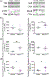
YAP/TAZ levels and phosphorylation in Piezo1cKO. (a) Western blots analysis was performed on Piezo1cKO sciatic nerves at 15 days of age. Calnexin (CNX) was used as a protein loading control. (b) YAP/pYAP and TAZ/pTAZ ratios are representative of YAP and TAZ activity, respectively. N = 8–10 animals per genotype. Data are represented in means ± SEM. Two-tailed unpaired Student’s t test. *p value <.05, **p value <.01, ***p value <.001
FIGURE 10
FIGURE 10
ERK and AKT levels and phosphorylation in PiezocKO mice. (a) Western blots analysis was performed on PiezocKO sciatic nerves at 15 days of age. Calnexin (CNX) was used as a protein loading control. (b) pERK/ERK and pAKT/AKT ratios are representative of ERK and AKT activity, respectively. N = 4 animals per genotype. Data are represented in means ± SEM. Two-tailed unpaired Student’s t test with Bonferroni postdoc test. **p value <.01, ***p value <.001, ****p value <.0001

References

    1. Acheta J, Stephens SBZ, Belin S, & Poitelon Y (2021). Therapeutic low-intensity ultrasound for peripheral nerve regeneration—A Schwann cell perspective. Frontiers in Cellular Neuroscience, 15, 812588. - PMC - PubMed
    1. Bae C, Sachs F, & Gottlieb PA (2011). The mechanosensitive ion channel Piezo1 is inhibited by the peptide GsMTx4. Biochemistry, 50, 6295–6300. - PMC - PubMed
    1. Bagriantsev SN, Gracheva EO, & Gallagher PG (2014). Piezo proteins: Regulators of mechanosensation and other cellular processes. The Journal of Biological Chemistry, 289, 31673–31681. - PMC - PubMed
    1. Baraban M, Koudelka S, & Lyons DA (2018). Ca (2+) activity signatures of myelin sheath formation and growth in vivo. Nature Neuroscience, 21, 19–23. - PMC - PubMed
    1. Battefeld A, Popovic MA, de Vries SI, & Kole MHP (2019). Highfrequency microdomain ca(2+) transients and waves during early myelin internode remodeling. Cell Reports, 26(182–91), e5. - PMC - PubMed

Publication types