. 2022 Aug 2;11(15):e026378.
doi: 10.1161/JAHA.122.026378. Epub 2022 Jul 29.

Endothelial Foxp1 Regulates Neointimal Hyperplasia Via Matrix Metalloproteinase-9/Cyclin Dependent Kinase Inhibitor 1B Signal Pathway

Affiliations

Endothelial Foxp1 Regulates Neointimal Hyperplasia Via Matrix Metalloproteinase-9/Cyclin Dependent Kinase Inhibitor 1B Signal Pathway

Xiaoli Chen et al. J Am Heart Assoc. .

Erratum in

Abstract

Background The endothelium is essential for maintaining vascular physiological homeostasis and the endothelial injury leads to the neointimal hyperplasia because of the excessive proliferation of vascular smooth muscle cells. Endothelial Foxp1 (forkhead box P1) has been shown to control endothelial cell (EC) proliferation and migration in vitro. However, whether EC-Foxp1 participates in neointimal formation in vivo is not clear. Our study aimed to investigate the roles and mechanisms of EC-Foxp1 in neointimal hyperplasia. Methods and Results The wire injury femoral artery neointimal hyperplasia model was performed in Foxp1 EC-specific loss-of-function and gain-of-function mice. EC-Foxp1 deletion mice displayed the increased neointimal formation through elevation of vascular smooth muscle cell proliferation and migration, and the reduction of EC proliferation hence reendothelialization after injury. In contrast, EC-Foxp1 overexpression inhibited the neointimal formation. EC-Foxp1 paracrine regulated vascular smooth muscle cell proliferation and migration via targeting matrix metalloproteinase-9. Also, EC-Foxp1 deletion impaired EC repair through reduction of EC proliferation via increasing cyclin dependent kinase inhibitor 1B expression. Delivery of cyclin dependent kinase inhibitor 1B-siRNA to ECs using RGD (Arg-Gly-Asp)-peptide magnetic nanoparticle normalized the EC-Foxp1 deletion-mediated impaired EC repair and attenuated the neointimal formation. EC-Foxp1 regulates matrix metalloproteinase-9/cyclin dependent kinase inhibitor 1B signaling pathway to control injury induced neointimal formation. Conclusions Our study reveals that targeting EC-Foxp1-matrix metalloproteinase-9/cyclin dependent kinase inhibitor 1B pathway might provide future novel therapeutic interventions for restenosis.

Keywords: cyclin dependent kinase inhibitor 1B (Cdkn1b); matrix metalloproteinase‐9 (MMP9); neointimal formation; transcription factor forkhead box protein P1 (Foxp1).


Figures
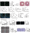
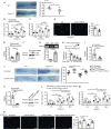
Figure 1
Figure 1. Genetic deletion of endothelial cell (EC)‐Foxp1 (forkhead box P1) increases neointimal formation through promoting vascular smooth muscle cell proliferation and migration.
A, Foxp1 immunostaining (left) in femoral artery, real‐time quantitative reverse transcription polymerase chain reaction (middle) and Western blot (right) in vascular ECs of EC‐Foxp1 deletion mice (Foxp1 ECKO , Foxp1 flox/flox ;Cdh5Cre ERT2 ) and wild‐type mice (n=7). White arrowheads indicating Foxp1 staining in ECs. B and C, Foxp1 ECKO mutant mice of both male and female sex exhibit increased neointimal formation at 28 days after femoral artery wire injury compared with wild‐type littermate mice, with representative images (B) and quantification of neointima area, intima to media ratio, percentage stenosis and media area (C) (n=7 females/7 males for each group). D, Foxp1 ECKO mutant mice exhibit significantly increased cell proliferation in the neointima by Ki67 immunostaining, with representative images (left) and quantification data (right) (n=7 females/7 males for each group). E, Significant reduction of Foxp1 expression in human umbilical vein ECs treated with lentiviral Foxp1‐shRNA is confirmed by real‐time quantitative reverse transcription (left) and Western blot (right) compared with human umbilical vein ECs with control scramble‐shRNA (n=5). F through H, Condition medium from Foxp1 knockdown human umbilical vein ECs significantly increases human aortic smooth muscle cell proliferation assessed by cell counting (F), 3‐(4, 5‐dimethylthiazol‐2‐yl)‐2, 5‐diphenyltetrazolium bromide (G), and Ki67 staining with quantification of Ki67‐positive cells on the right (H) (n=5). Statistical values of cell counting and 3‐(4, 5‐dimethylthiazol‐2‐yl)‐2, 5‐diphenyltetrazolium bromide are shown in Data S2. I and J, Condition medium from Foxp1 knockdown human umbilical vein endothelial cells significantly increases human aortic smooth muscle cell migration shown by wound healing (I) and transwell assay (J) with representative images (left) and quantification (right) (n=6). Data are means±SEM. EC indicates endothelial cell; Foxp1, forkhead box P1; OD, optical density; and WT, wild‐type. *P<0.05, **P<0.01. Scale bars: A=50 μm, B, D, H, I, J=100 μm.
Figure 2
Figure 2. Foxp1 directly regulate matrix metalloproteinase‐9 (MMP9) gene expression in endothelial cells (ECs) and in vivo MMP9 inhibition reverses the enhanced neointimal formation.
A through C, Foxp1 ECKO mutant mice exhibit increased expression and activity of MMP9 shown by real‐time quantitative reverse transcription polymerase chain reaction (A), Western blot (B), and MMP activity assay (C) in vascular ECs compared with wild‐type littermates (n=5). D, Chromatin immunoprecipitation assay shows that Foxp1 binds to the mouse MMP9 promoter by quantitative polymerase chain reaction and in agarose gel (n=6). E, The promoter region of MMP9, −4.0 kb to −3.5 kb before ATG translational site has 6 Foxp1 binding sites (left) and luciferase assay shows that Foxp1 expression vector dose‐dependent suppresses the luciferase reporter activity of this MMP9 promoter region (right) (n=5). F, Real‐time quantitative reverse transcription polymerase chain reaction and Western blot confirms the decreased expression of MMP9 in wire‐injured femoral artery after application of lentiviral MMP9‐shRNA compared with lentiviral scramble‐shRNA (n=6). G and H, MMP9 knockdown reverses the increased neointimal formation at 28 days after femoral artery wire injury caused by EC‐Foxp1 deletion, with representative images (G) and quantification of neointima area, intima‐to‐media ratio, percentage stenosis and media area (H) (n=6 for each group). I, MMP9 knockdown reverses EC‐Foxp1 deletion mediated increase of Ki67‐positive cells in neointima at 28 days after femoral artery wire injury, with representative images (left) and quantification data (right) (n=6 for each group). Data are means±SEM. EC indicates endothelial cell; Foxp1, forkhead box P1; IgG, immunoglobulin G; MMP9, matrix metalloproteinase‐9; OD, optical density; and WT, wild‐type. *P<0.05, **P<0.01. Scale bars: G, I=100 μm.
Figure 3
Figure 3. Endothelial matrix metalloproteinase‐9 (MMP9) knockdown reverses Foxp1 knockdown‐mediated increased vascular smooth muscle cell proliferation and migration in a paracrine manner.
A, MMP9 expression is significantly decreased in human umbilical vein endothelial cells treated with lentiviral MMP9‐shRNA compared with lentiviral scramble‐shRNA by real‐time quantitative reverse transcription polymerase chain reaction (left) and Western blot (right) (n=5). B through D, The condition medium of human umbilical vein endothelial cells treated with lentiviral MMP9‐shRNA reverses the Foxp1 knockdown‐mediated increase of vascular smooth muscle cell proliferation in comparison with that of human umbilical vein endothelial cells treated with lentiviral scramble‐shRNA, shown by cell counting (B), 3‐(4, 5‐dimethylthiazol‐2‐yl)‐2, 5‐diphenyltetrazolium bromide assay (C) and Ki67 staining with quantification on the right (D) (n=5). Statistical values of cell counting and MMT are shown in Data S2. E and F, Knockdown MMP9 in human umbilical vein endothelial cells reverses the Foxp1 knockdown mediated increase of vascular smooth muscle cell migration, shown by wound healing (E) and transwell assay (F), with representative images (left) and quantification data (right) (n=5). Data are means±SEM. Foxp1 indicates forkhead box P1; HASMCs, human aortic smooth muscle cells; MMP9, matrix metalloproteinase‐9; and NC, negative control. *P<0.05, **P<0.01. Scale bars: D, E, F=100 μm.
Figure 4
Figure 4. Endothelial cell (EC)‐Foxp1 (forkhead box P1) regulates cyclin dependent kinase inhibitor 1B (Cdkn1b) expression contributing to endothelial repair after injury denudation and neointimal formation.
A, EC‐Foxp1 deletion mice exhibit significant less EC coverage of whole mount femoral artery by in situ Evans blue staining at 7 days after wire injury compared with wild‐type littermate control mice, with representative images (left) and quantification (right) (n=6 for each group). B through D, Human umbilical vein endothelial cells (HUVECs) treated with lentiviral Foxp1‐shRNA display decreased cell proliferation in comparison with HUVECs treated with lentiviral scramble‐shRNA, shown by cell counting (B), 3‐(4, 5‐dimethylthiazol‐2‐yl)‐2, 5‐diphenyltetrazolium bromide assay (C) and Ki67 staining with quantification on the right (D) (n=5). Statistical values of cell counting and MMT are shown in Data S2. E and F, HUVECs treated with lentiviral Foxp1‐shRNA display increased expression of Cdkn1b shown by real‐time quantitative reverse transcription polymerase chain reaction (E) and Western blot (F) compared with HUVECs treated with lentiviral scramble‐shRNA (n=5). G, Chromatin immunoprecipitation assay shows that Foxp1 binds to the mouse Cdkn1b promoter by quantitative polymerase chain reaction (down) with agarose gel (up) (n=5). H, The promoter region of Cdkn1b, −1.4kb before Exon 1 has 3 Foxp1‐binding sites (left) and luciferase assay shows that Foxp1 expression vector dose‐dependent suppresses the luciferase reporter activity of this Cdkn1b promoter region (right) (n=5). I, The efficiency of decreased Cdkn1b expression following RGD (Arg‐Gly‐Asp)‐peptide magnetic nanoparticle application was confirmed by real‐time quantitative reverse transcription polymerase chain reaction in lung ECs (n=8). J, RGD (Arg‐Gly‐Asp)‐nanoparticles target delivery of Cdkn1b‐siRNA to endothelial cells (ECs) reverses the Foxp1 deletion‐mediated decreased EC repair following wire injury, shown by in situ Evans blue staining of femoral artery (n=8). K, Decreased expression of Cdkn1b in HUVECs treated with Cdkn1b‐siRNA is confirmed by real‐time quantitative reverse transcription polymerase chain reaction and Western blot (n=5). L through N, Cdkn1b‐siRNA knockdown HUVECs reverses the Foxp1 knockdown‐mediated decrease of cell proliferation in comparison with HUVECs treated with scramble‐siRNA, shown by cell counting (L), 3‐(4, 5‐dimethylthiazol‐2‐yl)‐2, 5‐diphenyltetrazolium bromide assay (M) and Ki67 staining with quantification on the right (N) (n=5). Statistical values of cell counting and MMT are shown in Data S2. Data are means±SEM. Cdkn1b indicates cyclin dependent kinase inhibitor 1B; Foxp1, forkhead box P1; HUVECs, human umbilical vein endothelial cells; IgG, immunoglobulin G; MMP9, matrix metalloproteinase‐9; NC, negative control; OD, optical density; and WT, wild‐type. *P<0.05, **P<0.01. Scale bars: A, J=1 mm; D, N=100 μm.
Figure 5
Figure 5. Endothelial cell (EC)‐Foxp1 (forkhead box P1) gain‐of‐function attenuates vascular neointimal formation through facilitation of EC repair and reduction of vascular smooth muscle cell proliferation and migration after wire injury.
A, EC‐Foxp1 gain‐of‐function mice (Foxp1 ECTg ) exhibit a significant increase of Foxp1 expression in vascular ECs compared with wild‐type mice by real‐time quantitative reverse transcription polymerase chain reaction (left) and Western blot (right) (n=7). B and C, Foxp1 ECTg mutant mice exhibit decreased neointimal formation at 28 days after femoral artery wire injury compared with wild‐type littermates, with representative images (B) and quantification of neointima area, intima to media ratio, percentage stenosis and media area (C) (n=7 for each group). D, Foxp1 ECTg mutant mice exhibit significantly decreased cell proliferation in neointima at 28 d after femoral artery wire injury, with representative images (left) and quantification data (right) (n=7 for each group). E, Foxp1 ECTg mutant mice exhibit a significant increase of EC coverage of femoral artery by in situ Evans blue staining at 7 days following wire injury compared with wild‐type littermates, with representative images (left) and quantification (right) (n=6 for each group). F, Real‐time quantitative reverse transcription polymerase chain reaction and Western blot confirm the significant increase of Foxp1 expression in human umbilical vein endothelial cells (HUVECs) treated with Foxp1 overexpression vector compared with empty control vector (n=5). G through I, Condition medium from Foxp1 overexpression HUVECs significantly inhibits human aortic smooth muscle cell proliferation compared with that from HUVECs of control vector, shown by cell counting (G), 3‐(4, 5‐dimethylthiazol‐2‐yl)‐2, 5‐diphenyltetrazolium bromide assay (H), and Ki67 staining with quantification of Ki67‐positive cells on the right (I) (n=5). Statistical values of cell counting and MMT are shown in Data S2. J and K, Condition medium from Foxp1 overexpression HUVECs significantly inhibits human aortic smooth muscle cell migration compared with that from HUVECs of control vector, shown by transwell (J) and wound healing assay (K), with representative images (left) and quantification (right) (n=5). L through N, Foxp1 overexpression in HUVECs increases cell proliferation in comparison with that of control vector, shown by cell counting (L), 3‐(4, 5‐dimethylthiazol‐2‐yl)‐2, 5‐diphenyltetrazolium bromide assay (M) and Ki67 staining with quantification on the right (N) (n=5). Statistical values of cell counting and MMT are shown in Data S2. Data are means±SEM. Foxp1 indicates forkhead box P1; HASMCs, human aortic muscle cells; HUVECs, human umbilical vein endothelial cells; MMP9, matrix metalloproteinase‐9; OD, optical density; and WT, wild‐type. *P<0.05, **P<0.01. Scale bars: B, D, I, J, K, N=100 μm; E=1 mm.
Figure 6
Figure 6. Working model of how endothelial cell‐Foxp1 (forkhead box P1) regulates neointimal hyperplasia through matrix metalloproteinase‐9/cyclin dependent kinase inhibitor 1B signal pathway following femoral artery injury.
Endothelial cell‐Foxp1 paracrine reduces matrix metalloproteinase‐9 expression to restrict vascular smooth muscle cells proliferation and migration and suppresses cyclin dependent kinase inhibitor 1B expression to promote endothelial cell proliferation for improvement of endothelial cell repair, and both work together leading to the attenuation of the injury induced neointimal hyperplasia. Cdkn1b indicates cyclin dependent kinase inhibitor 1B; EC, endothelial cells; Foxp1, forkhead box P1; MMP9, matrix metalloproteinase‐9; and WT, wild‐type.

Similar articles

Cited by

References

    1. Serruys PW, Ono M, Garg S, Hara H, Kawashima H, Pompilio G, Andreini D, Holmes DR Jr, Onuma Y, King Iii SB. Percutaneous coronary revascularization: JACC historical breakthroughs in perspective. J Am Coll Cardiol. 2021;78:384–407. doi: 10.1016/j.jacc.2021.05.024 - DOI - PubMed
    1. Chacko L, Howard JP, Rajkumar C, Nowbar AN, Kane C, Mahdi D, Foley M, Shun‐Shin M, Cole G, Sen S, et al. Effects of percutaneous coronary intervention on death and myocardial infarction stratified by stable and unstable coronary artery disease: a meta‐analysis of randomized controlled trials. Circ Cardiovasc Qual Outcomes. 2020;13:e006363. doi: 10.1161/CIRCOUTCOMES.119.006363 - DOI - PMC - PubMed
    1. Yang X, Yang Y, Guo J, Meng Y, Li M, Yang P, Liu X, Aung LHH, Yu T, Li Y. Targeting the epigenome in in‐stent restenosis: from mechanisms to therapy. Mol Ther Nucleic Acids. 2021;23:1136–1160. doi: 10.1016/j.omtn.2021.01.024 - DOI - PMC - PubMed
    1. Otsuka F, Finn AV, Yazdani SK, Nakano M, Kolodgie FD, Virmani R. The importance of the endothelium in atherothrombosis and coronary stenting. Nat Rev Cardiol. 2012;9:439–453. doi: 10.1038/nrcardio.2012.64 - DOI - PubMed
    1. Marx SO, Totary‐Jain H, Marks AR. Vascular smooth muscle cell proliferation in restenosis. Circ Cardiovasc Interv. 2011;4:104–111. doi: 10.1161/CIRCINTERVENTIONS.110.957332 - DOI - PMC - PubMed

Publication types

Substances

LinkOut - more resources