. 2022 Oct 5;12(10):2372-2391.
doi: 10.1158/2159-8290.CD-21-1026.

Modulation of BCL-2 in Both T Cells and Tumor Cells to Enhance Chimeric Antigen Receptor T-cell Immunotherapy against Cancer

Affiliations

Modulation of BCL-2 in Both T Cells and Tumor Cells to Enhance Chimeric Antigen Receptor T-cell Immunotherapy against Cancer

Yong Gu Lee et al. Cancer Discov. .

Abstract

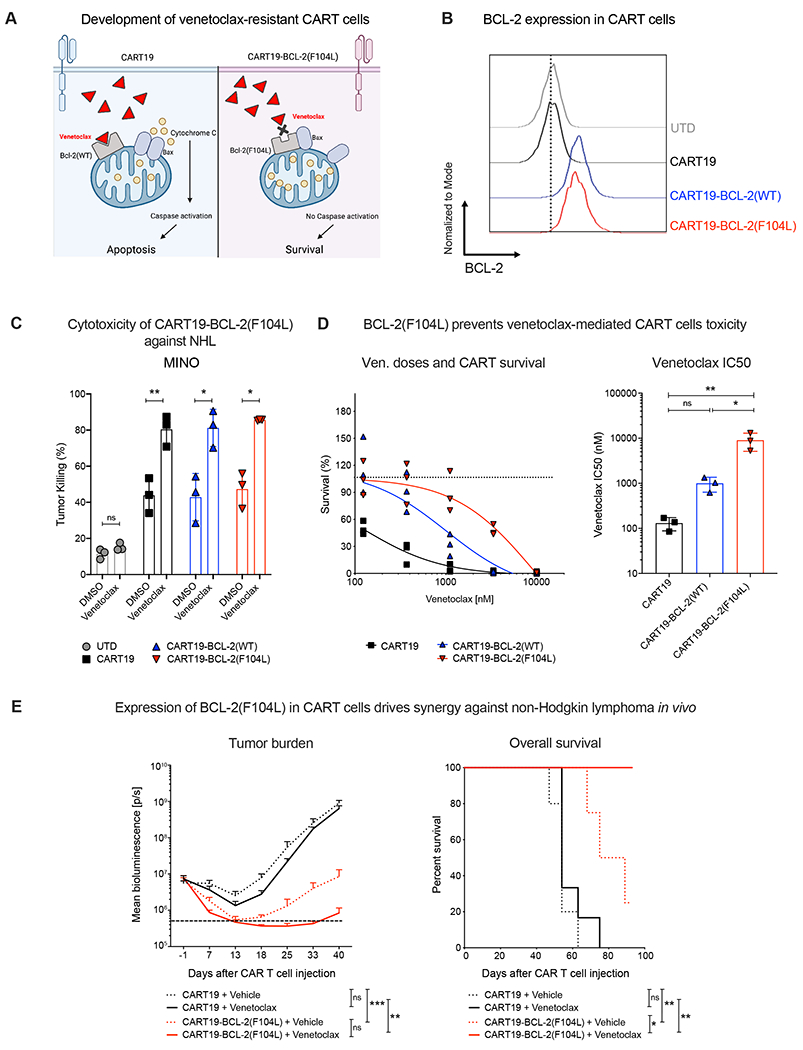
Chimeric antigen receptor T-cell (CART) immunotherapy led to unprecedented responses in patients with refractory/relapsed B-cell non-Hodgkin lymphoma (NHL); nevertheless, two thirds of patients experience treatment failure. Resistance to apoptosis is a key feature of cancer cells, and it is associated with treatment failure. In 87 patients with NHL treated with anti-CD19 CART, we found that chromosomal alteration of B-cell lymphoma 2 (BCL-2), a critical antiapoptotic regulator, in lymphoma cells was associated with reduced survival. Therefore, we combined CART19 with the FDA-approved BCL-2 inhibitor venetoclax and demonstrated in vivo synergy in venetoclax-sensitive NHL. However, higher venetoclax doses needed for venetoclax-resistant lymphomas resulted in CART toxicity. To overcome this limitation, we developed venetoclax-resistant CART by overexpressing mutated BCL-2(F104L), which is not recognized by venetoclax. Notably, BCL-2(F104L)-CART19 synergized with venetoclax in multiple lymphoma xenograft models. Furthermore, we uncovered that BCL-2 overexpression in T cells intrinsically enhanced CART antitumor activity in preclinical models and in patients by prolonging CART persistence.

Significance: This study highlights the role of BCL-2 in resistance to CART immunotherapy for cancer and introduces a novel concept for combination therapies-the engineering of CART cells to make them resistant to proapoptotic small molecules, thereby enhancing the therapeutic index of these combination therapies. This article is highlighted in the In This Issue feature, p. 2221.


Figures

Figure 1.
Figure 1.. Venetoclax enhances CART cell-mediated killing of venetoclax-sensitive lymphomas.
A. Drug screening of pro-apoptotic small molecules combined with CART19 against the B-cell leukemia cell line NALM6. Two concentrations of 29 drugs were used (100 nM and 1000 nM). Killing of NALM6 cells was assessed at 48 hours by luminescence. Two independent screenings, combined data. B. Proposal of CART/venetoclax combination therapy to enhance CART-mediated tumor killing. C. Half-maximal inhibitory concentration (IC50) of venetoclax against several lymphoid malignancy cell lines. Quantification of tumor killing by control untransduced control T cells (UTD) or CART19 in the presence of vehicle (DMSO) or venetoclax (48h). E:T ratios=0.125:1 (OCI-Ly18), 0.06:1 (MINO), 0.125:1 (NALM6) and 0.006:1 (primary MCL). Venetoclax concentration=10 nM (OCI-Ly18 and MINO), 250 nM (NALM6), and 3 nM (primary MCL). D. Tumor killing by the combination of venetoclax with CART19 cells that contain either CD28 or 4-1BB co-stimulation domains. E:T ratios=0.1:1. Venetoclax concentration=20 nM. E. Tumor killing by UTD or CART33 in the presence of vehicle (DMSO) or venetoclax (48h). E:T ratios=0.063:1 (MOLM-14), 0.15:1 (KG-1). Venetoclax concentration=125 nM (MOLM-14), 50 nM (KG-1). F. Caspase 3/7 activity by flow cytometry. E:T ratio=0.1:1. Venetoclax concentration=10 nM. G. Schematic of the xenograft model of venetoclax-sensitive lymphoma (OCI-Ly18). CART cells (2x10⁶) were infused via intravenous injection when tumor volume reached ~150 mm3. Either vehicle or venetoclax (25 mg/kg/daily) was administrated for 3 weeks via oral gavage. Tumor burden over time was measured by caliper. Overall survival was also monitored. All data represent mean±SD. A two-tailed unpaired Student t-test with Welch’s correction was performed (panels C, D, E, and F). In panel G, tumor volume was compared with one-way ANOVA with posthoc Tukey tests, and overall survival was analyzed using the log-rank (Mantel-Cox) test. All data presented are representative of at least two independent experiments. ns: not significant, *p < 0.05, **p < 0.01. UTD: untransduced T cells; CART19: anti-CD19 CAR T cell; MCL: mantle cell lymphoma; E:T=ratio of effector to target; IAP: Inhibition of apoptosis protein.
Figure 2.
Figure 2.. Venetoclax treatment induces CART cell toxicity.
A. Schematic of the in vivo xenograft model of venetoclax-resistant tumors. For the MINO model, CART cells (5x10⁴) were infused 14 days after luciferase+ MINO cells were implanted (i.v. injection). For the NALM6 model, CART cells (5x10⁵) were infused 3 - 4 days after luciferase+ NALM6 cells were implanted (i.v. injection). Either vehicle or venetoclax (50 mg/kg/daily) was administrated for 5 weeks via oral gavage. B and C. Tumor progression of mice bearing MINO (B) or NALM6 (C) cells treated with UTD or CART19 plus either vehicle or venetoclax. D. In vivo CART cell expansion. To quantify CART cell expansion in the NALM6 xenograft model, peripheral mouse blood was harvested on day 10 after CART cell infusion and analyzed by flow cytometry. E. Quantification of venetoclax-induced CART cell toxicity upon treatment of various doses of venetoclax in vitro (110 nM ~ 10000 nM). Each dot indicates CART cells generated from different healthy donors (n=8). E:T ratio=0.25:1. Venetoclax concentration=1100 nM. All data represent mean±SD. One-way ANOVA with posthoc Tukey tests was performed (panels B and C). A two-tailed unpaired Student t-test with Welch’s correction was performed (panels D and F). All data presented are representative of at least two independent experiments. ns: not significant, *p < 0.05; **p < 0.01; UTD: untransduced T cells; CART19: anti-CD19 CAR T cells; E:T=ratio of effector to target.
Figure 3.
Figure 3.. Expression of mutant BCL-2 prevents venetoclax-mediated CART cell toxicity.
A. Strategy to develop venetoclax-resistant CART cells. B. BCL-2 expression in CART cells measured by flow cytometry. C. Quantification of tumor (MINO) killing by untransduced control T cells (UTD) or CART19, CART19-BCL-2(WT), or CART19-BCL-2(F104L) in the presence of vehicle (DMSO) or venetoclax. E:T ratio=0.06:1. Venetoclax concentration=10 nM. D. Evaluation of venetoclax-mediated toxicity on either CART19, CART19-BCL-2(WT) or CART19-BCL-2(F104L). CART cell survival (left panel) and IC50 value (right panel). Each dot indicates CART cells generated from different healthy donors (n=3). E. Tumor progression and survival of xenografted mice bearing MINO. Treated with CART19 or CART19-BCL-2(F104L) plus either vehicle or venetoclax. All data represent mean±SD. One-way ANOVA with posthoc Tukey tests was performed (panels C and D). In panel E, tumor volume was compared with one-way ANOVA with post-hoc Tukey tests, and survival was analyzed using the log-rank (Mantel-Cox) test. All data presented are representative of at least two independent experiments: ns: not significant, *p < 0.05, **p < 0.01. UTD: untransduced T cells; CART19: anti-CD19 CAR T cells; CART19-BCL-2(WT): BCL-2(WT)-expressing CART19; CART19-BCL-2(F104L): BCL-2(F104L)-expressing CART19; E:T=ratio of effector to target.
Figure 4.
Figure 4.. Chromosomal alterations of BCL-2 in lymphoma patients associate with poor prognosis of CART therapy.
A. Schematic description of the strategy to investigate whether genetic alteration of BCL-2 affects CART’s anti-tumor clinical response. Pre-CART biopsies from patients with LCL were analyzed by fluorescence in situ hybridization (FISH) to search for BCL-2 chromosomal aberration. B. Best overall response rate of 87 LCL patients treated with CART19 according to the presence of BCL-2 chromosomal alteration (gain or translocation). C. Overall survival of 87 LCL patients treated with CART19 according to the presence of BCL-2 chromosomal alteration (gain or translocation). D. Best overall response of 37 DLBCL patients treated with CART19 according to the presence of BCL-2 chromosomal alteration (gain or translocation). E. Overall survival of 37 DLBCL patients treated with CART19 according to the presence of BCL-2 chromosomal alteration (gain or translocation). F. Schematic description of the strategy to investigate the impact of venetoclax bridging therapy on CART19’s clinical response in MCL patients. G. Best overall response rate of 18 MCL patients treated with CART19 according to bridging therapy including venetoclax or not. H. Event-free survival of MCL patients treated with CART19 after bridging therapy with (YES) or without (NO) venetoclax. Comparisons between the groups were performed with the chi-square test for categorical variables and t Student’s test for continuous variables, as appropriate. Survival analysis was performed by the Kaplan-Meier estimation and compared with log-rank test. All statistical tests were two-sided and statistical significance was defined as p-value <0.05. Analysis was performed with the Statistical Package for the Social Sciences software v.22.0 (Chicago, IL, USA). CR: Complete response; PR: Partial response; SD: Stable disease; PD: Progress disease. LCL: Large B cell lymphoma; DLBCL: Diffuse large B cell lymphoma; MCL: mantle cell lymphoma.
Figure 5.
Figure 5.. Overexpression of BCL-2(WT) in CART cells enhances their anti-tumor efficacy.
A. Schematic of the in vivo xenograft model to study the effect of BCL-2 overexpression on CART’s anti-tumor activity. For the MINO model (B), CART cells (5x10⁴) were infused 14 days after luciferase+ MINO cell i.v. injection. For the NALM6 model (C), CART cells (5x10⁵) were infused 3 - 4 days after luciferase+ NALM6 cell i.v. injection. B and C. Tumor progression and overall survival over time in mice bearing MINO (B) and NALM6 (C) treated with CART19 or CART19-BCL2(WT) (representative of 2 replicate experiments, n=5). D. Quantification of CART cells peak expansion in mouse blood collected from CART-treated mouse bearing NALM6 on day 10 after CART cell infusion by flow cytometry. E. CART cell persistence in CART-treated mouse blood over time by flow cytometry (NALM6 model). F. Fold change of CART cell upon stimulation with irradiated MINO (representative of 2 replicate experiments). G. Volcano plot showing differentially expressed genes in CART19-BCL2(WT) compared to CART19 on day 18 after stimulation with irradiated MINO. H. Gene Set Enrichment Analysis (GSEA) of differentially expressed genes in CART19-BCL2(WT) compared to CART19 on day 18 after stimulation with irradiated MINO. I. Survival of CART cell after withdrawal cytokines. CART cells were stimulated with irradiated MINO for 48 hours and culture media were replaced with fresh media to withdraw cytokines. Survival of CART cells was monitored by flow cytometry 48 hours after adding fresh media. All data represent mean±SD. One-way ANOVA with posthoc Tukey tests was performed for all comparisons. Overall survival was analyzed using the log-rank (Mantel-Cox) test (panels B and C). All data presented are representative of at least two independent experiments except bulk RNA-seq (performed once with two biological replicates). ns: not significant, *p < 0.05, **p < 0.05. UTD: untransduced T cells; CART19: anti-CD19 CAR T cells; CART19-BCL-2(WT): BCL-2(WT)-expressing CART19; E:T=ratio of effector to target
Figure 6.
Figure 6.. Increased BCL-2 expression in T cells from CART apheretic products is associated with positive clinical outcomes in lymphoma patients at long term.
A. Schematic description of the approach taken to investigate the relationship between the level of BCL-2 and CART’s clinical response. RNA was extracted from T cells from apheretic products of 38 lymphoma patients who received CART19 immunotherapy (CTL019, now known as tisagenleucleucel) in the clinical trial (NCT02030834). Next, BCL-2 mRNA expression was quantified via the nCounter analysis system (NanoString Technologies, Inc, Seattle, WA). B. Volcano plot showing differential gene expression in T cells based on best overall response (CR or NR). C. Comparison of BCL-2 expression in T cell apheretic products of CART19-treated patients in CR/PR vs. NR. D. Correlation of BCL-2 expression in T cell apheretic products with CART persistence. E. Correlation of BCL-2 expression in T cell apheretic products with overall survival. F. Monitoring of abnormal CART expansion mediated by constant overexpression of BCL-2 (left panel: CART expansion (fold change), right panel: frequency of CART (%) G. Cytotoxicity on CART19 and CART19-BCL-2(WT) 24-hour after treatment of chemotherapy (doxorubicin, 300 and 1000 nM). H. Cytotoxicity of CART19-tEGFR and CART19-BCL2(WT)-tEGFR after 24h treatment with either isotype control or anti-EGFR antibody (Cetuximab). All data represent mean±SD. Linear regression analysis was performed (D and E). A two-tailed unpaired Student t test with Welch’s correction was performed (panels C, F, H, and I). *P < 0.05. ns: not significant, *p < 0.05, **p < 0.05. UTD: untransduced T cells; CART19: anti-CD19 CAR T cells; CART19-BCL-2(WT): BCL-2(WT)-expressing CART19; CART19-tEGFR: anti-CD19 CAR T cells expressing truncated EGFR; CART19-BCL-2(WT): CART19 expressing BCL-2(WT) and truncated EGFR; E:T=ratio of effector to target. EGFR: Epidermal growth factor receptor.

References

    1. Spiegel JY, Patel S, Muffly L, Hossain NM, Oak J, Baird JH, et al. CAR T cells with dual targeting of CD19 and CD22 in adult patients with recurrent or refractory B cell malignancies: a phase 1 trial. Nature medicine 2021;27(8):1419–31. - PMC - PubMed
    1. Melenhorst JJ, Chen GM, Wang M, Porter DL, Chen C, Collins MA, et al. Decade-long leukaemia remissions with persistence of CD4+ CAR T cells. Nature 2022;602(7897):503–9. - PMC - PubMed
    1. Chong EA, Ruella M, Schuster SJ. Five-year outcomes for refractory B-cell lymphomas with CAR T-cell therapy. New England Journal of Medicine 2021;384(7):673–4. - PubMed
    1. Ruella M, Gill S. How to train your T cell: genetically engineered chimeric antigen receptor T cells versus bispecific T-cell engagers to target CD19 in B acute lymphoblastic leukemia. Expert opinion on biological therapy 2015;15(6):761–6. - PubMed
    1. Turtle CJ, Hanafi L-A, Berger C, Hudecek M, Pender B, Robinson E, et al. Immunotherapy of non-Hodgkin’s lymphoma with a defined ratio of CD8+ and CD4+ CD19-specific chimeric antigen receptor–modified T cells. Science translational medicine 2016;8(355):355ra116–355ra116. - PMC - PubMed

Publication types

MeSH terms