Combatting the effect of image reconstruction settings on lymphoma [18F]FDG PET metabolic tumor volume assessment using various segmentation methods
- PMID: 35904645
- PMCID: PMC9338209
- DOI: 10.1186/s13550-022-00916-9
Combatting the effect of image reconstruction settings on lymphoma [18F]FDG PET metabolic tumor volume assessment using various segmentation methods
Abstract
Background: [18F]FDG PET-based metabolic tumor volume (MTV) is a promising prognostic marker for lymphoma patients. The aim of this study is to assess the sensitivity of several MTV segmentation methods to variations in image reconstruction methods and the ability of ComBat to improve MTV reproducibility.
Methods: Fifty-six lesions were segmented from baseline [18F]FDG PET scans of 19 lymphoma patients. For each scan, EARL1 and EARL2 standards and locally clinically preferred reconstruction protocols were applied. Lesions were delineated using 9 semiautomatic segmentation methods: fixed threshold based on standardized uptake value (SUV), (SUV = 4, SUV = 2.5), relative threshold (41% of SUVmax [41M], 50% of SUVpeak [A50P]), majority vote-based methods that select voxels detected by at least 2 (MV2) and 3 (MV3) out of the latter 4 methods, Nestle thresholding, and methods that identify the optimal method based on SUVmax (L2A, L2B). MTVs from EARL2 and locally clinically preferred reconstructions were compared to those from EARL1. Finally, different versions of ComBat were explored to harmonize the data.
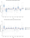
Results: MTVs from the SUV4.0 method were least sensitive to the use of different reconstructions (MTV ratio: median = 1.01, interquartile range = [0.96-1.10]). After ComBat harmonization, an improved agreement of MTVs among different reconstructions was found for most segmentation methods. The regular implementation of ComBat ('Regular ComBat') using non-transformed distributions resulted in less accurate and precise MTV alignments than a version using log-transformed datasets ('Log-transformed ComBat').
Conclusion: MTV depends on both segmentation method and reconstruction methods. ComBat reduces reconstruction dependent MTV variability, especially when log-transformation is used to account for the non-normal distribution of MTVs.
Keywords: Lymphoma; Metabolic tumor volume; Reconstruction; Segmentation; [18F]FDG PET.
© 2022. The Author(s).
Conflict of interest statement
All authors declare no competing financial interest.
Figures


Similar articles
-
Quantitative and clinical implications of the EARL2 versus EARL1 [18F]FDG PET-CT performance standards in head and neck squamous cell carcinoma.EJNMMI Res. 2023 Oct 25;13(1):91. doi: 10.1186/s13550-023-01042-w. EJNMMI Res. 2023. PMID: 37878160 Free PMC article.
-
Interobserver Agreement on Automated Metabolic Tumor Volume Measurements of Deauville Score 4 and 5 Lesions at Interim 18F-FDG PET in Diffuse Large B-Cell Lymphoma.J Nucl Med. 2021 Nov;62(11):1531-1536. doi: 10.2967/jnumed.120.258673. Epub 2021 Mar 5. J Nucl Med. 2021. PMID: 33674403 Free PMC article.
-
Automated Segmentation of Baseline Metabolic Total Tumor Burden in Diffuse Large B-Cell Lymphoma: Which Method Is Most Successful? A Study on Behalf of the PETRA Consortium.J Nucl Med. 2021 Mar;62(3):332-337. doi: 10.2967/jnumed.119.238923. Epub 2020 Jul 17. J Nucl Med. 2021. PMID: 32680929 Free PMC article.
-
The Impact of Semiautomatic Segmentation Methods on Metabolic Tumor Volume, Intensity, and Dissemination Radiomics in 18F-FDG PET Scans of Patients with Classical Hodgkin Lymphoma.J Nucl Med. 2022 Sep;63(9):1424-1430. doi: 10.2967/jnumed.121.263067. Epub 2022 Jan 6. J Nucl Med. 2022. PMID: 34992152 Free PMC article.
-
Sensitivity of an AI method for [18F]FDG PET/CT outcome prediction of diffuse large B-cell lymphoma patients to image reconstruction protocols.EJNMMI Res. 2023 Sep 28;13(1):88. doi: 10.1186/s13550-023-01036-8. EJNMMI Res. 2023. PMID: 37758869 Free PMC article.
Cited by
-
Advances in positron emission tomography and radiomics.Hematol Oncol. 2023 Jun;41 Suppl 1(Suppl 1):11-19. doi: 10.1002/hon.3137. Hematol Oncol. 2023. PMID: 37294959 Free PMC article. Review.
-
Multicenter development of a PET-based risk assessment tool for product-specific outcome prediction in large B-cell lymphoma patients undergoing CAR T-cell therapy.Eur J Nucl Med Mol Imaging. 2024 Apr;51(5):1361-1370. doi: 10.1007/s00259-023-06554-0. Epub 2023 Dec 20. Eur J Nucl Med Mol Imaging. 2024. PMID: 38114616 Free PMC article.
-
Quantitative and clinical implications of the EARL2 versus EARL1 [18F]FDG PET-CT performance standards in head and neck squamous cell carcinoma.EJNMMI Res. 2023 Oct 25;13(1):91. doi: 10.1186/s13550-023-01042-w. EJNMMI Res. 2023. PMID: 37878160 Free PMC article.
-
Metabolic bulk volume predicts survival in a homogeneous cohort of stage II/III diffuse large B-cell lymphoma patients undergoing R-CHOP treatment.Front Oncol. 2023 Jun 13;13:1186311. doi: 10.3389/fonc.2023.1186311. eCollection 2023. Front Oncol. 2023. PMID: 37384292 Free PMC article.
-
Optimization and validation of 18F-DCFPyL PET radiomics-based machine learning models in intermediate- to high-risk primary prostate cancer.PLoS One. 2023 Nov 9;18(11):e0293672. doi: 10.1371/journal.pone.0293672. eCollection 2023. PLoS One. 2023. PMID: 37943772 Free PMC article.
References
-
- Wahl RL. Principles and practice of PET and PET/CT. Lippincott Williams & Wilkins; 2008.
-
- Eertink JJ, van de Brug T, Wiegers SE, Zwezerijnen GJC, Pfaehler EAG, Lugtenburg PJ, et al. (18)F-FDG PET baseline radiomics features improve the prediction of treatment outcome in diffuse large B-cell lymphoma. Eur J Nucl Med Mol Imaging. 2022;49(3):932–942. doi: 10.1007/s00259-021-05480-3. - DOI - PMC - PubMed
LinkOut - more resources
Full Text Sources

