Review
. 2022 Sep;12(9):1967-1988.
doi: 10.1007/s13555-022-00779-x. Epub 2022 Jul 29.

Update on Melasma-Part I: Pathogenesis

Affiliations
Review

Update on Melasma-Part I: Pathogenesis

Ana Cláudia C Espósito et al. Dermatol Ther (Heidelb). 2022 Sep.

Abstract

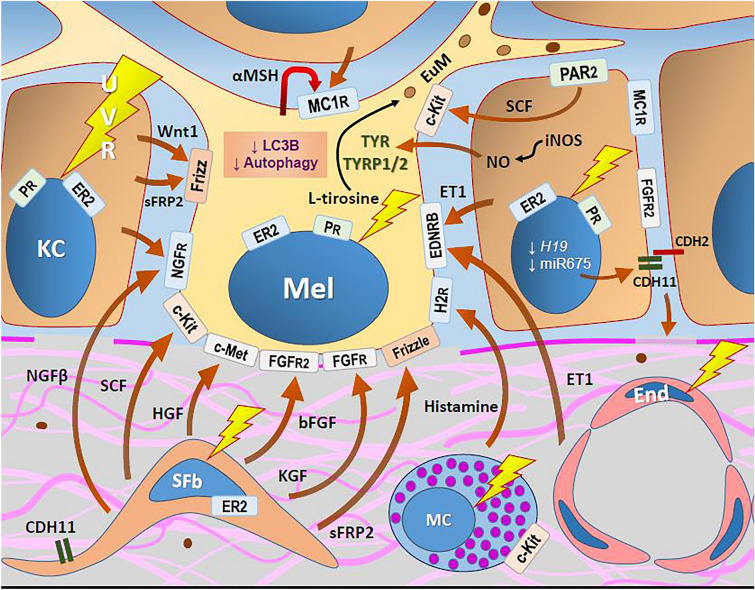
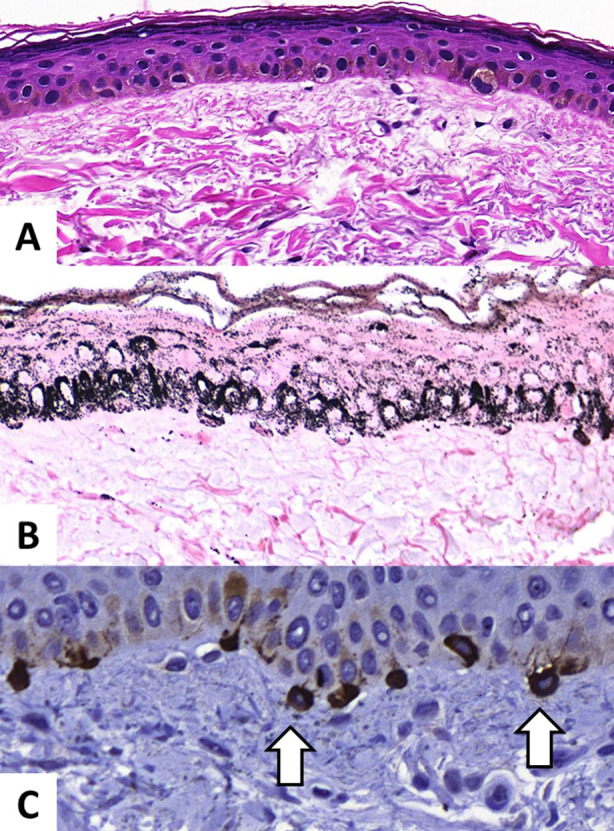
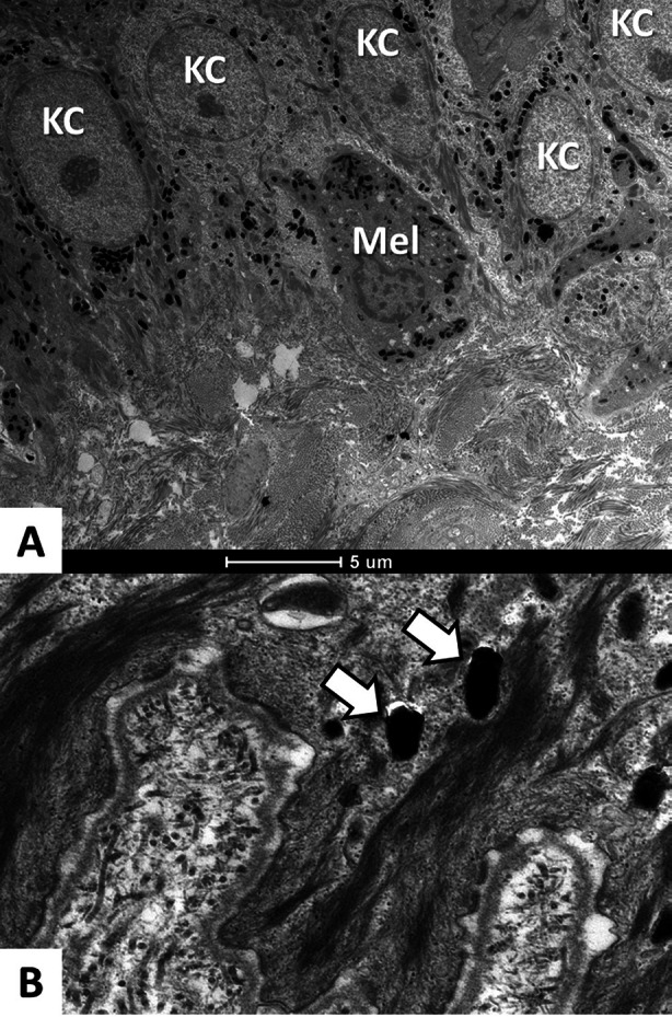
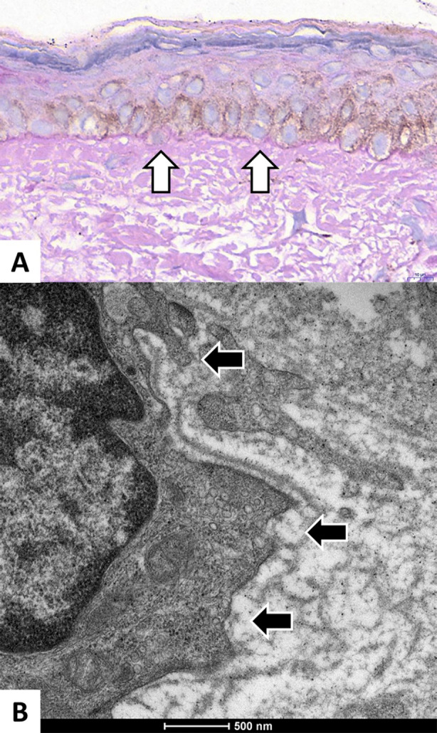
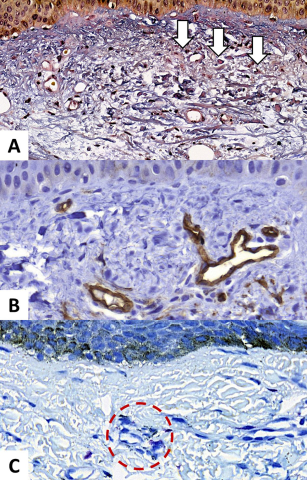
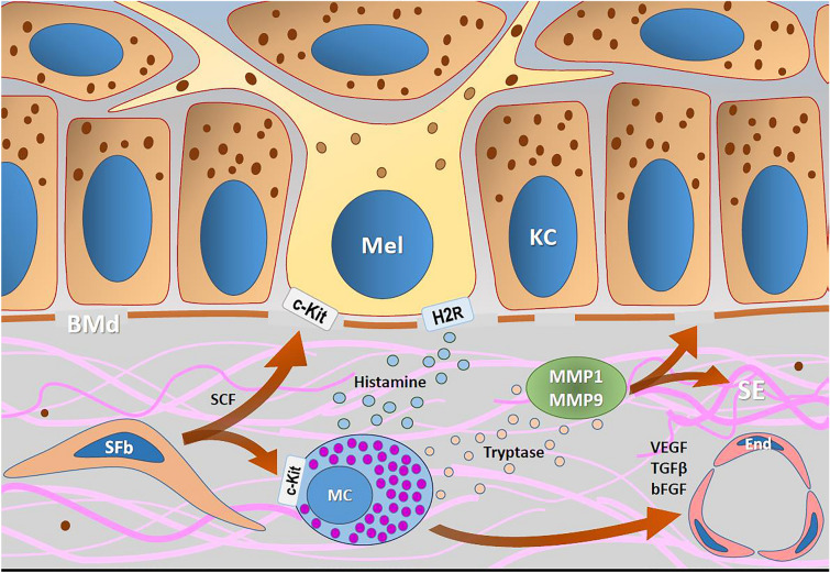
Melasma is a multifactorial dyschromia that results from exposure to external factors (such as solar radiation) and hormonal factors (such as sex hormones and pregnancy), as well as skin inflammation (such as contact dermatitis and esthetic procedures), in genetically predisposed individuals. Beyond hyperfunctional melanocytes, skin with melasma exhibits a series of structural and functional alterations in the epidermis, basement membrane, and upper dermis that interact to elicit and sustain a focal hypermelanogenic phenotype. Evolution in the knowledge of the genetic basis of melasma and the cutaneous response to solar radiation, as well as the roles of endocrine factors, antioxidant system, endothelium proliferation, fibroblast senescence, mast cell degranulation, autophagy deficits of the melanocyte, and the paracrine regulation of melanogenesis, will lead to the development of new treatments and preventive strategies. This review presents current knowledge on these aspects of the pathogenesis of melasma and discusses the effects of specific treatments and future research on these issues.

Keywords: Melanin; Melanocytes; Melasma; Pathogenesis; Photoaging; UV radiation.


Figures

Fig. 1
Fig. 1
Theoretic model of melanogenic pathways involved in melasma. Melanocytes (Mels) are hyperfunctional, promoting eumelanogenesis (Eum) due to paracrine and autocrine stimuli. UVR elicits melanogenic, oxidative, and inflammatory responses in the epidermis and upper dermis. Melanocortin (αMSH) and its receptor (MC1R) are increased in keratinocytes (KCs) and Mels. Hormonal stimuli mediate melanogenesis through the nuclear receptors of estrogen-β (ER2) and progesterone (PR). Several growth factors, which are also melanogenic, are actively released by senescent fibroblasts (SFbs), including nerve growth factor type β (NGFβ), SCF, HGF, bFGF, KGF, and sFRP2. Endothelin-1 (ET1) is secreted by the endothelium (End) and KCs after UVR exposure. Mast cells (MCs) release histamine under paracrine stimulation and UVR. Protease-activated receptor-2 (PAR2) stimulates melanocyte dendricity and melanosome phagocytosis by KCs and induces the release of SCF. In melasma, Mels present diminished autophagy (↓ LC3B–microtubule-associated proteins 1A/1B light chain 3B), which stimulates melanogenesis. In addition, the lower expression of miR-675, a MITF-targeted micro-RNA, is associated with greater expression of cadherin-11 (CDH11) in KCs and fibroblasts, which contributes to basement membrane and upper dermal damage. Nitric oxide (NO), produced by inducible nitrogen oxide synthase (iNOS) and Wnt1, is increased in the epidermis in melasma
Fig. 2
Fig. 2
Histologic images of facial melasma. A Atrophic epidermis with a thin stratum corneum, hypogranulosis, and polarization loss of the nuclei in the basal layer. Upper dermis revealing solar elastosis and overall unstructured collagen fibers (hematoxylin and eosin, 100×). B Dense and homogeneous melanin pigmentation with coarse melanosomes in all epidermal layers, including the stratum corneum, and extracellular melanin granules in the upper dermis (Fontana–Masson, 400×). C Atrophic epidermis with hypertrophic melanocytes (in brown) with prominent dendrites and melanocytes protruding into the dermis (pendulum melanocytes, arrows) and losing contact with the basal layer (Melan-A, 400×)
Fig. 3
Fig. 3
Transmission electronic microscopy of facial melasma. A Intense distribution of mature melanosomes in the epidermis (KC, keratinocyte; Mel, Melanocyte). Sparse extracellular melanosomes in the upper dermis. B Mature and large melanosomes (type IV) in the cytoplasm of a keratinocyte from the basal layer (white arrows)
Fig. 4
Fig. 4
Facial melasma. A Periodic acid–Schiff staining (400×) evidencing thinning and several discontinuities in the basement membrane (white arrows). B Transmission electronic microscopy of the dermoepidermal junction under a melanocyte revealing complete interruption of the lamina densa, structural damage, and loss of anchoring fibrils in the lamina lucida (black arrows)
Fig. 5
Fig. 5
Facial melasma. A Histologic image (Herovici, 200×) revealing upper dermis collagen fiber fragmentation with loss of structure and elastonization (solar elastosis, arrows). B Immunohistochemistry image of facial melasma (CD34, 400×) evidencing upper dermis endothelial proliferation (brown structures). C Histologic image (Toluidine Blue, 400×) evidencing mast cells in the upper dermis, especially in the perivascular areas (dashed circle)
Fig. 6
Fig. 6
Schematic representation of the interaction between senescent fibroblasts (SFbs) and mast cells (MCs) in melasma. Histamine stimulates melanogenesis directly through H2-receptors (H2Rs) in melanocytes (Mels). SCF is overexpressed in melasma, which influences MC survival, migration, and activation; it binds to the c-KIT receptor, inducing melanogenesis and the Mel cell cycle. Tryptase activates metalloproteinases (MMP1 and MMP9), which degrade type I and IV collagens, leading to extracellular matrix degradation (solar elastosis; SE) and basement membrane damage (BMd). MCs also induce endothelial (End) proliferation by secreting VEGF, bFGF, and TGF-β

Similar articles

Cited by

References

    1. Esposito ACC, Brianezi G, de Souza NP, Miot LDB, Miot HA. Exploratory study of epidermis, basement membrane zone, upper dermis alterations and Wnt pathway activation in melasma compared to adjacent and retroauricular skin. Ann Dermatol. 2020;32(2):101–108. doi: 10.5021/ad.2020.32.2.101. - DOI - PMC - PubMed
    1. Tamega Ade A, Miot LD, Bonfietti C, Gige TC, Marques ME, Miot HA. Clinical patterns and epidemiological characteristics of facial melasma in Brazilian women. J Eur Acad Dermatol Venereol. 2013;27(2):151–156. doi: 10.1111/j.1468-3083.2011.04430.x. - DOI - PubMed
    1. Ishiy PS, Silva LR, Penha MA, Handel AC, Miot HA. Skin diseases reported by workers from UNESP campus at Rubiao Jr, Botucatu-SP (Brazil) An Bras Dermatol. 2014;89(3):529–531. doi: 10.1590/abd1806-4841.20142875. - DOI - PMC - PubMed
    1. Ortonne JP, Arellano I, Berneburg M, Cestari T, Chan H, Grimes P, et al. A global survey of the role of ultraviolet radiation and hormonal influences in the development of melasma. J Eur Acad Dermatol Venereol. 2009;23(11):1254–1262. doi: 10.1111/j.1468-3083.2009.03295.x. - DOI - PubMed
    1. Handel AC, Lima PB, Tonolli VM, Miot LD, Miot HA. Risk factors for facial melasma in women: a case-control study. Br J Dermatol. 2014;171(3):588–594. doi: 10.1111/bjd.13059. - DOI - PubMed

LinkOut - more resources