. 2022 Jul 29;17(7):e0272090.
doi: 10.1371/journal.pone.0272090. eCollection 2022.

Antioxidative enzyme NAD(P)H quinone oxidoreductase 1 (NQO1) modulates the differentiation of Th17 cells by regulating ROS levels

Affiliations

Antioxidative enzyme NAD(P)H quinone oxidoreductase 1 (NQO1) modulates the differentiation of Th17 cells by regulating ROS levels

Kyoko Nishida-Tamehiro et al. PLoS One. .

Abstract

NAD(P)H quinone oxidoreductase 1 (NQO1) is a flavoprotein that catalyzes two-electron reduction of quinone to hydroquinone by using nicotinamide adenine dinucleotide (NADPH), and functions as a scavenger for reactive oxygen species (ROS). The function of NQO1 in the immune response is not well known. In the present study, we demonstrated that Nqo1-deficient T cells exhibited reduced induction of T helper 17 cells (Th17) in vitro during Th17(23)- and Th17(β)- skewing conditions. Nqo1-deficient mice showed ameliorated symptoms in a Th17-dependent autoimmune Experimental autoimmune encephalomyelitis (EAE) model. Impaired Th17-differentiation was caused by overproduction of the immunosuppressive cytokine, IL-10. Increased IL-10 production in Nqo1-deficient Th17 cells was associated with elevated intracellular Reactive oxygen species (ROS) levels. Furthermore, overproduction of IL-10 in Th17 (β) cells was responsible for the ROS-dependent increase of c-avian musculoaponeurotic fibrosarcoma (c-maf) expression, despite the lack of dependency of c-maf in Th17(23) cells. Taken together, the results reveal a novel role of NQO1 in promoting Th17 development through the suppression of ROS mediated IL-10 production.


Conflict of interest statement

The authors have declared that no competing interests exist.

Figures
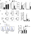
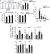
Fig 1
Fig 1. NQO1 promotes Th17 differentiation in vitro.
Isolated CD4+ CD62Lhi naïve T cells were stimulated with anti-CD3 (2 μg/ml) and anti-CD28 (2 μg/ml) antibodies and cultured with Th17(β) (stimulated with IL-6 +TGFβ) and Th17(23) (stimulated with IL-1β +IL-6 +IL-23) skewing conditions for 3 days. (A) Real-time PCR analysis of Nqo1 gene expression in WT (n = 3). (B) IL-17A and IL-22 production in the cell culture supernatant was measured by ELISA. (n = 5) (C) On days 3, the differentiated cells were stimulated with PMA, Ionomycin, and BFA for 4 h. The plot indicates frequencies of CD4+ IL-17A+ T cells (n = 3). (D) Histogram showing internal staining of CD4+ RORγt+ expression (n = 3). Each graph indicates the mean ± SE, One way ANOVA.
Fig 2
Fig 2. NQO1 promotes Th17 differentiation by inhibiting IL-10 production.
(A, C) Purified naïve CD4+ T cells from WT, Nqo1 KO, Il-10 KO, or Nqo1/Il-10 double knock-out were stimulated with anti-CD3/CD28 antibody in each differentiation conditions for 3 days. Cytokines secreted into the cell culture supernatant were measured by ELISA (n = 4–8). (B) Naïve CD4+ T cells from Il-10 venus reporter mice, and Nqo1 KO/Il-10 venus reporter mice were differentiated into Th17 (β) or Th17 (23) cells (n = 3). The ratio of IL-10 expressing CD4+ cells cultured under each condition for 3 days was determined. Data are representative of three independent experiments. Graphs indicate the mean ± SE, One way ANOVA.
Fig 3
Fig 3. Nqo1 deficiency ameliorates EAE severity depend on IL-10.
(A, B) The graph shows clinical scores and induction ratio for EAE in WT, Nqo1 KO mice after immunization with a MOG-CFA mixture and p-toxin. Mice were sacrificed under anesthesia at end of experiment (n = 4). (C) Absolute numbers of CNS infiltrating cells were analyzed. Total CD4+ cells are presented in left panel and IL-17 expression CD4+ cells are shown in right panel (n = 4). (D) Splenocytes from immunized mice were re-stimulated with 50 μM MOG peptide for 72 h. Frequencies of CD4+ IL-17+, IFNγ+ cells (%) were measured by FACS (n = 4). (E, F) The graph indicates clinical scores for adoptive transfer EAE in recipient mice (n = 5). Mice were sacrificed under anesthesia at 23 days (end of experiment) or at the humane endpoints. Data are representative of three independent experiments. Graphs indicate the mean ± SE. (A-C) Unpaired student T-test, (D-F) One way ANOVA.
Fig 4
Fig 4. NQO1 inhibits IL-10 production by regulating ROS.
(A-C) Naïve CD4+ T cells from Nqo1 KO and WT mice were differentiated into Th0, Th17(β), and Th17(23) cells with the indicated cytokines for 1 day. Cells were loaded with the ROS indicator, DCFDA, DHE, or mtROS detecting reagent and the fluorescence intensities were detected by flow cytometry (n = 3). (D) IL-10 secretion in the cell culture supernatant with or without NAC (5 mM) as measured by ELISA (n = 4). (D) The expressions of a lot of antioxidative enzymes were measured by RT-PCR (n = 4). These graphs indicate the mean ± SE, One way ANOVA.
Fig 5
Fig 5. NQO1 inhibits IL-10 production by controlling c-maf upregulation by ROS in Th17(β) cells.
(A) Isolated naïve CD4+ T cells from Nqo1KO or WT mice stimulated with Th0, Th17(β), and Th17(23) skewing cytokine conditions for 3 days. The expression of c-maf was determined by RT-PCR (n = 5). (B) Naïve CD4+ T cells from CD4cre/+ or c-mafFlox/Flox, CD4cre/+ mice were differentiated into Th0, Th17(β), or Th17(23) cells for 3 days. Secreted IL-10 in the cell culture supernatant was measured by ELISA (n = 4). (C) Naïve CD4+ T cells from CD4cre/+ or Nqo1 KO, CD4cre/+ or c-mafFlox/Flox, CD4cre/+ or Nqo1 KO, c-mafFlox/Flox, CD4cre/+ mice were cultured in Th17(β) skewing conditions for 3 days. The secretion of IL-10 in the supernatant was measured by ELISA (n = 5). (D) c-maf gene expression with or without the antioxidant NAC (5 mM) was measured by RT-PCR (n = 4). Graphs indicate the mean ± SE, One way ANOVA.

References

    1. Wu B, Wan YS. Molecular control of pathogenic Th17 cells in autoimmune diseases. International Immunopharmacology. 2020;80. doi: 10.1016/j.intimp.2020.106187 . - DOI - PMC - PubMed
    1. Bauquet AT, Jin HL, Paterson AM, Mitsdoerffer M, Ho IC, Sharpe AH, et al.. The costimulatory molecule ICOS regulates the expression of c-Maf and IL-21 in the development of follicular T helper cells and T-H-17 cells. Nature Immunology. 2009;10(2):167–175. doi: 10.1038/ni.1690 . - DOI - PMC - PubMed
    1. Lee Y, Awasthi A, Yosef N, Quintana FJ, Xiao S, Peters A, et al.. Induction and molecular signature of pathogenic T(H)17 cells. Nature Immunology. 2012;13(10):991–999. doi: 10.1038/ni.2416 . - DOI - PMC - PubMed
    1. McGeachy MJ, Bak-Jensen KS, Chen Y, Tato CM, Blumenschein W, McClanahan T, et al.. TGF-beta and IL-6 drive the production of IL-17 and IL-10 by T cells and restrain TH-17 cell-mediated pathology. Nature Immunology. 2007;8(12):1390–1397. doi: 10.1038/ni1539 . - DOI - PubMed
    1. Heo YJ, Joo YB, Oh HJ, Park MK, Heo YM, Cho ML, et al.. IL-10 suppresses Th17 cells and promotes regulatory T cells in the CD4(+) T cell population of rheumatoid arthritis patients. Immunology Letters. 2010;127(2):150–156. doi: 10.1016/j.imlet.2009.10.006 . - DOI - PubMed

Publication types