. 2022 Jul 29;18(7):e1010583.
doi: 10.1371/journal.ppat.1010583. eCollection 2022 Jul.

Engineered disulfide reveals structural dynamics of locked SARS-CoV-2 spike

Affiliations

Engineered disulfide reveals structural dynamics of locked SARS-CoV-2 spike

Kun Qu et al. PLoS Pathog. .

Abstract

The spike (S) protein of SARS-CoV-2 has been observed in three distinct pre-fusion conformations: locked, closed and open. Of these, the function of the locked conformation remains poorly understood. Here we engineered a SARS-CoV-2 S protein construct "S-R/x3" to arrest SARS-CoV-2 spikes in the locked conformation by a disulfide bond. Using this construct we determined high-resolution structures confirming that the x3 disulfide bond has the ability to stabilize the otherwise transient locked conformations. Structural analyses reveal that wild-type SARS-CoV-2 spike can adopt two distinct locked-1 and locked-2 conformations. For the D614G spike, based on which all variants of concern were evolved, only the locked-2 conformation was observed. Analysis of the structures suggests that rigidified domain D in the locked conformations interacts with the hinge to domain C and thereby restrains RBD movement. Structural change in domain D correlates with spike conformational change. We propose that the locked-1 and locked-2 conformations of S are present in the acidic high-lipid cellular compartments during virus assembly and egress. In this model, release of the virion into the neutral pH extracellular space would favour transition to the closed or open conformations. The dynamics of this transition can be altered by mutations that modulate domain D structure, as is the case for the D614G mutation, leading to changes in viral fitness. The S-R/x3 construct provides a tool for the further structural and functional characterization of the locked conformations of S, as well as how sequence changes might alter S assembly and regulation of receptor binding domain dynamics.


Conflict of interest statement

The authors have declared that no competing interests exist.

Figures

Fig 1
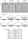
Fig 1. Design of the x3 disulfide bond to stabilise the SARS-CoV-2 spike in the “locked” state.
(A) Structure of the S-R/PP spike in the locked state (PDB: 6ZP2). Structural domains are coloured and labelled. The box indicates the location of the zoomed-in view in the right panel. Residues 427 and 987 that were mutated to cysteine to form the x3 disulfide bond are indicated. (B) Coomassie-stained SDS-PAGE gels to assess expression of the S-R/x3 and S-R/x3/D614G S proteins and confirm formation of a disulphide bond. NR or R indicates protein samples were prepared in non-reducing or reducing conditions. (C) Negative stain EM images of the purified S-R/x3 and S-R/x3/D614G spikes.
Fig 2
Fig 2. Biochemical properties of purified x3 spikes comparing to x2 spikes.
(A) Reduction of x3 and x2 disulfide bonds under native conditions by 5 min incubation with indicated concentrations of DTT, reactions were stopped by excess amount of iodoacetamide before SDS-PAGE. (B) binding of ACE2-Fc to different spike proteins in the absence and presence of 20 mM DTT. Spike proteins were serial diluted to 1500, 500, 166.7, 55.6, 18.5, 6.17, 2.06 and 0 nM and used as analytes in the assays. Kinetic parameters (kon, koff) and dissociation constants derived from kinetic analyses (KDkin) are summarised in the table.
Fig 3
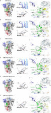
Fig 3. Structural features of spike proteins in different conformations as determined by cryo-EM.
(A)—(F) the left panels show side views of indicated spike proteins and conformational states, the structural domains are coloured as in Fig 1. The numbered boxes indicate the zoomed-in views as shown in the middle and right panels, dashed boxes indicate absence of bound factors. The middle panels show cryo-EM densities of bound factors; the right panels show cryo-EM densities of structural regions around domain D. Disordered loops are indicated by dashed lines, glycans are shown as stick representations in blue. The region between C617 and N641 (marked with asterisks) changes conformation between the different structures.
Fig 4
Fig 4. Structural changes in domain D between spikes in locked-1, locked-2 and closed conformations.
(A)—(F) structures of domain D in S-R/x3 and S-R/x3/D614G spikes of different conformations. Domain C is in light blue, Domain D green, FPPR yellow. Selected amino acid sidechains are shown and key amino acid sidechains are marked. The disordered loops are represented by dashed lines. C617 and N641 are marked with stars to highlight the dynamic region in between. π-π and cation-π interactions stabilising locked conformations are highlighted with dashed lines. Positions of hydrophobic residues with omitted side chains interacting with the domain D hydrophobic core are marked by black dots. The hydrophobic core formed by the domain D beta sheet is shown as a transparent molecular surface. (G) overlay of the S1 backbone from different conformations of the S-R/x3 spike. (H) the region within the box in panel (G) is zoomed to highlight the movements of the domain C/D junction and domain C between locked and closed conformations.
Fig 5
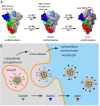
Fig 5. SARS-CoV-2 virus particle release and the accompanied structural transitions of surface spike protein.
(A) the three prefusion conformational states observed for the SARS-CoV-2 spike protein and factors that influence the structural transitions between the conformational states. Data in this study suggests that the D614G mutation may modulate locked to closed transitions. Data in the literature suggests that the D614G mutation may also modulate closed to open transitions and the stability of the open form (see main text). (B) schematic diagram illustrating the release of nascent SARS-CoV-2 virus particles from the cell. Green, blue or red triangles indicate the predicted conformational states of spike proteins as the virus travel through the acidic (pink) intracellular compartment before it was released into the neutral (blue) extracellular environment.

References

    1. Li F., Structure, Function, and Evolution of Coronavirus Spike Proteins. Annu Rev Virol. 2016;3: 237–261. doi: 10.1146/annurev-virology-110615-042301 - DOI - PMC - PubMed
    1. Krammer F. SARS-CoV-2 vaccines in development. Nature. 2020;586: 516–527. doi: 10.1038/s41586-020-2798-3 - DOI - PubMed
    1. Klasse P, Moore JP. Antibodies to SARS-CoV-2 and their potential for therapeutic passive immunization. Elife. 2020;9. doi: 10.7554/eLife.57877 - DOI - PMC - PubMed
    1. Xiong X, Qu K, Ciazynska KA, Hosmillo M, Carter AP, Ebrahimi S, et al.. A thermostable, closed SARS-CoV-2 spike protein trimer. Nat Struct Mol Biol. 2020;27: 934–941. doi: 10.1038/s41594-020-0478-5 - DOI - PMC - PubMed
    1. Wrapp D, Wang N, Corbett KS, Goldsmith JA, Hsieh C-L, Abiona O, et al.. Cryo-EM structure of the 2019-nCoV spike in the prefusion conformation. Science. 2020;367: 1260–1263. doi: 10.1126/science.abb2507 - DOI - PMC - PubMed

Publication types