

doi: 10.2460/javma.20.12.0688.
Pathology in Practice
Affiliations
- PMID: 35905169
- PMCID: PMC11472644
- DOI: 10.2460/javma.20.12.0688
Item in Clipboard
Pathology in Practice
J Am Vet Med Assoc.
.
Abstract
In collaboration with the American College of Veterinary Pathologists.
Figures
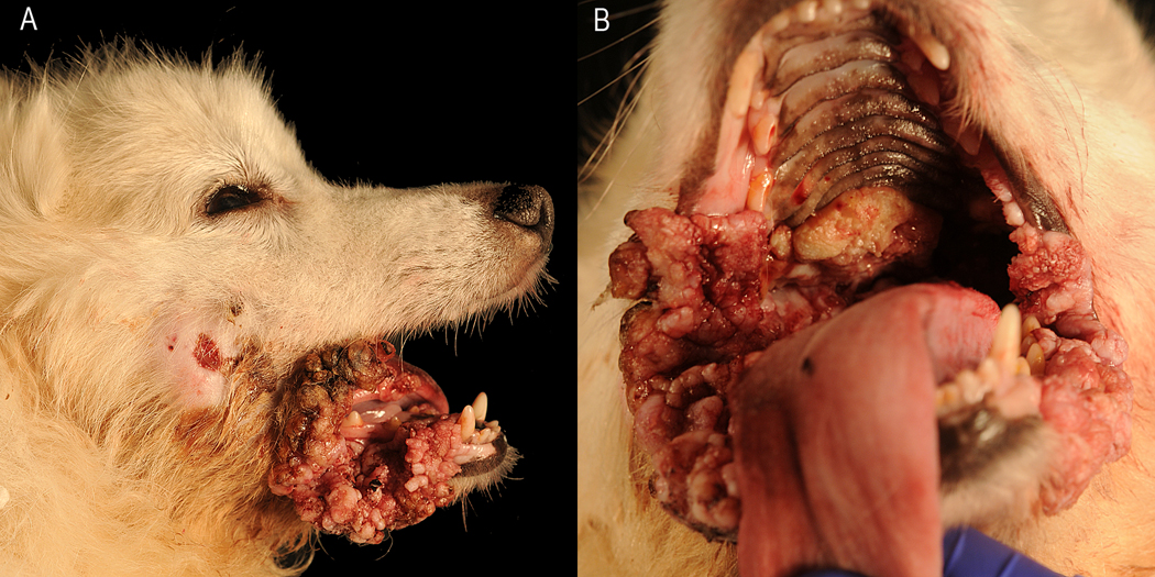
Photographs of the head (A) and oral cavity (B) of an 8-year-old American Eskimo Dog that was presented for evaluation of multiple oral and mucocutaneous junction masses, a ruptured facial abscess, and lethargy. A—Caudal to the right lip commissure is a draining tract. B—Numerous multifocal to coalescing papillomatous masses ranging from 0.5 to 4.0 cm in diameter are present on the oral mucosa and along the mucocutaneous junction of the lips, with a larger mass at the right lip commissure.
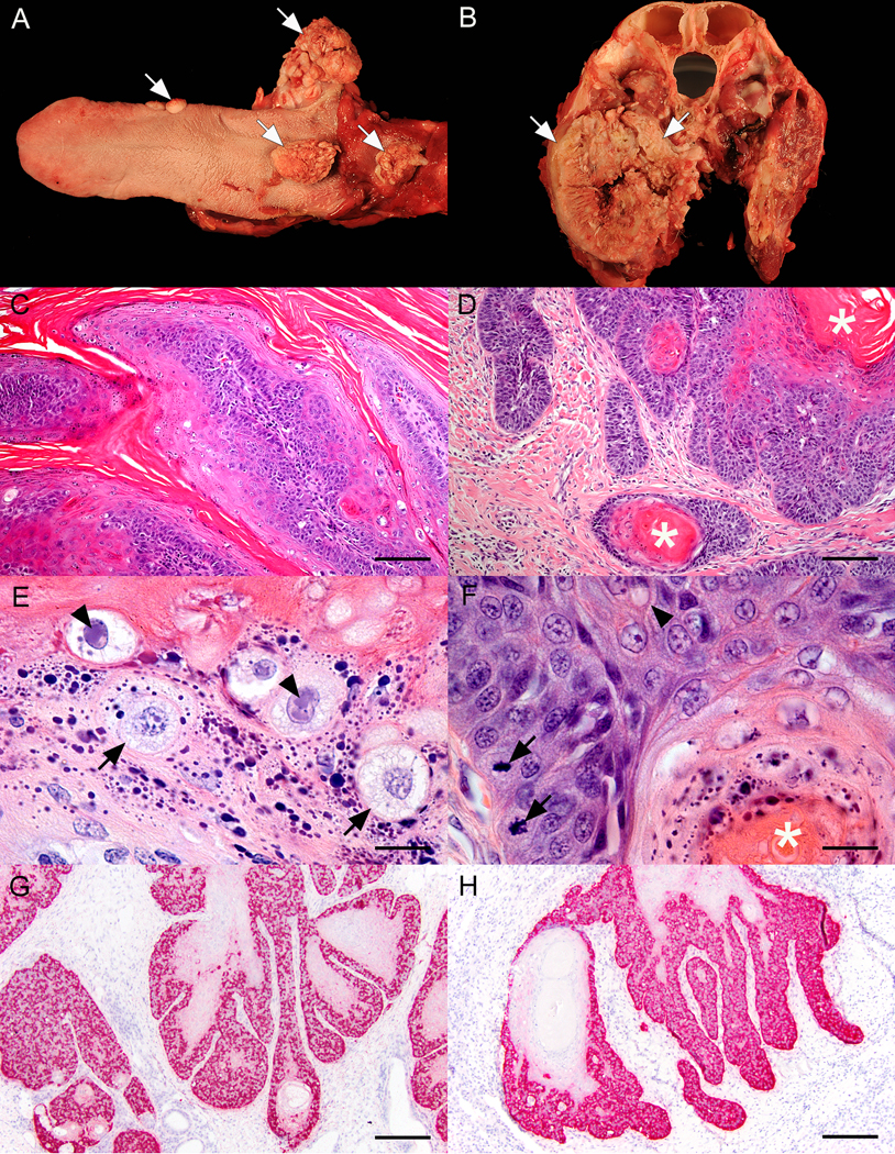
Postmortem photographs of the tongue (A) and a cross-section of the head (B), along with photomicrographs of sections of the masses (C–H). A—Widespread papillomatosis (arrows) of the caudal aspect of the tongue, the right laryngeal saccule, and the ventral aspect of the epiglottis partially occludes the larynx. B—An invasive, large mass (arrows) nearly completely effaces the caudal portion of the right mandibular body and ramus and infiltrates most of the right masseter muscle. C—Photomicrograph of an oral papilloma reveals papillary exophytic projections of hyperplastic epithelium with hyperkeratosis. H&E stain; bar = 100 μm. D—Photomicrograph of a section of the large invasive mass reveals a well-differentiated squamous cell carcinoma with keratin pearl formation (asterisks). H&E stain; bar = 100 μm. E—Higher-magnification image of a papilloma, taken at the stratum granulosum–stratum corneum interface, illustrates typical viral papilloma features of koilocytes without inclusion bodies (arrows) and koilocytes with magenta intranuclear inclusion bodies (arrowheads). Numerous, large, blue-black keratohyalin granules are visible (hypergranulosis). H&E stain; bar = 25 μm. F—Higher-magnification image of a section of the squamous cell carcinoma reveals mitotic figures (arrows), intercellular spinous projections (arrowheads), and a portion of a keratin pearl (asterisk). Blue-black keratohyalin granules are present at the margin of the keratin pearl. H&E stain; bar = 25 μm. G—Photomicrograph of a papilloma after in situ hybridization for canine papillomavirus 1 shows strong hybridization signals in the cytoplasm and nucleus of epithelial cells. Fast red chromogen stain; bar = 100 μm. H—Photomicrograph of a section of the squamous cell carcinoma after in situ hybridization for canine papillomavirus 1 shows strong hybridization signals in the cytoplasm and nucleus of epithelial cells. The morphology at the base of the papilloma resembles the morphology of the squamous cell carcinoma, suggesting an association between these lesions. Fast red chromogen stain; bar = 100 μm.
Similar articles
-
Pathology in Practice.J Am Vet Med Assoc. 2022 May 20;259(S2):1-4. doi: 10.2460/javma.21.04.0201. J Am Vet Med Assoc. 2022. PMID: 35587911
-
Pathology in Practice.J Am Vet Med Assoc. 2022 May 20;259(S2):1-4. doi: 10.2460/javma.21.04.0191. J Am Vet Med Assoc. 2022. PMID: 35587910
-
Pathology in Practice.J Am Vet Med Assoc. 2022 May 20;259(S2):1-5. doi: 10.2460/javma.21.05.0253. J Am Vet Med Assoc. 2022. PMID: 35587909
-
Pathology in Practice.J Am Vet Med Assoc. 2022 May 20;259(S2):1-4. doi: 10.2460/javma.21.04.0209. J Am Vet Med Assoc. 2022. PMID: 35587908
-
Forensic pathology of companion animal abuse and neglect.Vet Pathol. 2013 Nov;50(6):994-1006. doi: 10.1177/0300985813488895. Epub 2013 May 17. Vet Pathol. 2013. PMID: 23686766 Review.
References
-
- Gil da Costa RM, Peleteiro MC, Pires MA, et al. An Update on Canine, Feline and Bovine Papillomaviruses. Transbound Emerg Dis 2017;64:1371–1379. - PubMed
-
- Delius H, Van Ranst MA, Jenson AB, et al. Canine oral papillomavirus genomic sequence: a unique 1.5-kb intervening sequence between the E2 and L2 open reading frames. Virology. 1994;204:447–452. - PubMed
-
- Lange CE, Tobler K, Brandes K, et al. Canine inverted papillomas associated with DNA of four different papillomaviruses. Vet Dermatol 2010;21:287–291. - PubMed
-
- Falk E, Lange CE, Jennings S, et al. Two cutaneous horns associated with canine papillomavirus type 1 infection in a pit bull dog. Vet Dermatol 2017;28:420–421. - PubMed
-
- Nicholls PK, Klaunberg BA, Moore RA, et al. Naturally occurring, nonregressing canine oral papillomavirus infection: Host immunity, virus characterization, and experimental infection. Virology 1999;265:365–374. - PubMed
MeSH terms
Grants and funding
LinkOut - more resources
Full Text Sources
