

Proximal fibular osteotomy relieves pain in spontaneous osteonecrosis of the knee: A retrospective study
- PMID: 35905203
- PMCID: PMC9333514
- DOI: 10.1097/MD.0000000000029585
Proximal fibular osteotomy relieves pain in spontaneous osteonecrosis of the knee: A retrospective study
Abstract
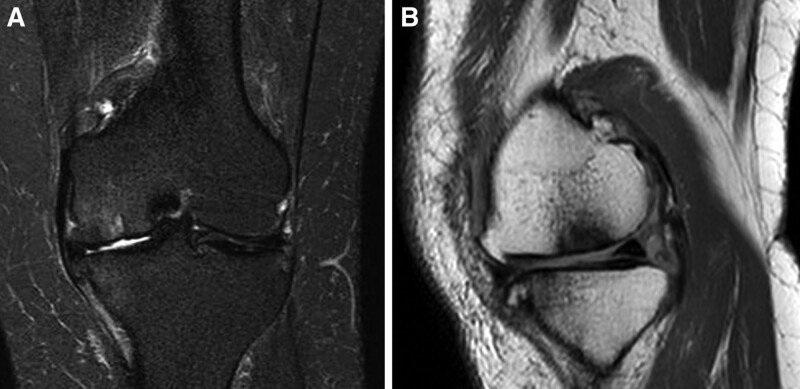
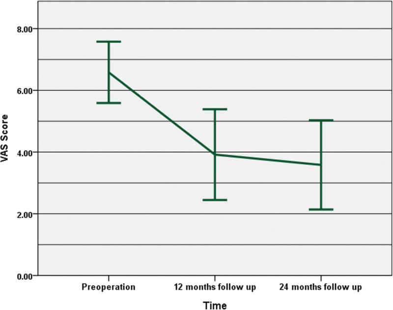
Spontaneous osteonecrosis of the knee (SONK) causes knee pain and joint motion limitation. Ischemia or insufficiency fracture may be the cause, but no consensus has been developed. Proximal fibular osteotomy (PFO) has been reported to relieve pain from osteoarthritis through medial compartment decompression. We reviewed the effect of this procedure on medial compartment SONK patients and explored clinical and radiological results. Since January 2018 to January 2020, the data of 12 knees (8 right and 4 left) from 11 SONK patients (9 women and 2 men) who received PFO were analyzed. The average age was 61.5 years. The diagnosis of SONK was established through weight-bearing anterior-posterior radiographs or magnetic resonance imaging (MRI). Visual analog scale (VAS) scores, Oxford knee score (OKS), Femorotibial angle (FTA), medial joint space, and lateral joint space were documented preoperatively and at follow up visits. Outcome assessment for the clinical and radiographic data was reviewed at 12- and 24-month follow-up visits. The mean follow up period was 33 months. All patients were able to walk with or without cane assistance the day after surgery. Both VAS score and OKS (preoperative: 6.6 ± 0.9 and 24.7 ± 3.8, respectively) improved significantly at the 12-month follow-up, and to 24-month follow-up (3.6 ± 1.3 and 35.6 ± 4.5, respectively, P < .05). Medial joint space ratio increased from 0.36 to 0.50 (P < .05). Changes of FTA were insignificant at any point of follow up. Four patients underwent follow-up MRI, and a decrease in the osteonecrotic area was clearly observed in 2 patients. By achieving medial knee decompression, PFO allowed quick weight-bearing recovery, pain relief, and improvement in knee function in SONK patients.
Copyright © 2022 the Author(s). Published by Wolters Kluwer Health, Inc.
Conflict of interest statement
The authors have no funding and conflict of interest to disclose.
Figures


References
-
- Pape D, Seil R, Fritsch E, et al. . Prevalence of spontaneous osteonecrosis of the medial femoral condyle in elderly patients. Knee Surg Sports Traumatol Arthrosc. 2002;10:233–40. - PubMed
-
- Mont MA, Marker DR, Zywiel MG, et al. . Osteonecrosis of the knee and related conditions. J Am Acad Orthop Surg. 2011;19:482–94. - PubMed
-
- Reddy AS, Frederick RW. Evaluation of the intraosseous and extraosseous blood supply to the distal femoral condyles. Am J Sports Med. 1998;26:415–9. - PubMed
-
- Yamamoto T, Bullough PG. Spontaneous osteonecrosis of the knee: the result of subchondral insufficiency fracture. J Bone Joint Surg Am. 2000;82:858–66. - PubMed
-
- Ramnath RR, Kattapuram SV. MR appearance of SONK-like subchondral abnormalities in the adult knee: SONK redefined. Skeletal Radiol. 2004;33:575–81. - PubMed
Publication types
MeSH terms
LinkOut - more resources
Full Text Sources
Medical
