. 2022 Jul 26;40(4):111144.
doi: 10.1016/j.celrep.2022.111144.

Rapid GPR183-mediated recruitment of eosinophils to the lung after Mycobacterium tuberculosis infection

Affiliations

Rapid GPR183-mediated recruitment of eosinophils to the lung after Mycobacterium tuberculosis infection

Andrea C Bohrer et al. Cell Rep. .

Abstract

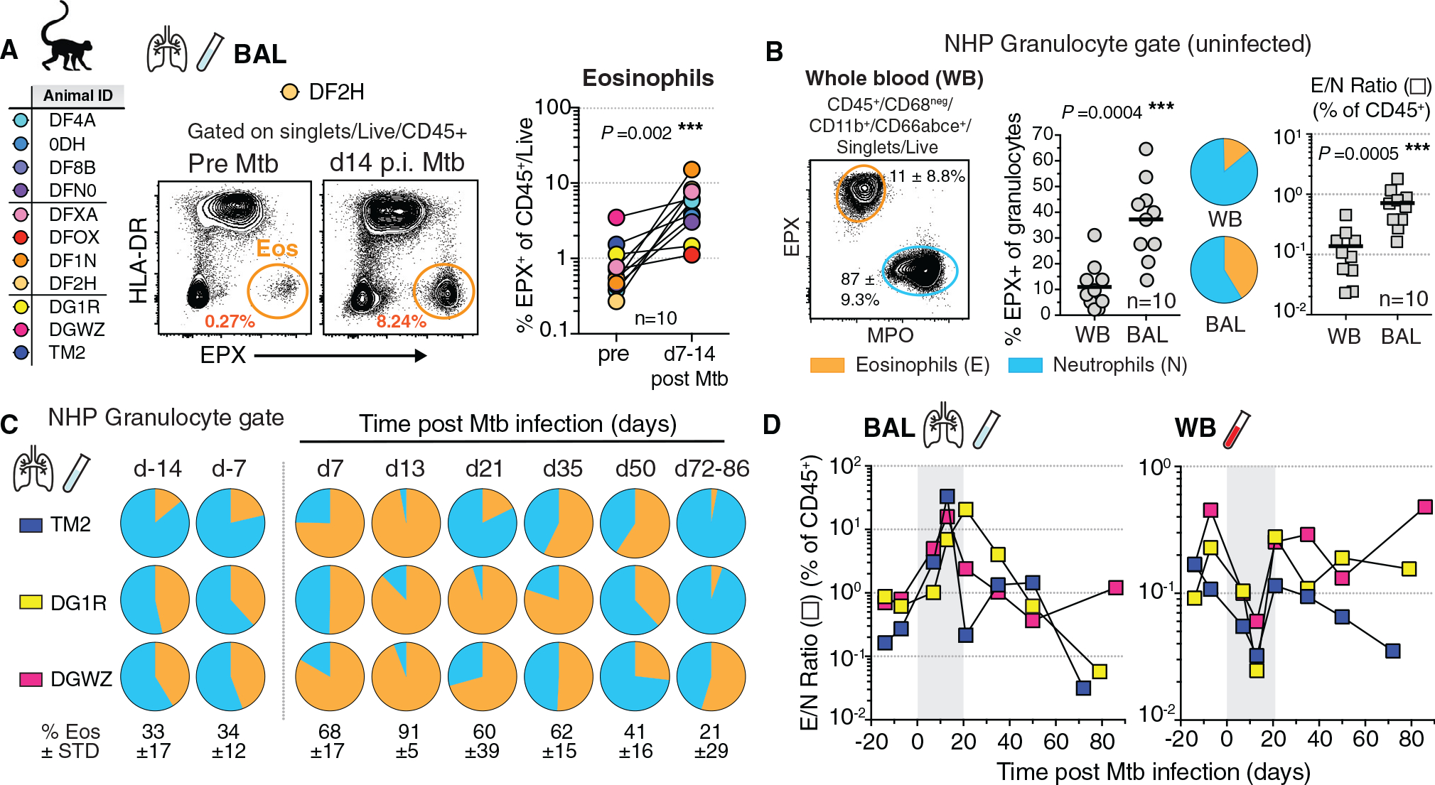
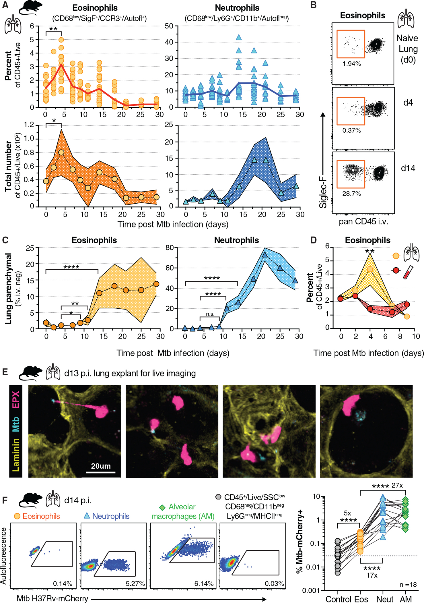
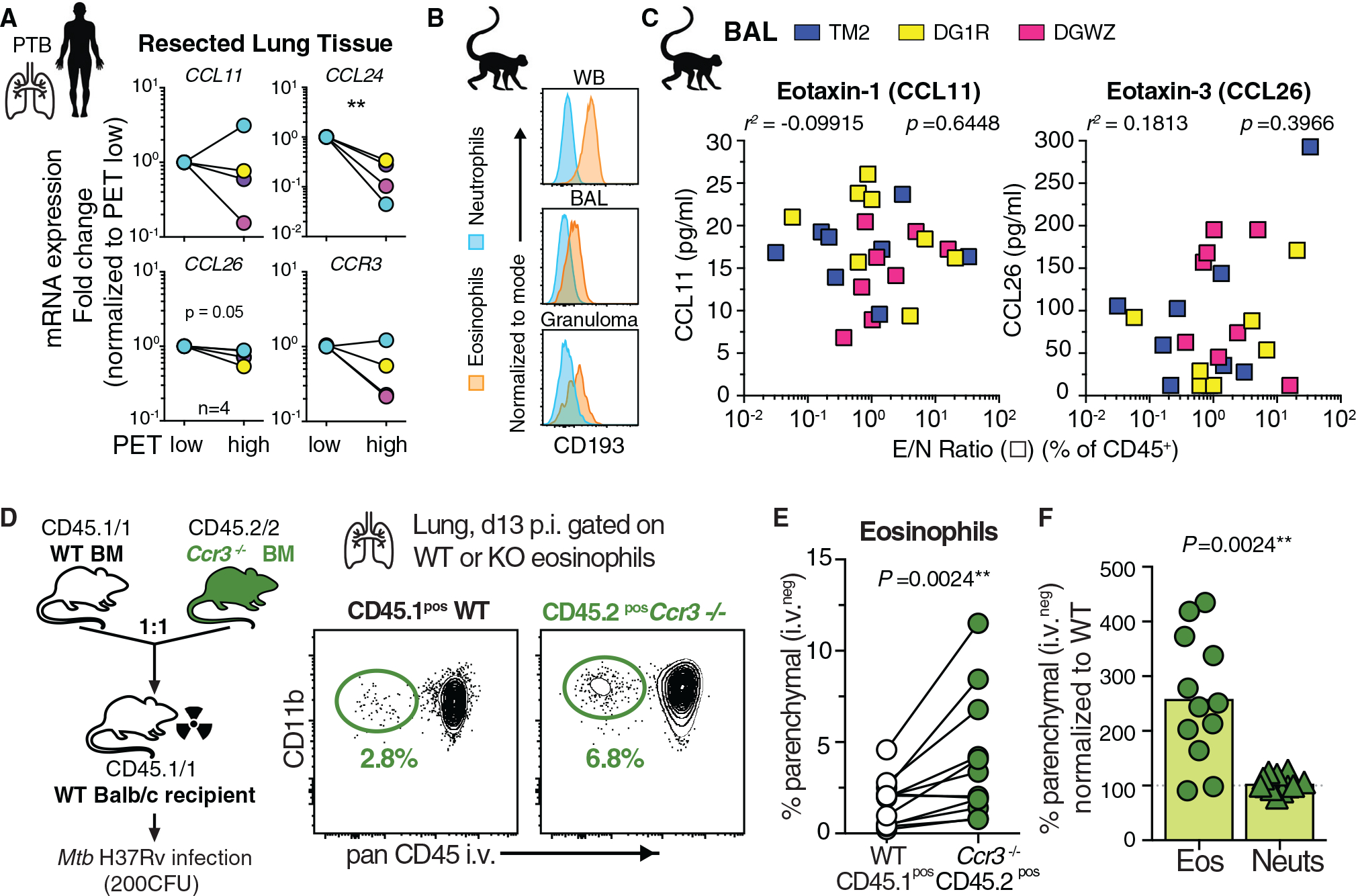
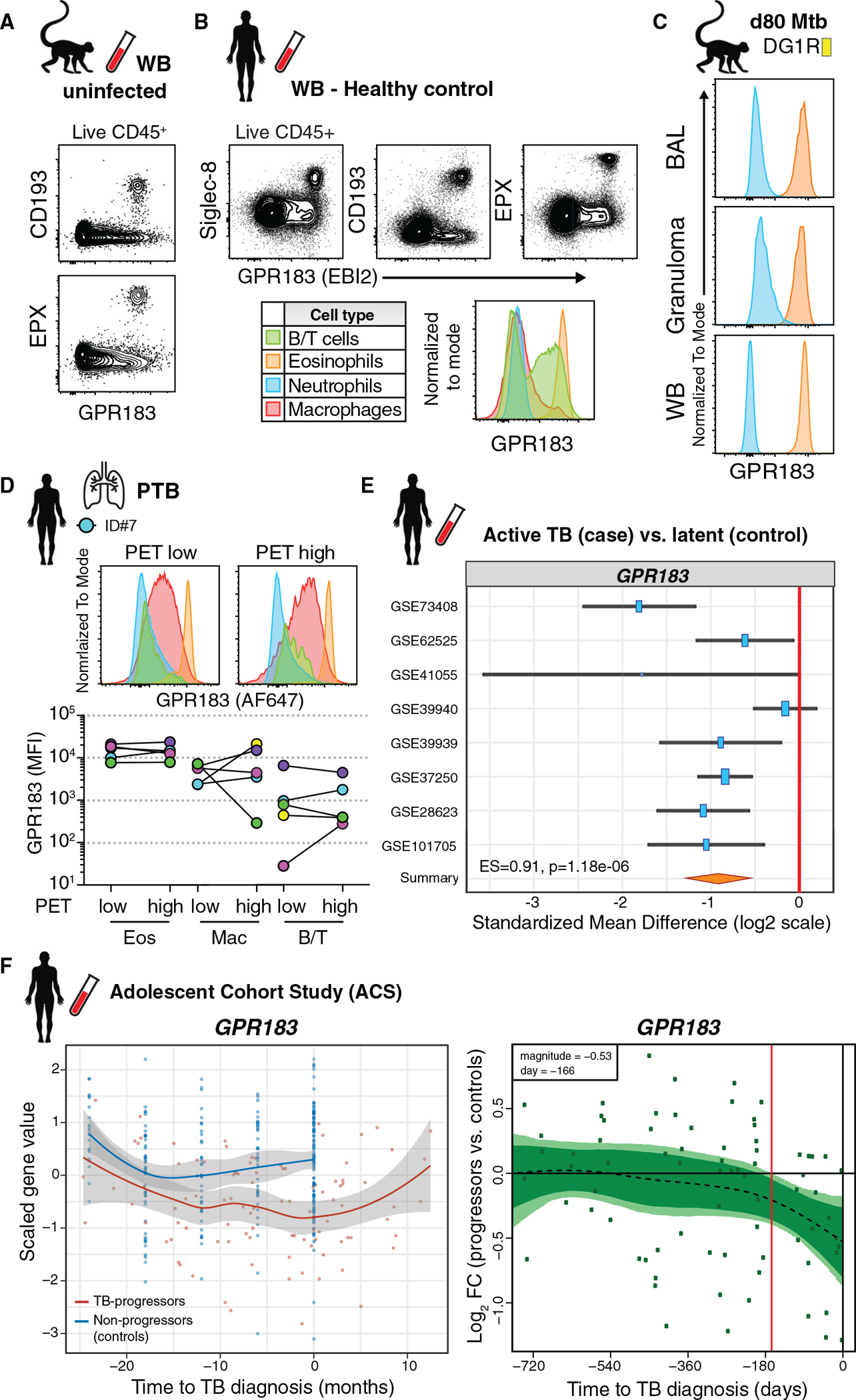
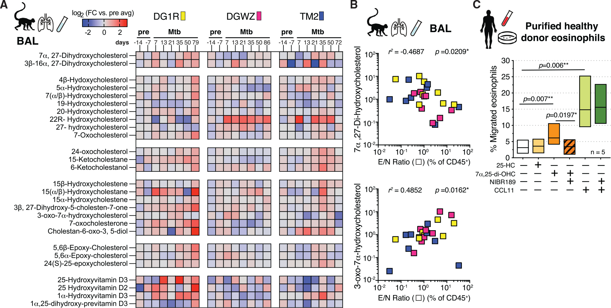
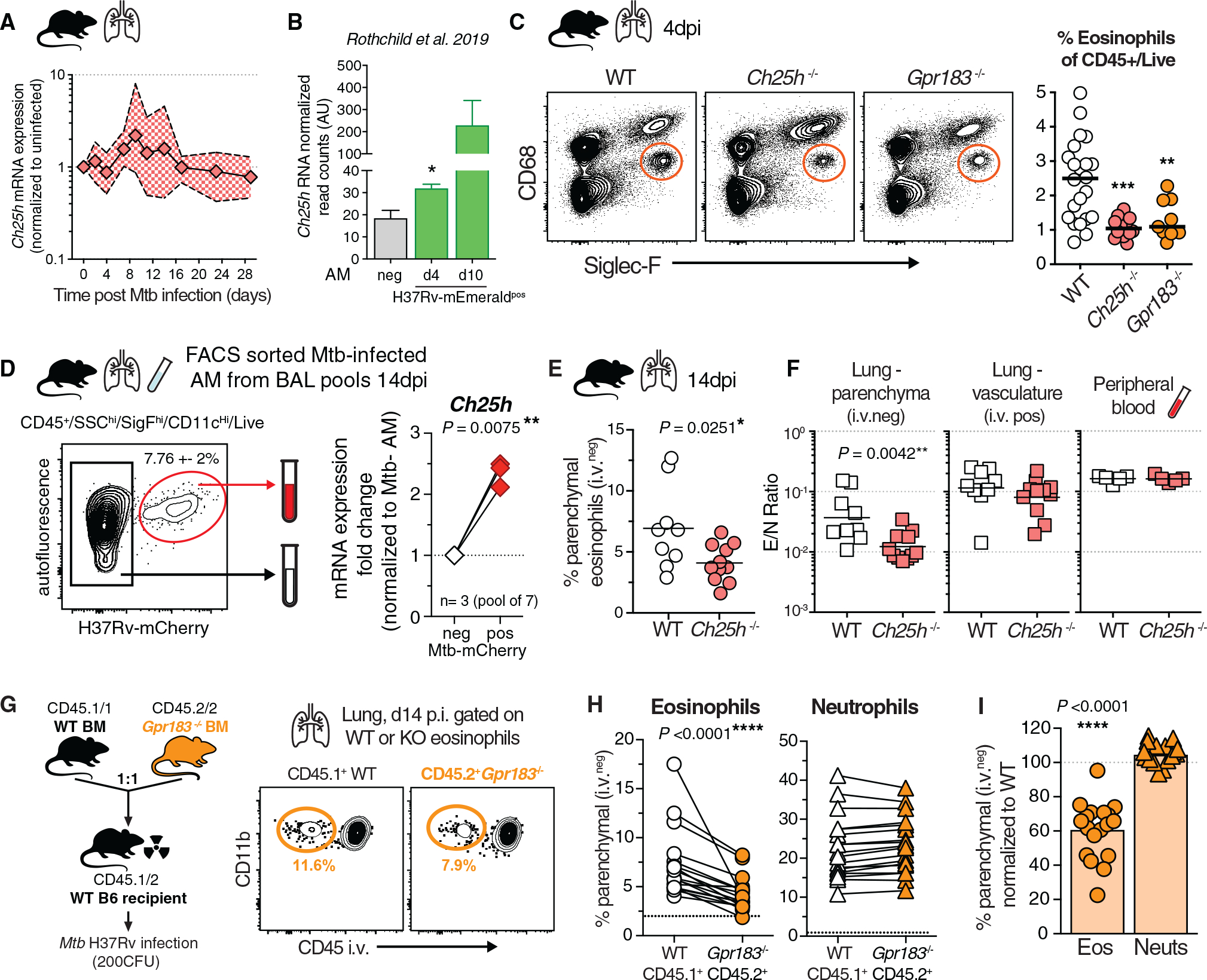
Influx of eosinophils into the lungs is typically associated with type II responses during allergy and fungal and parasitic infections. However, we previously reported that eosinophils accumulate in lung lesions during type I inflammatory responses to Mycobacterium tuberculosis (Mtb) in humans, macaques, and mice, in which they support host resistance. Here we show eosinophils migrate into the lungs of macaques and mice as early as one week after Mtb exposure. In mice this influx is CCR3 independent and instead requires cell-intrinsic expression of the oxysterol receptor GPR183, which is highly expressed on human and macaque eosinophils. Murine eosinophils interact directly with bacilli-laden alveolar macrophages, which upregulate the oxysterol-synthesizing enzyme Ch25h, and eosinophil recruitment is impaired in Ch25h-deficient mice. Our findings show that eosinophils are among the earliest cells from circulation to sense and respond to Mtb infection of alveolar macrophages and reveal a role for GPR183 in the migration of eosinophils into lung tissue.

Keywords: CCR3; CP: Immunology; CP: Microbiology; Ch25h; GPR183; Mycobacterium tuberculosis; NHP; alveolar macrophages; bacterial infection; eosinophils; eotaxin; granulocytes; lung; neutrophils; nonhuman primate; oxysterols; rhesus macaque.


Conflict of interest statement

Declaration of interests The authors declare no competing interests.

Figures

Figure 1.
Figure 1.. Eosinophils dominate the early granulocytic response in the airways of Mtb-infected rhesus macaques
(A) Animal ID list of Mtb infections in rhesus macaques with representative FACS plots of EPX in BAL and quantification of eosinophils before and 1–2 weeks p.i. (n = 10, male [M] + female [F], matched Wilcoxon). (B) NHP granulocyte gating strategy and representative FACS plot with proportions of eosinophils (orange) and neutrophils (blue), alongside eosinophil/neutrophil ratio (E/N) in WB and BAL of uninfected rhesus macaques (n = 10, M + F, matched Wilcoxon). (C) NHP granulocyte gate frequency of eosinophils in BAL prior to and throughout Mtb infection (n = 3, ±SD). (D) E/N ratio in NHP BAL and WB throughout Mtb infection (n = 3; d7–d14 time points are highlighted in gray).
Figure 2.
Figure 2.. Eosinophils are recruited to lungs early after Mtb infection in mice and interact with Mtb-infected cells in the airways
(A) Percentage and cell number of total lung eosinophils and neutrophils over time after aerosol infection (100–300 colony-forming units [CFU]) in WT mice (n = 6–25 per time point, M + F, 95% confidence interval [CI], SEM, 2 or 3 experiments per time point, *p = 0.04 and **p = 0.0032, unpaired t test). (B) Representative FACS plot of intravascular staining of eosinophils over time in mouse lung. (C) Percentage of lung parenchymal (CD45 i.v. negative) eosinophils and neutrophils over time after Mtb infection (n = 6–25 per time point, M + F, 95% CI, SEM, 2 or 3 experiments per time point, Mann-Whitney with indicated comparisons, *p = 0.0109, **p = 0.0032, and ****p < 0.0001). (D) Frequency of eosinophils in lung and whole blood after Mtb infection (n = 6–10 per time point, M + F, 95% CI, SEM, **p = 0.007, Mann-Whitney, 2 experiments). (E) Lung explant model of d13 Mtb-CFP (blue) infected mice expressing an EPX-eosinophil reporter (pink), representative still images (20 μm) from supplemental movie (2 experiments). (F) Representative FACS plots and quantification of H37Rv-mCherry-containing cells in lung on d14 (n = 18, M + F) of eosinophils (orange), neutrophils (blue), AMs (green) or gated on non-phagocytic, non-myeloid-lineage control cells (gray); dotted line is average signal of control cells (Wilcoxon-matched pairs test for eosinophil comparisons, ****p < 0.0001, 4 experiments).
Figure 3.
Figure 3.. CCR3 expression on eosinophils is dispensable for their migration into the lung parenchyma after Mtb infection
(A) qRT-PCR of indicated genes in 18FDG PET/CT low- or high-signal intense human TB lung lesions (n = 4, Wilcoxon-matched pairs, **p < 0.01). (B) Example CD193/CCR3 expression of granulocytes after Mtb infection in rhesus macaques. (C) Eotaxin levels in BAL of Mtb-infected rhesus macaques across all time points correlated with E/N ratio in same samples (Spearman). (D) Balb/c Ccr3−/− competitive mixed BM chimeric mice (mBM) and representative FACS plot of lung parenchymal eosinophils after Mtb infection (2 experiments). (E) Quantification of CD45 i.v. negative granulocytes in mBM lung (M + F, n = 12, Wilcoxon-matched pairs). (F) Migration efficiency of granulocytes normalized to WT cells in same lung (Wilcoxon-matched pairs).
Figure 4.
Figure 4.. Human and rhesus macaque eosinophils express uniformly high levels of the oxysterol receptor GPR183
(A) Representative FACS plots of GPR183 expression on eosinophils in WB of uninfected macaques. (B) Representative FACS plots of GPR183 expression on leukocytes and eosinophils in WB from healthy donors. (C) Example GPR183 expression of granulocytes after Mtb infection in BAL, WB, and granuloma of macaque DG1R. (D) GPR183 expression by flow cytometry on immune cells in 18FDG PET/CT low or high FDG uptake in human TB lung lesions (n = 5). (E) GPR183 expression from transcriptome profiles of LTBI controls (n = 433) or ATB (n = 528) blood samples across 8 indicated cohorts (z-test on summary effect size computed as Hedges’ g). (F) GPR183 expression kinetics over time from adolescent cohort study. Left-side graphs depicts treatment follow-up from 42 subjects that progressed to active TB (red) and 109 latently infected controls (blue) (local regression model, Hedge’s g) and GPR183 mRNA expression over time, expressed as log2 fold change (FC) between bin-matched progressors (n = 44) and controls (n = 106) and modeled as nonlinear splines (dotted line). Right-side graph shows progressors, with light green shading representing 99% CI and dark green shading 95% CI temporal trends, computed by performing 2,000 spline-fitting iterations after bootstrap resampling from the full dataset. Deviation time (day −166) is calculated as the time point at which the 99% CI deviates from a log2 fold change of 0, indicated by the vertical red line.
Figure 5.
Figure 5.. Oxysterol GPR183 ligands are dynamically regulated in rhesus macaque airways after Mtb infection and trigger migration of eosinophils in vitro
(A) Oxysterol metabolite signals in BAL of Mtb-infected rhesus macaques over time displayed as log2 fold change versus average pre-infection time point intensity measured using LC-MS/MS (n = 3). (B) 7α,27-di-OHC and 3-oxo-7α-OHC levels in BAL of Mtb-infected rhesus macaques across all time points correlated with E/N ratio in same samples (Spearman). (C) In vitro migration assay of purified human eosinophils to oxysterol ligands 25-OHC (100 nM) and 7α,25-di-OHC (100 nM) or CCL11 (eotaxin-1, 100 ng/mL) in the presence or absence of NIBR189 (50 nM), a selective GPR183 inhibitor (n = 5, Wilcoxon-matched pairs).
Figure 6.
Figure 6.. Ch25h-derived oxysterols mediate eosinophil-intrinsic GPR183-dependent extravasation into Mtb-infected lungs in vivo
(A) qRT-PCR Ch25h expression in lung tissue after aerosol Mtb infection in WT mice (n = 5–12; 2 experiments). (B) Ch25h mRNA read arbitrary units (AU) on the basis of data published by Rothchild et al. (2019), normalized to uninfected bystander reads, after H37Rv-mEmerald high dose (2,000–4,000 CFU) Mtb infection in mice (two-tailed unpaired t test). (C) Representative FACS plots of eosinophils in the lungs of Gpr183−/−, Ch25h−/−, or WT B6 mice at 4 d p.i. and quantification (M + F, n = 10–20, **p < 0.01 AND ***p < 0.001, Mann-Whitney, 3 experiments). (D) Representative FACS plots of low-dose aerosol Mtb-infected (100–300 CFU) WT B6 AM at 14 d p.i. from BAL and Ch25h gene expression in purified AM populations (M + F, 3 experiments with a pool of 7 mice, Wilcoxon-matched pairs). (E) Percentage lung parenchymal eosinophils d14 p.i. in Ch25h−/− mice (M + F, n = 9 or 10, Mann-Whitney, 2 experiments). (F) E/N ratio in lung parenchymal and lung vascular CD45+ immune cells compared with the peripheral blood at 14 d p.i. in Ch25h−/− mice (M + F, n = 9 or 10, Mann-Whitney, 2 experiments). (G) WT and Gpr183−/− competitive mixed BM chimeric mice and representative FACS at 14 d p.i. Mtb infection. (H) Quantification of CD45 i.v. negative granulocytes in mBM lung (M + F, n = 18, 3 experiments, Wilcoxon-matched pairs, dotted black line shows average CD45 i.v. negative eosinophils or neutrophils in uninfected mixed BM chimeric mice). (I) Lung migration normalized to WT cells from the same lung (Wilcoxon-matched pairs).

Similar articles

Cited by

References

    1. Ahsan F, Maertzdorf J, Guhlich-Bornhof U, Kaufmann SHE, and Moura-Alves P (2018). IL-36/LXR axis modulates cholesterol metabolism and immune defense to Mycobacterium tuberculosis. Sci. Rep 8, 1520. 10.1038/s41598-018-19476-x. - DOI - PMC - PubMed
    1. Anderson KG, Mayer-Barber K, Sung H, Beura L, James BR, Taylor JJ, Qunaj L, Griffith TS, Vezys V, Barber DL, and Masopust D (2014). Intravascular staining for discrimination of vascular and tissue leukocytes. Nat. Protoc 9, 209–222. 10.1038/nprot.2014.005. - DOI - PMC - PubMed
    1. Baptista AP, Gola A, Huang Y, Milanez-Almeida P, Torabi-Parizi P, Urban JF Jr., Shapiro VS, Gerner MY, and Germain RN (2019). The chemoattractant receptor Ebi2 drives intranodal naive CD4(+) T cell peripheralization to promote effective adaptive immunity. Immunity 50, 1188–1201.e6. 10.1016/j.immuni.2019.04.001. - DOI - PubMed
    1. Barington L, Wanke F, Niss Arfelt K, Holst PJ, Kurschus FC, and Rosenkilde MM (2018). EBI2 in splenic and local immune responses and in autoimmunity. J. Leukoc. Biol 104, 313–322. 10.1002/jlb.2vmr1217-510r. - DOI - PubMed
    1. Bartlett S, Gemiarto AT, Ngo MD, Sajiir H, Hailu S, Sinha R, Foo CX, Kleynhans L, Tshivhula H, Webber T, et al. (2020). GPR183 regulates interferons, autophagy, and bacterial growth during Mycobacterium tuberculosis infection and is associated with TB disease severity. Front. Immunol 11, 601534. 10.3389/fimmu.2020.601534. - DOI - PMC - PubMed

Publication types

Substances

LinkOut - more resources