. 2022 Aug 23;67(17):10.1088/1361-6560/ac8593.
doi: 10.1088/1361-6560/ac8593.

A multi-layer strip ionization chamber (MLSIC) device for proton pencil beam scan quality assurance

Affiliations

A multi-layer strip ionization chamber (MLSIC) device for proton pencil beam scan quality assurance

Shuang Zhou et al. Phys Med Biol. .

Abstract

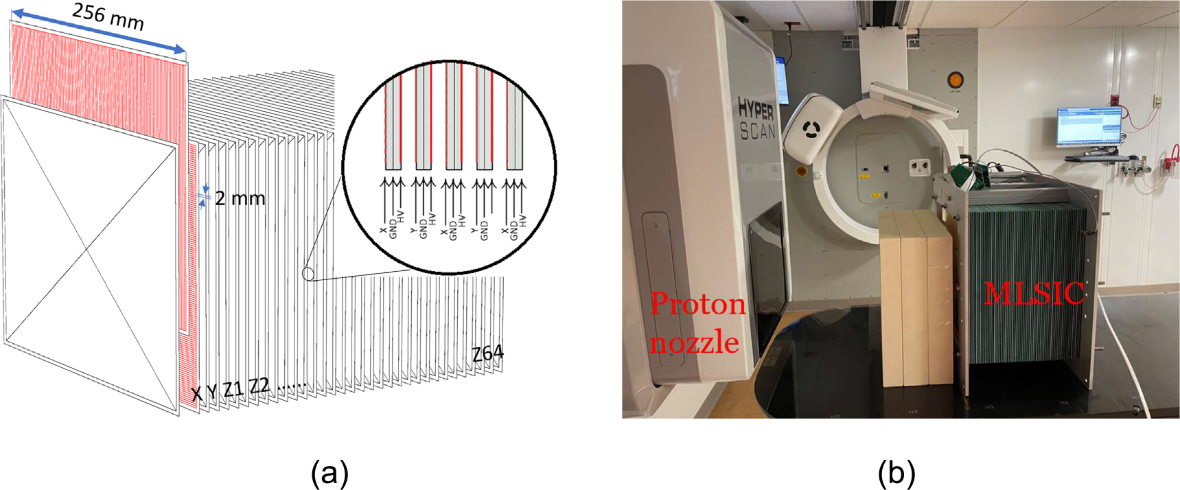
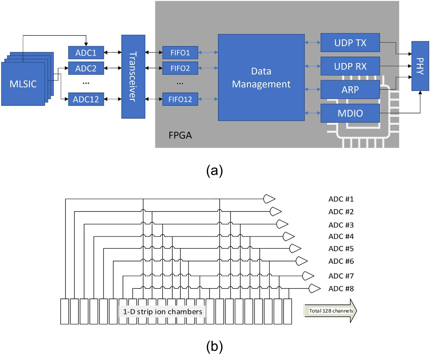
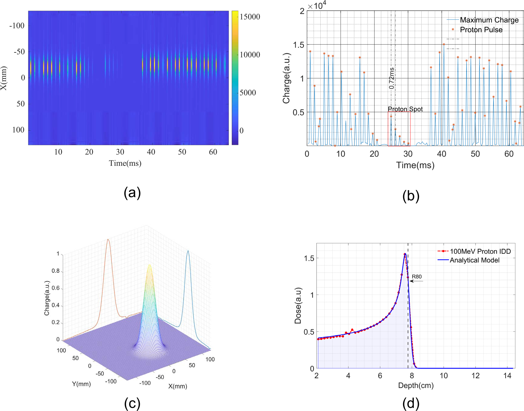
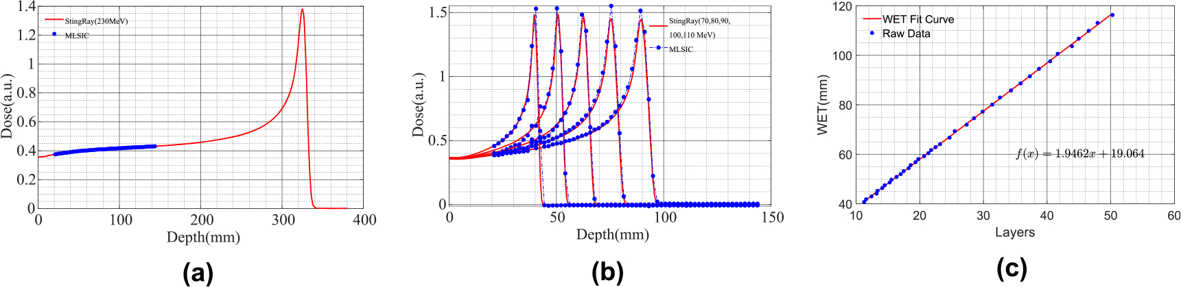
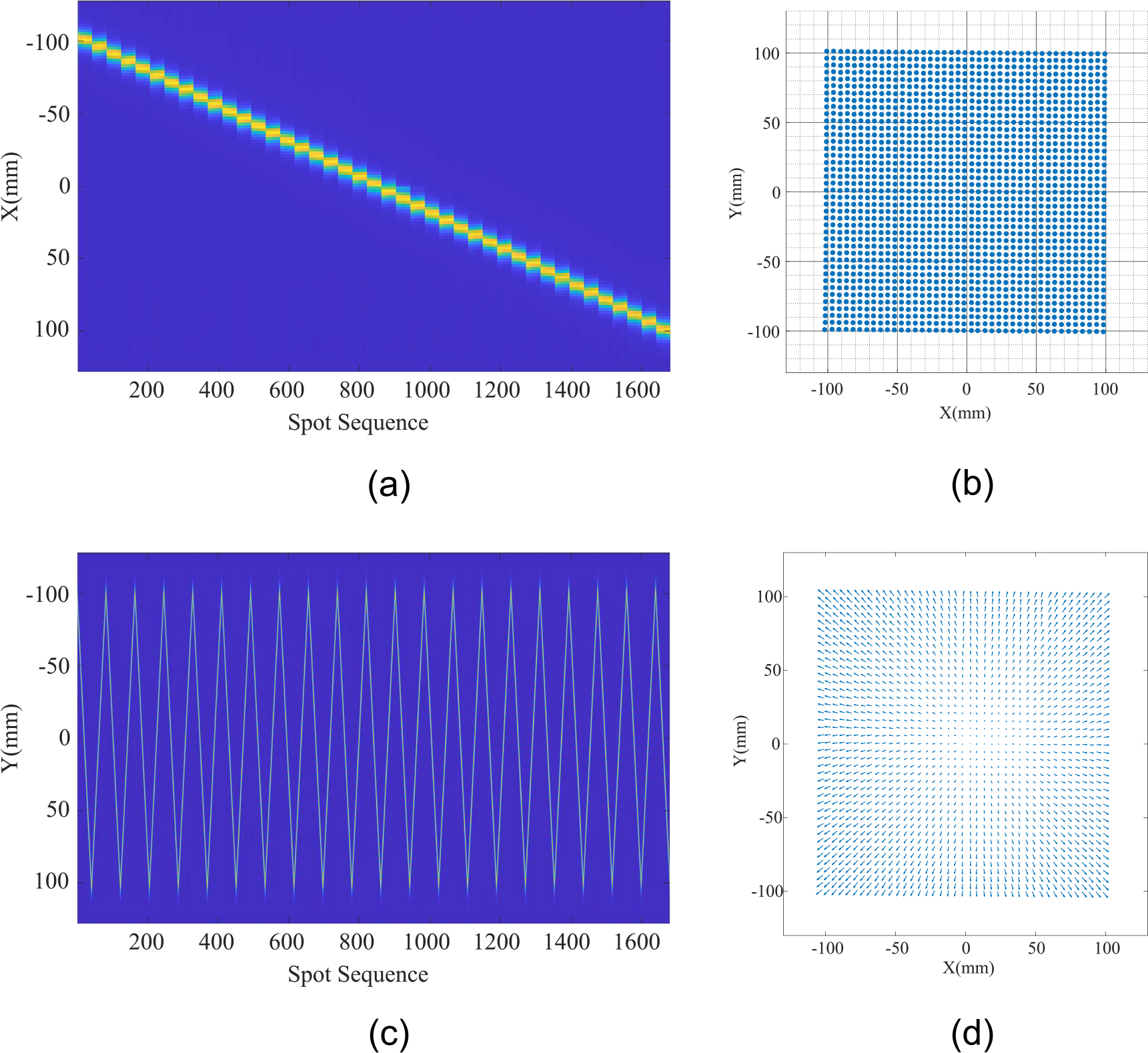
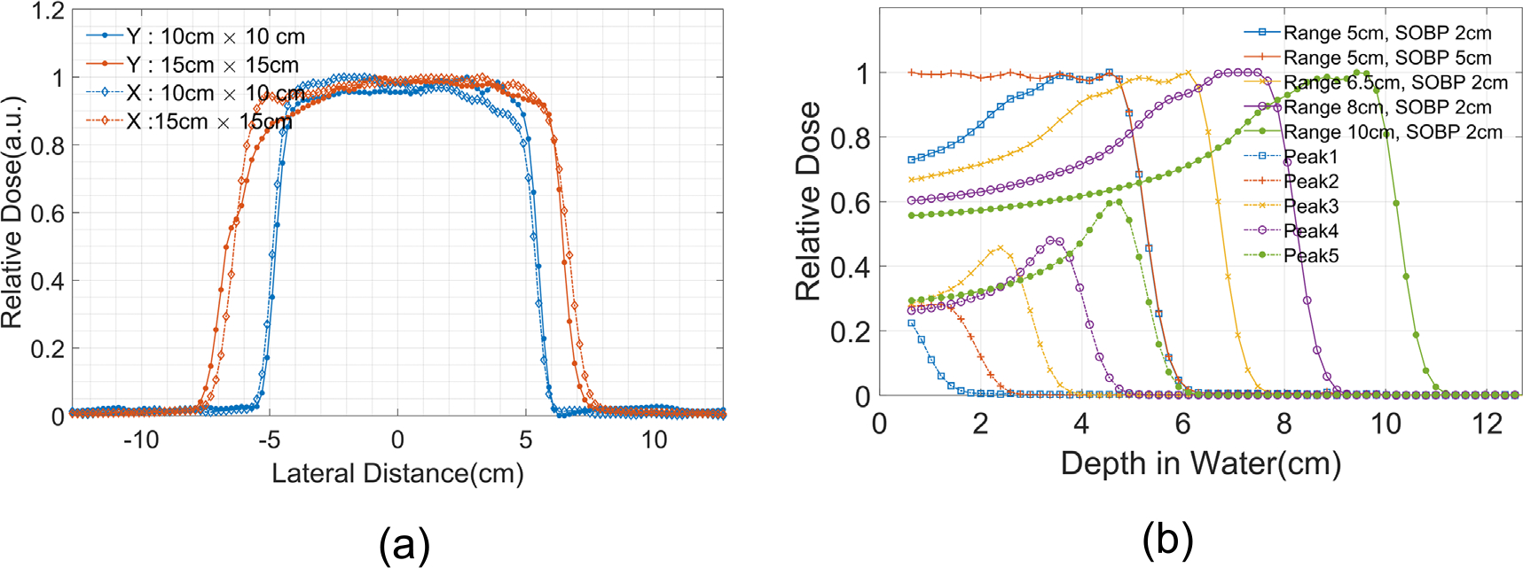
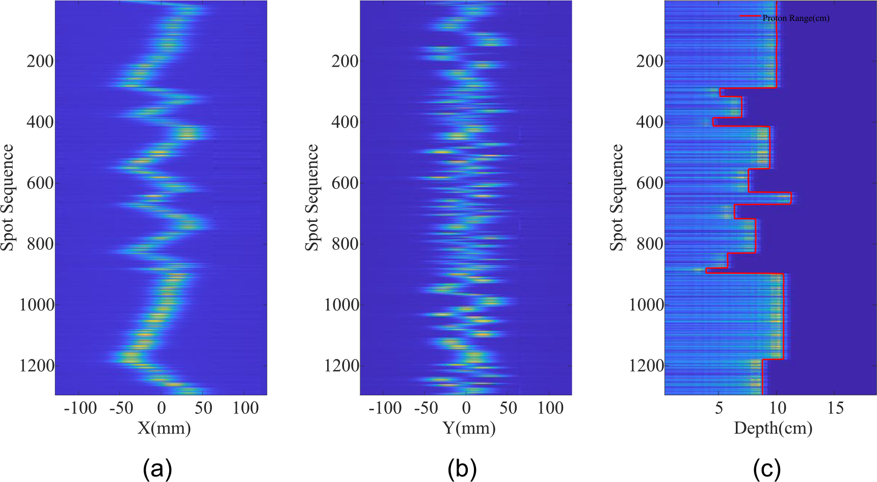
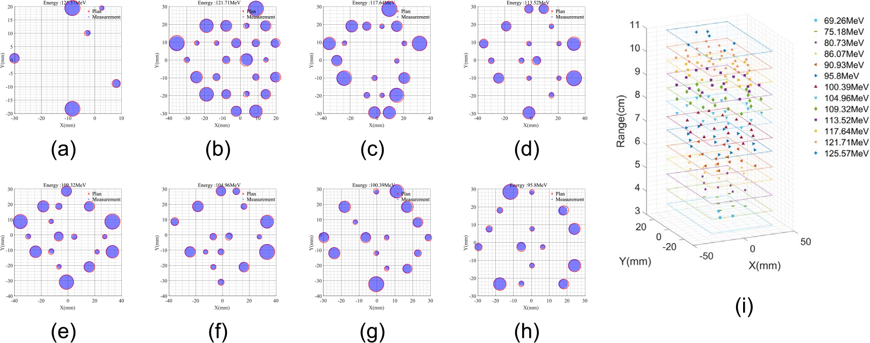
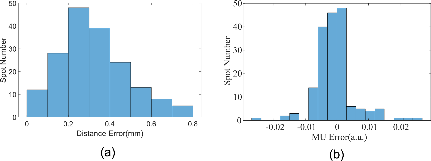
Objective. Proton pencil beam scanning (PBS) treatment fields needs to be verified before treatment deliveries to ensure patient safety. In current practice, treatment beam quality assurance (QA) is measured at a few selected depths using film or a 2D detector array, which is insensitive and time-consuming. A QA device that can measure all key dosimetric characteristics of treatment beams spot-by-spot within a single beam delivery is highly desired.Approach. We developed a multi-layer strip ionization chamber (MLSIC) prototype device that comprises of two layers of strip ionization chambers (IC) plates for spot position measurement and 64 layers of plate IC for beam energy measurement. The 768-channel strip ion chamber signals are integrated and sampled at a speed of 3.125 kHz. It has a 25.6 cm × 25.6 cm maximum measurement field size and 2 mm spatial resolution for spot position measurement. The depth resolution and maximum depth were 2.91 mm and 18.6 cm for 1.6 mm thick IC plate, respectively. The relative weight of each spot was determined from total charge by all IC detector channels.Main results. The MLSIC is able to measure ionization currents spot-by-spot. The depth dose measurement has a good agreement with the ground truth measured using a water tank and commercial one-dimensional (1D) multi-layer plate chamber. It can verify the spot position, energy, and relative weight of clinical PBS beams and compared with the treatment plans.Significance. The MLSIC is a highly efficient QA device that can measure the key dosimetric characteristics of proton treatment beams spot-by-spot with a single beam delivery. It may improve the quality and efficiency of clinical proton treatments.

Keywords: intensity modulated proton therapy; multilayer ionization chamber; proton therapy; quality assurance.


Figures

Figure 1.
Figure 1.
(a) The diagram and (b) the prototype of MLSIC device, dimensions 26 cm (L) × 38 cm (W) × 40 cm (H), weight 29 kg. It comprises 66 layers of ionization chamber arrays: two layers of strip ionization chambers X and Y at the entrance measure the spot position, and the following 64 layers of plate ionization chamber (Z1-Z64) measure the range of a proton beam.
Figure 2.
Figure 2.
(a) Architecture of data acquisition (DAQ) system of the MLSIC device; (b) internal connectivity of the Z-channels. 768 total channels of ionization currents are integrated and sampled at a frequency of 3.125 kHz at 16-bit resolution. The Z plates were also made of the same strip chambers as X–Y plates, except every eighth channel was interconnected, ADC1(1:8:128), ADC2(2:8:128), etc.
Figure 3.
Figure 3.
Measurement of a PBS beam and extract dosimetric information spot-by-spot. (a) Raw data of proton pulses measured by the X-channel plate; (b) classification results of the proton pulse from raw data; (c) determination of spot position from X–Y measurement; (d) determination of spot range and relative weight from Z measurement. Range is calculated by the depth at 80% of the maximum dose.
Figure 4.
Figure 4.
(a) IDD (shown in red) of 230 MeV proton beam measured with StingRay device in water tank. Segment of IDD (shown in blue) measured by MLSIC that was used in Z-channel relative sensitivity calibration; (b) IDD curves of 70, 80, 90, 100, and 110 MeV beams after Z-channel sensitivity calibration and comparison with StingRay measurement; (c) the relation between measured range and nominal range of proton beams in water. The WET of Z-channel board was measured to be 1.95mmby fitting the data with a linear function.
Figure 5.
Figure 5.
(a) The measured spot positions by the X channels of the MLSIC in which 41 groups of spots with the same x-values can be seen; (b) the spot location calculated from X–Y channels; (c) the measured spot position of Y-channels of the MLSIC. The raster scan pattern is clearly shown in this image (21 peaks and 20 valleys); (d) measured beam divergence of the cone beam geometry. The arrow denotes the vector-connected beam spot from the first layer to the last layer in relative scale.
Figure 6.
Figure 6.
(a) Beam profile of double-scattering proton beams without buildup; (b) IDD measurements of several selected double-scattering beams. One beam (Range 5 cm, SOBP 5 cm) was decomposed into five Bragg peaks (Peak 1, 2K5) generated by the modulation wheel.
Figure 7.
Figure 7.
(a) and (b) the spot trajectories of the brain case on X and Y channels, respectively; (c) the nominal proton range measured by the MLSIC. The gaps between subsequent signals have been eliminated.
Figure 8.
Figure 8.
(a)–(h) Measurement of several selected energy layers and (i) all spot ranges and position information. Blue filled circles are measured spots and red circles are planned spots. Circle size denotes the relative dose in (0,1].
Figure 9.
Figure 9.
(a) Euclidean distance difference between measured and planned spots; (b) MU difference between measured and planned spots. The MU values were normalized to (0,1].

Similar articles

Cited by

References

    1. Arjomandy B et al. 2019. AAPM task group 224: comprehensive proton therapy machine quality assurance Med. Phys. 46 6–8 - PubMed
    1. Arjomandy B, Sahoo N, Ding X and Gillin M 2008. Use of a two-dimensional ionization chamber array for proton therapy beam quality assurance Med. Phys. 35 3889–94 - PubMed
    1. Baumer C, Koska B, Lambert J, Timmermann B, Mertens T and Talla PT 2015. Evaluation of detectors for acquisition of pristine depth-dose curves in pencil beam scanning J. Appl. Clin. Med. Phys. 16 151–63 - PMC - PubMed
    1. Besl PJ and McKay ND 1992. A method for registration of 3D shapes IEEE Trans. Pattern Anal. Mach. Intell. 14 239–56
    1. Bizzocchi N, Fracchiolla F, Schwarz M and Algranati C 2017. A fast and reliable method for daily quality assurance in spot scanning proton therapy with a compact and inexpensive phantom Med. Dosim. 42 238–46 - PubMed

Publication types

LinkOut - more resources