. 2022 Oct;81(10):1409-1419.
doi: 10.1136/annrheumdis-2021-222069. Epub 2022 Jul 29.

Cross-species transcriptome analysis for early detection and specific therapeutic targeting of human lupus nephritis

Affiliations

Cross-species transcriptome analysis for early detection and specific therapeutic targeting of human lupus nephritis

Eleni Frangou et al. Ann Rheum Dis. 2022 Oct.

Abstract

Objectives: Patients with lupus nephritis (LN) are in urgent need for early diagnosis and therapeutic interventions targeting aberrant molecular pathways enriched in affected kidneys.

Methods: We used mRNA-sequencing in effector (spleen) and target (kidneys, brain) tissues from lupus and control mice at sequential time points, and in the blood from 367 individuals (261 systemic lupus erythematosus (SLE) patients and 106 healthy individuals). Comparative cross-tissue and cross-species analyses were performed. The human dataset was split into training and validation sets and machine learning was applied to build LN predictive models.

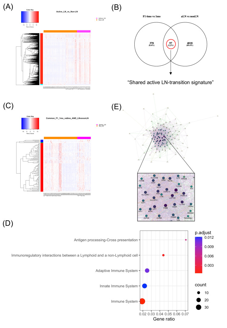
Results: In murine SLE, we defined a kidney-specific molecular signature, as well as a molecular signature that underlies transition from preclinical to overt disease and encompasses pathways linked to metabolism, innate immune system and neutrophil degranulation. The murine kidney transcriptome partially mirrors the blood transcriptome of patients with LN with 11 key transcription factors regulating the cross-species active LN molecular signature. Integrated protein-to-protein interaction and drug prediction analyses identified the kinases TRRAP, AKT2, CDK16 and SCYL1 as putative targets of these factors and capable of reversing the LN signature. Using murine kidney-specific genes as disease predictors and machine-learning training of the human RNA-sequencing dataset, we developed and validated a peripheral blood-based algorithm that discriminates LN patients from normal individuals (based on 18 genes) and non-LN SLE patients (based on 20 genes) with excellent sensitivity and specificity (area under the curve range from 0.80 to 0.99).

Conclusions: Machine-learning analysis of a large whole blood RNA-sequencing dataset of SLE patients using human orthologs of mouse kidney-specific genes can be used for early, non-invasive diagnosis and therapeutic targeting of LN. The kidney-specific gene predictors may facilitate prevention and early intervention trials.

Keywords: Autoimmune Diseases; Autoimmunity; Lupus Nephritis; Systemic Lupus Erythematosus; Therapeutics.


Conflict of interest statement

Competing interests: ED is an employee of GSK. His contribution was performed before he joined GSK.

Figures

Figure 1
Figure 1
Mouse kidney-specific transcriptome of lupus mice between the clinical (nephritic) and the preclinical (prepuberty) stage of the lupus. (A) Venn diagram demonstrating the comparison between differentially expressed genes (DEGs) within the spleen, the kidneys and the brain from NZB/W-F1 lupus mice at the clinical (nephritic) versus the preclinical (prepuberty) stage of lupus. The kidney-specific gene signature is defined by 507 genes that are differentially expressed only within kidneys but not in other tissues, (B) Heatmap of the 507 kidney-specific DEGs (316 upregulated, 191 downregulated), (C) Dot-plot diagram demonstrating functionally enriched REACTOME pathways of the 507 kidney-specific DEGs, (D) gene network representation of the 507 kidney-specific DEGs. Hub genes that correspond to lupus risk loci are depicted by larger size fonds. ROS, reactive oxygen species; TCR, T cell receptor.
Figure 2
Figure 2
Common genes between the kidney-specific gene expression profile from lupus mice at the symptomatic (nephritic) versus the asymptomatic (prepuberty) stage and the whole-blood gene expression profile from active LN (aLN) patients versus SLE patients without history of kidney involvement (non-LN) define a ‘shared active LN-transition signature’. (A) Heatmap of the 4119 differentially expressed genes (DEGs) in the whole-blood from aLN patients versus non-LN patients, (B) Venn diagram demonstrating the comparison between the orthologous genes of the mouse kidney-specific DEGs from NZB/W-F1 lupus mice at the symptomatic (nephritic) versus the asymptomatic (prepuberty) stage and the whole-blood gene expression profile from aLN versus non-LN SLE patients. The ‘shared active LN-transition signature’ is defined by the union of the Venn diagram, corresponding to 97 common genes, (C) Heatmap of the ‘shared active LN-transition signature’, composed of 97 genes (67 upregulated, 30 downregulated), (D) Dot-plot diagram demonstrating functionally enriched REACTOME pathways of the ‘shared active LN-transition signature’, (E) gene network representation of the ‘shared active LN-transition signature’. Hub genes that correspond to lupus risk loci are depicted by characters of a larger size. LN, lupus nephritis; SLE, systemic lupus erythematosus.
Figure 3
Figure 3
Upstream regulators of the ‘shared active LN signature’ and the ‘shared active LN-transition signature’. (A) Dot-plot diagram demonstrating the transcription factors (TF) that are predicted to reverse the common genes between the kidney-specific gene expression profile from lupus vs healthy mice at the clinical (nephritic) stage and the whole-blood gene expression profile from active LN (aLN) patients vs healthy individuals (HI). The x-axis represents the hypergeometric p value and dots correspond to the number of enriched targets of the TF, (B) Dot-plot diagram demonstrating the TF that are predicted to reverse the common genes between the kidney-specific gene expression profile from lupus mice at the clinical (nephritic) versus the preclinical (prepuberty) stage and the whole-blood gene expression profile from patients with active LN (aLN) versus SLE patients without history of kidney involvement (non-LN). The x-axis represents the hypergeometric p- value and dots correspond to the number of enriched targets of the TF. LN, lupus nephritis; SLE, systemic lupus erythematosus.
Figure 4
Figure 4
Schematic overview of the machine-learning approach. RNA-sequencing data from the two human cohorts were combined and then split in training to test sets at 70:30 ratio. For each outcome measure, a corresponding gene list derived from mouse data was used. The training set was used to develop a prediction model and the test set was used to validate the results. Using the training set, feature selection was applied to remove noise and keep the smallest set of genes which best predicts each outcome based on accuracy. Then, different prediction models were fit to identify which performs best using the gene signature selected in the previous step. Once the best model was selected based on accuracy, sensitivity and specificity, the addition of age, gender and the presence of anti-dsDNA as predictors were tested if they could improve the model. The final model was validated in the test set. AUC, area under the curve; CV, cross-validation; dsDNA, double-stranded DNA; ROC, receiver operating characteristic curve.
Figure 5
Figure 5
Machine-learning modelling of the human whole-blood RNA-sequencing data, using mouse kidney-specific genes as predictors, distinguishes patients with active lupus nephritis (active LN) from healthy individuals (H) in a non-invasive manner and defines a LN prognostic gene signature. (A) The 18 predictors of the glmnet model distinguishing patients with active LN from healthy individuals based on their importance, as evidenced by their absolute coefficient. Gene predictors in green fonts indicate that the higher their expression the higher the probability of being a patient with active LN compared with being a healthy individual; while gene predictors in red fonts indicate that the lower their expression the higher the probability of being a patient with active LN, (B) Characteristics of the prediction model of patients with active LN from healthy individuals, (C) Receiver operating characteristic curve (ROC) analysis of the glmnet model in the validation set reveals an area under the curve (AUC) of 0.99, (D) principal component analysis (PCA) using the 18 genes.
Figure 6
Figure 6
Machine-learning modelling of the human whole-blood RNA-sequencing data using mouse kidney-specific LN-transition genes as predictors distinguishes patients with active lupus nephritis (active LN) from SLE patients without history of kidney disease, non-invasively. (A) The 23 predictors of the glm model distinguishing patients with active LN (active LN) from SLE patients without kidney disease (non-LN) based on their importance, as evidenced by absolute z value. Gene predictors in green fonts indicate that the higher their expression the higher the probability of being a patient with active LN compared with being non-LN patient, while gene predictors in red fonts indicate that the lower their expression the higher the probability of being a patient with active LN. The presence of anti-dsDNA (indicated in green fonts) is associated with a higher the probability of being a patient with active LN and the older age and female gender (indicated in red fonts) are associated with a lower probability of being a patient with active LN, (B) Characteristics of the prediction model of active LN patients from non-LN patients, (C) Receiver operating characteristic curve analysis of the glm model in the validation set reveals an area under the curve (AUC) of 0.8, (D) Principal component analysis (PCA) using the 20 gene-predictors. LN, lupus nephritis; SLE, systemic lupus erythematosus;

References

    1. Malvar A, Pirruccio P, Alberton V, et al. . Histologic versus clinical remission in proliferative lupus nephritis. Nephrol Dial Transplant 2017;32:1338–44. 10.1093/ndt/gfv296 - DOI - PMC - PubMed
    1. Tektonidou MG, Dasgupta A, Ward MM. Risk of end-stage renal disease in patients with lupus nephritis, 1971-2015: a systematic review and Bayesian meta-analysis. Arthritis Rheumatol 2016;68:1432–41. 10.1002/art.39594 - DOI - PMC - PubMed
    1. Hill GS, Delahousse M, Nochy D, et al. . Predictive power of the second renal biopsy in lupus nephritis: significance of macrophages. Kidney Int 2001;59:304–16. 10.1046/j.1523-1755.2001.00492.x - DOI - PubMed
    1. Merrill JT, Neuwelt CM, Wallace DJ, et al. . Efficacy and safety of rituximab in moderately-to-severely active systemic lupus erythematosus: the randomized, double-blind, phase II/III systemic lupus erythematosus evaluation of rituximab trial. Arthritis Rheum : 2010;62:222–33. 10.1002/art.27233 - DOI - PMC - PubMed
    1. Rovin BH, Furie R, Latinis K, et al. . Efficacy and safety of rituximab in patients with active proliferative lupus nephritis: the lupus nephritis assessment with rituximab study. Arthritis Rheum : 2012;64:1215–26. 10.1002/art.34359 - DOI - PubMed

Publication types