Review
. 2022 Jul 29;7(1):258.
doi: 10.1038/s41392-022-01102-y.

Radiotherapy combined with immunotherapy: the dawn of cancer treatment

Affiliations
Review

Radiotherapy combined with immunotherapy: the dawn of cancer treatment

Zengfu Zhang et al. Signal Transduct Target Ther. .

Abstract

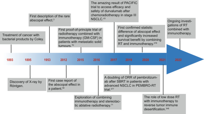
Radiotherapy (RT) is delivered for purposes of local control, but can also exert systemic effect on remote and non-irradiated tumor deposits, which is called abscopal effect. The view of RT as a simple local treatment has dramatically changed in recent years, and it is now widely accepted that RT can provoke a systemic immune response which gives a strong rationale for the combination of RT and immunotherapy (iRT). Nevertheless, several points remain to be addressed such as the interaction of RT and immune system, the identification of the best schedules for combination with immunotherapy (IO), the expansion of abscopal effect and the mechanism to amplify iRT. To answer these crucial questions, we roundly summarize underlying rationale showing the whole immune landscape in RT and clinical trials to attempt to identify the best schedules of iRT. In consideration of the rarity of abscopal effect, we propose that the occurrence of abscopal effect induced by radiation can be promoted to 100% in view of molecular and genetic level. Furthermore, the "radscopal effect" which refers to using low-dose radiation to reprogram the tumor microenvironment may amplify the occurrence of abscopal effect and overcome the resistance of iRT. Taken together, RT could be regarded as a trigger of systemic antitumor immune response, and with the help of IO can be used as a radical and systemic treatment and be added into current standard regimen of patients with metastatic cancer.


Conflict of interest statement

The authors declare no competing interests.

Figures

Fig. 1
Fig. 1
Historical timeline of some important developments regarding the iRT
Fig. 2
Fig. 2
Immunomodulatory effect of radiation on Treg cells. Radiation promotes the conversion from CD4+ T cells to Treg cells and enhances Treg function through IL-10R-mediated STAT3 signaling pathway. Radiation also increases the secretion of IL-10 which can bind to IL-10 receptors. This binding activates JAKs and activated JAKs, such as JAK1 and JAK2, phosphorylate STAT3 at Tyr705, resulting in translocation of activated STAT3 dimers to the nucleus. STAT3, as a cotranscription factor with FOXP3, promotes expansion, differentiation, and T cell suppression and increases CTLA-4 expression of Treg cells. In addition, miR-10a induced by radiation can enhance the expression level of FOXP3 and promote the differentiation of Treg from naive CD4+ T cell. Radiation increases the level of TGF-β in the TME greatly and TGF-β recognizes and binds TGFβRII, which then phosphorylates TGFβRI. TGF-β activates Smad2 and Smad3 and promotes the formation of a heterotrimer with Smad4. Smads are recruited to the CNS1 region which has been identified at the Foxp3 gene locus. The CNS1 promotes generation, expansion, differentiation, and development of Treg cells. Parts of this figure were drawn with aid of Servier Medical Art (http://www.servier.com), licensed under a Creative Commons Attribution 3.0 Unported License. Parts of this figure were drawn with aid of Servier Medical Art (http://www.servier.com), licensed under a Creative Commons Attribution 3.0 Unported License
Fig. 3
Fig. 3
Immunomodulatory effect of radiation on TANs. Radiation shows complexity regarding the immunomodulatory effect on TANs. On the one hand, radiation may induce TANs to exhibit the antitumor characteristic (N1) by IFN-β. N1 phenotype induces tumor cells cytotoxicity/apoptosis through ROS and activates CD8+ T cells and M1 macrophage. On the other hand, radiation may induce TANs to exhibit the pro-tumor characteristic through TGF-β. N2 phenotype promotes genetic instability by ROS, cancer proliferation, and immunosuppression effect by inhibiting CD8+ T cells and NK cells and enhancing Treg cells. Parts of this figure were drawn with aid of Servier Medical Art (http://www.servier.com), licensed under a Creative Commons Attribution 3.0 Unported License
Fig. 4
Fig. 4
Immunomodulatory effect of radiation on TAMs. Under the effect of p50–p50 NFκB homodimer induced by radiation, M2 macrophages acquired their phenotype. Meanwhile, increased ROS caused by radiation also promotes the polarization to M2 macrophage. The activation of p50–p50 dimer promotes the conversion towards M2 phenotype, leading to the secretion of IL-10 and TGF-β which inhibit DCs. And CCL22 secreted by M2 macrophage also recruits Treg cells to exert immunosuppressive function. Radiation elicits a high recruitment of TAMs through CCL2/CCR2 and CSF1/CSF1R pathways. And it can also recruit TAMs to infiltrate tumor sites, especially hypoxia sites through SDF-1/CXCR4-dependent signaling pathways. Moreover, M1 macrophage ban be activated by CD4+ cells through TNF and IFN-γ and then kill tumor cells via phagocytosis which plays a crucial role in abscopal effect. Parts of this figure were drawn with aid of Servier Medical Art (http://www.servier.com), licensed under a Creative Commons Attribution 3.0 Unported License
Fig. 5
Fig. 5
Signaling pathways of radiation on TAMs. Radiation increases the level of CXCL12 which then binds to CXCR4, a kind of G-protein-coupled receptor (GPCR). Similarly, CCL2 and its receptor, CCR2 are also activated by radiation. Next, the receptor undergoes a second conformational change that activates the intracellular trimeric G protein by the dissociation of Gα subunit from the Gβ/Gγ dimer. Then, the phosphatidylinositide 3-kinases (PI3Ks) can be activated by both Gβ/γ and Gα subunits. PI3Ks regulate gene transcription, migration, and adhesion of TAMs by the phosphorylation of AKT and of several focal adhesion components. In addition, Gα subunit also activates the Ras and Rac/Rho pathways, leading to the phosphorylation of ERK. Activated ERK can phosphorylate and regulate other cellular proteins, as well as translocate into the nucleus and phosphorylate and regulate transcription factors, leading to migration, proliferation, and cytokines expression of TAMs. Gβ/Gγ dimer also activates JAK/STAT signaling pathway to promote changes in cell morphology leading to chemotactic responses. Of note, CSF-1/CSF-1R activates all the three signaling pathways we discussed above. Parts of this figure were drawn with aid of Servier Medical Art (http://www.servier.com), licensed under a Creative Commons Attribution 3.0 Unported License
Fig. 6
Fig. 6
Immunomodulatory effect of radiation on MDSCs. DNA damage resulted from radiation may activate two signaling pathways in MDSCs: cGAS-STING and JAK/STAT signaling pathways. Similar to signaling pathways in TAMs, JAK/STAT signaling and PI3K/AKT signaling pathway are activated in MDSCs caused by CSF-1/CSF-1R and CCL2/CCR2. Upon DNA damage, all of these damaged DNA including cytoplasmic DNA and mtDNA induced by IR can be recognized by cGAS, which then oligomerizes with DNA in the form of a 2:2 complex. After binding to DNA, cGAS then exerts a catalytic role to promote the synthesis of the second messenger 2′3′-cyclic GMP–AMP (cGAMP). Binding of 2′3′-cGAMP stimulates STING and promotes the translocation to the Golgi which acivates TANK-binding kinase 1 (TBK1). TBK1 phosphorylates STING and promote the interferon regulatory factor 3 (IRF3) to translocate to the nucleus which triggers the expression of IFN-β gene. Parts of this figure were drawn with aid of Servier Medical Art (http://www.servier.com), licensed under a Creative Commons Attribution 3.0 Unported License
Fig. 7
Fig. 7
Immunomodulatory effect of radiation on DCs. Radiation on DCs significantly increases the expression of chemokines CCL19 and CCL21 which then bind to CCR7 and mediate migration of DCs. In addition, immunogenic cell death caused by radiation may release large amounts of antigens and DAMPs including HMGB1, ATP, calreticulin, heat shock proteins, and other cellular factors which bind specific pattern-recognition receptors on the dendritic cell, including Toll-like receptors, RIG1-like receptors, and NOD-like receptors. And radiation also results in the release of TAAs which promotes DCs activation, migration, and proliferation of T cells. Parts of this figure were drawn with aid of Servier Medical Art (http://www.servier.com), licensed under a Creative Commons Attribution 3.0 Unported License
Fig. 8
Fig. 8
Immunomodulatory effect of radiation on NK cells. On the one hand, radiation inhibits NK cells through increasing TGF-β. Moreover, the exposure of MHC-I molecules on the surface of tumor cells caused by radiation also inactivates NK cells through KIR. In addition, radiation induces the upregulation of activating receptors NKG2D and NKG2D ligands (NKG2DLs) to enhance the function of NK cells via granzyme B and perforin, FAS and FAS ligand and ADCC. Parts of this figure were drawn with aid of Servier Medical Art (http://www.servier.com), licensed under a Creative Commons Attribution 3.0 Unported License
Fig. 9
Fig. 9
Macro, molecular, and genetic abscopal effect. Abscopal effect in the traditional sense refers to the tumor regression at distant non-irradiated sites which can be observed in clinic. However, when the primary tumor is irradiated, cytokines and immune cells at distant non-irradiated sites also changes due to the systemic immune response caused by IR. In addition, there are alterations of gene expression at distant non-irradiated sites. Parts of this figure were drawn with aid of Servier Medical Art (http://www.servier.com), licensed under a Creative Commons Attribution 3.0 Unported License

Similar articles

Cited by

References

    1. Atun R, et al. Expanding global access to radiotherapy. Lancet Oncol. 2015;16:1153–1186. doi: 10.1016/S1470-2045(15)00222-3. - DOI - PubMed
    1. Citrin DE. Recent developments in radiotherapy. N. Engl. J. Med. 2017;377:1065–1075. doi: 10.1056/NEJMra1608986. - DOI - PubMed
    1. Mole RH. Whole body irradiation; radiobiology or medicine? Br. J. Radiol. 1953;26:234–241. doi: 10.1259/0007-1285-26-305-234. - DOI - PubMed
    1. Delaney G, Jacob S, Featherstone C, Barton M. The role of radiotherapy in cancer treatment: estimating optimal utilization from a review of evidence-based clinical guidelines. Cancer. 2005;104:1129–1137. doi: 10.1002/cncr.21324. - DOI - PubMed
    1. Caudell JJ, et al. The future of personalised radiotherapy for head and neck cancer. Lancet Oncol. 2017;18:e266–e273. doi: 10.1016/S1470-2045(17)30252-8. - DOI - PMC - PubMed

Publication types