. 2022 Jul 29;13(1):4420.
doi: 10.1038/s41467-022-31974-1.

Neprilysins regulate muscle contraction and heart function via cleavage of SERCA-inhibitory micropeptides

Affiliations

Neprilysins regulate muscle contraction and heart function via cleavage of SERCA-inhibitory micropeptides

Ronja Schiemann et al. Nat Commun. .

Abstract

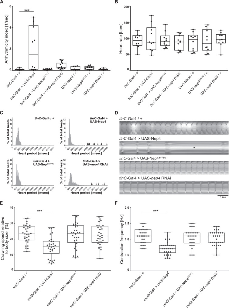
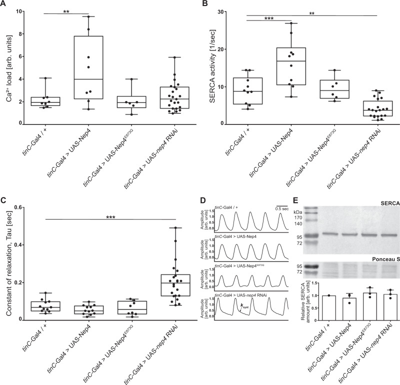
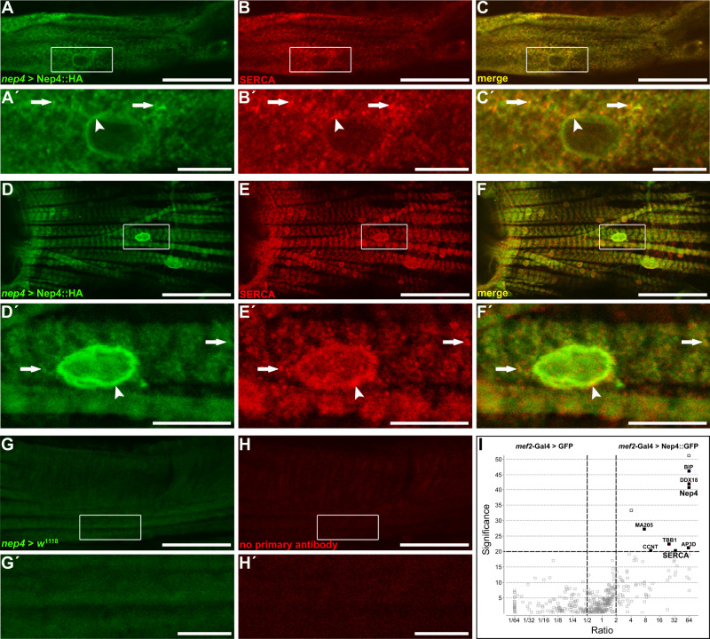
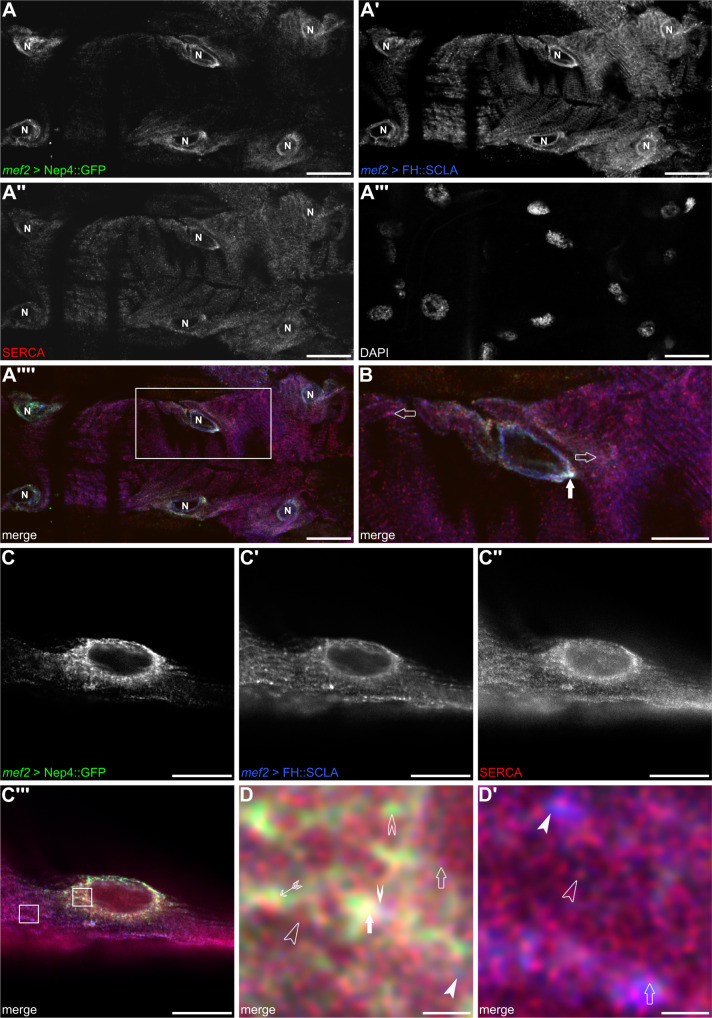
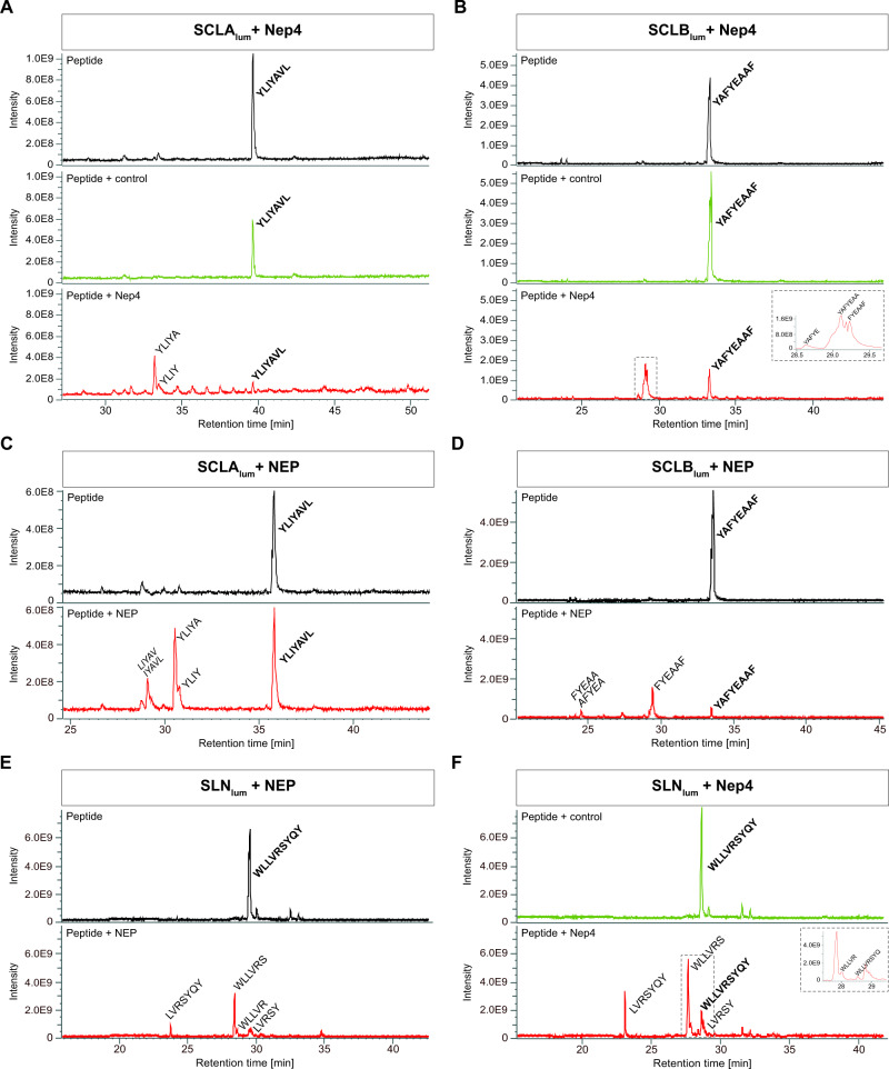
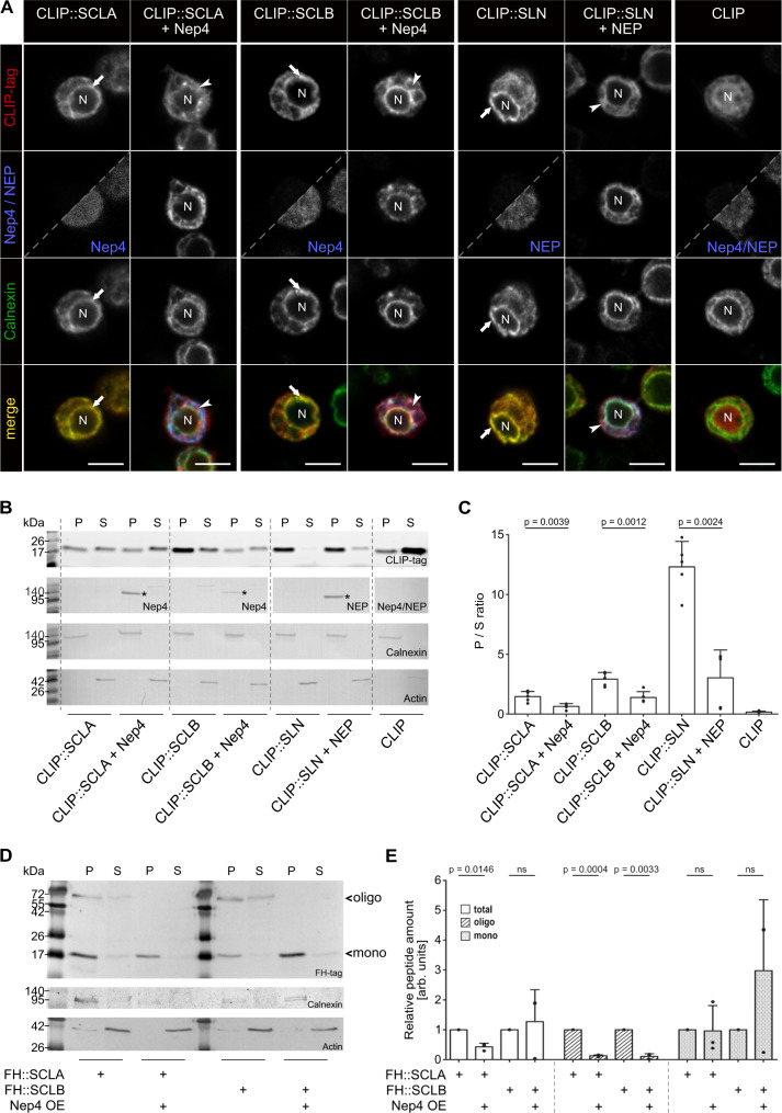
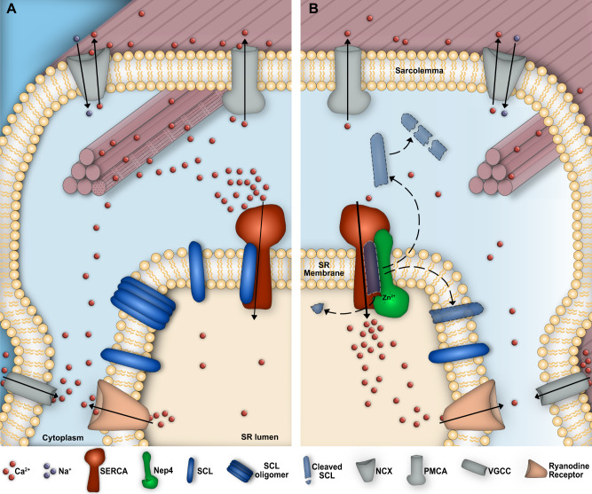
Muscle contraction depends on strictly controlled Ca2+ transients within myocytes. A major player maintaining these transients is the sarcoplasmic/endoplasmic reticulum Ca2+ ATPase, SERCA. Activity of SERCA is regulated by binding of micropeptides and impaired expression or function of these peptides results in cardiomyopathy. To date, it is not known how homeostasis or turnover of the micropeptides is regulated. Herein, we find that the Drosophila endopeptidase Neprilysin 4 hydrolyzes SERCA-inhibitory Sarcolamban peptides in membranes of the sarcoplasmic reticulum, thereby ensuring proper regulation of SERCA. Cleavage is necessary and sufficient to maintain homeostasis and function of the micropeptides. Analyses on human Neprilysin, sarcolipin, and ventricular cardiomyocytes indicates that the regulatory mechanism is evolutionarily conserved. By identifying a neprilysin as essential regulator of SERCA activity and Ca2+ homeostasis in cardiomyocytes, these data contribute to a more comprehensive understanding of the complex mechanisms that control muscle contraction and heart function in health and disease.


Conflict of interest statement

The authors declare no competing interests.

Figures

Fig. 1
Fig. 1. Nep4 activity is critical to heart and body wall muscle function.
A Relative to control animals (tinC-Gal4/+, n = 10), cardiomyocyte-specific overexpression of catalytically active Nep4 (tinC-Gal4>UAS-Nep4, n = 10) causes arrhythmia. Overexpression of inactive Nep4 (tinC-Gal4>UAS-Nep4E873Q, n = 10) or reduced expression of the peptidase (tinC-Gal4>UAS-nep4 RNAi, n = 11) do not affect rhythmicity. UAS controls (UAS-Nep4, n = 10; UAS-Nep4E873Q, n = 10; UAS-nep4 RNAi, n = 10) are also without any effect. B Relative to control animals (tinC-Gal4/+, n = 10), neither knockdown of nep4 (tinC-Gal4>UAS-nep4 RNAi, n = 11) nor increased expression of active (tinC-Gal4>UAS-Nep4, n = 10) or inactive Nep4 (tinC-Gal4>UAS-Nep4E873Q, n = 10) affect heart rate. C Combined histograms showing the distribution of heart periods (HP) from flies of the indicated genotypes. Overexpression of catalytically active Nep4 (tinC-Gal4>UAS-Nep4, n = 10, 964 recorded beats) as well as reduced expression of the peptidase (tinC-Gal4>UAS-nep4 RNAi, n = 11, 881 recorded beats) result in the occurrence of abnormally long HPs (arrows). Control hearts (tinC-Gal4/+, n = 10, 897 recorded beats) or hearts overexpressing inactive Nep4 (tinC-Gal4>UAS-Nep4E873Q, n = 10, 929 recorded beats) do not exhibit such impairments. D Representative 10 s M-mode traces depict heart contractions from flies of the indicated genotypes. Increased expression of catalytically active Nep4 (tinC-Gal4>UAS-Nep4) causes arrhythmia with prolonged periods of diastolic heart arrest (asterisk). E Relative to control animals (mef2-Gal4/+, n = 31), muscle-specific Nep4 overexpression (mef2-Gal4>UAS-Nep4, n = 30) decreases crawling speed by 34%. Increased expression of catalytically inactive Nep4 (mef2-Gal4>UAS-Nep4E873Q, n = 30) or knockdown of the peptidase (mef2-Gal4>UAS-nep4 RNAi, n = 32) has no effect. F Relative to control animals (mef2-Gal4/+, median: 1.1 Hz, n = 31), muscle-specific Nep4 overexpression (mef2-Gal4>UAS-Nep4, n = 30) decreases muscle contraction frequency (median: 0.7 Hz). Increased expression of catalytically inactive Nep4 (mef2-Gal4>UAS-Nep4E873Q, n = 30) or knockdown of the peptidase (mef2-Gal4>UAS-nep4 RNAi, n = 32) has no significant effect (median: 1.1 and 1.1 Hz, respectively). Asterisks indicate statistically significant deviations from respective controls (p < 0.001, one-way ANOVA followed by Dunnett’s Multiple Comparison Test). For all experiments, at least 10 individual animals were analyzed per genotype. Source data are provided as a Source data file.
Fig. 2
Fig. 2. Cardiac Ca2+ homeostasis is affected by altered nep4 expression levels.
A Relative to control animals (tinC-Gal4/+, n = 8), sarcoplasmic reticulum Ca2+ load is increased by 105% in cardiomyocytes overexpressing active Nep4 (tinC-Gal4 > UAS-Nep4, n = 8). Overexpression of catalytically inactive Nep4 (tinC-Gal4 > UAS-Nep4E873Q, n = 6) or knockdown of the peptidase (tinC-Gal4 > UAS-nep4 RNAi, n = 23) has no significant effect. B Relative to control animals (tinC-Gal4/+ n = 10), overexpression of active Nep4 (tinC-Gal4 > UAS-Nep4, n = 10) results in an increase in SERCA activity by 91%, while knockdown of the peptidase (tinC-Gal4 > UAS-nep4 RNAi, n = 20) reduces SERCA activity by 57%. Overexpression of catalytically inactive Nep4 (tinC-Gal4 > UAS-Nep4E873Q, n = 6) has no significant effect. C The constant of relaxation (Tau) is only affected by the knockdown of nep4. Relative to controls (tinC-Gal4/+, n = 11), corresponding animals (tinC-Gal4 > UAS-nep4 RNAi, n = 22) exhibit a 2.9-fold increase in Tau. Overexpression of active (tinC-Gal4 > UAS-Nep4, n = 12) or inactive Nep4 (tinC-Gal4 > UAS-Nep4E873Q, n = 8) has no significant effects. Asterisks indicate statistically significant deviations from respective controls (**p < 0.01, ***p < 0.001, one-way ANOVA followed by Dunnett’s Multiple Comparison Test). Each dot represents one analyzed animal. D Representative Ca2+ traces indicating the decay rate constant of the systolic Ca2+ transients (ksyst). Traces from 1-week-old adult Drosophila hearts of the depicted genotypes are shown. E Representative Western blot of total protein extracts isolated from adult flies of the indicated genotypes. For quantification, pixel intensity measurements were normalized to corresponding loading controls (Ponceau S). The resulting values are shown relative to the control (tinC-Gal4/+). The lower panel depicts the mean values (+SD) of three individual biological replicates. SERCA protein levels are not affected by altered nep4 expression (paired t test, two-tailed). Source data are provided as a Source data file.
Fig. 3
Fig. 3. Nep4 partially colocalizes with SERCA in heart tissue.
A, D Nep4::HA was expressed under the control of the native nep4 enhancer and labeled with a monospecific anti-HA antibody (nep4 > Nep4::HA). B, E SERCA was labeled with a monospecific antibody detecting the endogenous protein (SERCA). Optical slices of adult heart muscle fibers (AC) or ventral longitudinal muscle fibers (DF) are shown. Scale bars: 50 µm; ventral view, anterior left. Boxes indicate areas of higher magnification, as depicted in () and (). Scale bars: 10 µm. Nep4::HA colocalizes with SERCA in membranes contiguous with the nuclear membrane (, , arrowheads). In addition, both proteins partially colocalize in a punctate manner along the muscle fibers (, , arrows). Control stainings lacking the UAS-Nep4::HA construct (nep4 > w1118, G, ) or primary SERCA antibodies (H, ) do not exhibit any signal above background. I Volcano plot depicting the results of pull-down assays using Nep4::GFP as bait and free cytoplasmic GFP as control. Bait proteins were expressed in third instar larval muscle tissue. SERCA coprecipitates with Nep4::GFP. Data are based on three individual biological replicates. Open outlined squares depict proteins with quantification being based on only one detected peptide. Corresponding candidates were excluded from further analysis. A significance value of 20 corresponds to p < 0.01 (one-way ANOVA).
Fig. 4
Fig. 4. Nep4 partially colocalizes with Sarcolamban A.
GFP-tagged Nep4 (mef2 > Nep4::GFP, A, A´´´´, B, C, C´´´, D, ) and FH-tagged Sarcolamban A (mef2 > FH::SCLA, , A´´´´, B, , C´´´, D, ) were expressed under the control of the muscle-specific mef2 enhancer and labeled with monospecific antibodies against the GFP- or the FH-tag. SERCA was labeled with a monospecific antibody detecting the endogenous protein (SERCA, A´´, A´´´´, B, C´´, C´´´, D, ). DAPI was used as a nuclear marker (A´´´). Optical slices of third instar larval heart tissue are shown. The box in A´´´´ indicates an area of higher magnification as depicted in (B). (AA´´´´, B) Nep4, SERCA, and SCLA signals colocalize predominantly around the nuclei (B, solid arrow). More distant from the nuclei, only SERCA and SCLA signals are visible (B, open arrows). Scale bars: 20 µm (AA´´´´); 10 µm (B); ventral view, anterior left. (C) STED images of analogously stained cardiac tissue. Boxes in C´´´ indicate areas of higher magnification as depicted in (D, ). Overlap between Nep4, SERCA, and SCLA signals is visible predominantly around the nucleus (D, solid arrow). Individual colocalization between SCLA and SERCA (D, open arrow), Nep4 and SERCA (D, feathered arrow), or Nep4 and SCLA (D, solid winged arrowhead) occurs occasionally. Individual signals of SERCA (D, open arrowhead), Nep4 (D, open winged arrowhead), and SCLA (D, solid arrowhead) are present as well. More distant from the nucleus, only SERCA and SCLA are detected, while Nep4 is largely absent (). For SERCA and SCLA, again a colocalizing portion (, open arrow) as well as individual SERCA (, open arrowhead) and SCLA signals (, solid arrowhead) are present. Scale bars: 10 µm (C-C´´´); 1 µm (D, ).
Fig. 5
Fig. 5. Neprilysins hydrolyze the luminal domain of SERCA-inhibitory micropeptides.
Depicted are total ion chromatograms of the luminal part of Drosophila Sarcolamban A (SCLAlum, A, C), Drosophila Sarcolamban B (SCLBlum, B, D), and human sarcolipin (SLNlum, E, F). Full-length peptides (bold) are detected under all applied experimental conditions (peptide only, black chromatograms; peptide incubated with control preparation, green chromatograms; peptide incubated with purified enzyme, red chromatograms). Specific cleavage fragments are detected only after the addition of enzyme. A Incubation of SCLAlum (YLIYAVL) with Drosophila Nep4 results in the formation of YLIYA and YLIY fragments (red chromatogram). C The same fragments are generated by human neprilysin (NEP)-mediated hydrolysis of SCLAlum. An additional peak corresponds to either LIYAV or IYAVL. B Incubation of SCLBlum (YAFYEAAF) with Drosophila Nep4 results in the formation of FYEAAF, YAFYEAA, and YAFYE fragments (red chromatogram). D Human NEP-mediated hydrolysis of SCLBlum generates predominantly FYEAAF. In addition, minor amounts of FYEAA or AFYEA are produced. E Incubation of SLNlum (WLLVRSYQY) with human NEP results in the formation of WLLVRS, LVRSYQY, LVRSY, and WLLVR fragments (red chromatogram). F Incubation of SLNlum (WLLVRSYQY) with Drosophila Nep4 results in the formation of WLLVRS, LVRSYQY, and LVRSY fragments (red chromatogram). Insets depict areas of magnification indicated by the dashed boxes. Italicized fragments could not be assigned to one exclusive peptide sequence. Y-axes show absolute peak intensities, X-axes depict retention times. Individual cleavage assays were repeated at least three times.
Fig. 6
Fig. 6. Neprilysin-hydrolyzed SERCA-inhibitory micropeptides exhibit reduced membrane anchoring.
A All constructs were expressed in Drosophila S2 cells as full-length peptides (CLIP::SCLA; CLIP::SCLB; CLIP::SLN), either with or without co-expression of Nep4 (SCLA, SCLB) or human NEP (SLN). CLIP-tag and Nep4 or NEP were visualized by immunostainings as indicated. Split images are overexposed in the lower right part to confirm the presence of cells. Anti-Calnexin antibodies were applied as ER membrane marker. Cells expressing the free CLIP-tag were used as a soluble control. While full-length SCL/SLN peptides mainly localize to the ER (arrows), co-expression of Nep4 or NEP results in reduced signal overlap of the peptides with the ER marker (arrowheads). Scale bars: 5 µM. B Subcellular fractions of Drosophila S2 cells expressing the indicated CLIP-tagged SCL or SLN constructs, with or without co-expression of Nep4 or NEP. Western blot analysis was performed with anti-Nep4 and anti-NEP antibodies, as indicated, to confirm peptidase expression (asterisks), and with anti-Calnexin antibodies (a marker for ER membranes) and anti-Actin antibodies (cytosolic marker) to confirm the identity of the individual fractions. P = pellet (membrane-enriched); S = supernatant. C Peptide-specific ratios between membrane-enriched (P) and soluble (S) fractions were determined by pixel intensity measurements. The diagram depicts the resultant mean values (+ SD) of five individual biological replicates. Significant differences between the individual peptide-specific ratios are indicated (paired t-test, two-tailed). D Subcellular fractions of Drosophila third instar larvae expressing the indicated SCL constructs in a muscle-specific manner (mef2-Gal4) were analyzed by Western blot. Peptides were detected with anti-HA antibodies. Calnexin or Actin signals were used to confirm the identity of the individual fractions. Actin signals were used for normalization. P = pellet (membrane-enriched); S = supernatant; mono = peptide monomer; oligo = peptide oligomer. E Relative amounts of peptide oligomers (oligo), peptide monomers (mono), and the sum of both (total) were determined by pixel intensity measurements. For each of the indicated SCL/Nep4 combinations, the combined signals from pellet and supernatant fractions, as depicted in D, were evaluated. Resultant data represent mean values (+SD) of three individual biological replicates (paired t-test, two-tailed). Source data are provided as a Source data file.
Fig. 7
Fig. 7. Neprilysin-mediated hydrolysis of SERCA-inhibitory micropeptides is essential to control SERCA activity.
A Without Nep4, Sarcolamban (SCL) peptides occur in a monomeric and an oligomeric state and accumulate within the sarcoplasmic reticulum (SR) membrane, resulting in abnormal binding and inhibition of SERCA and a consequential reduction in the SERCA-mediated Ca2+ transport. B Nep4-mediated SCL hydrolysis reduces the ability of the peptides to oligomerize and releases them from the SR membrane, thus preventing SCL accumulation and excessive SERCA inhibition. Released peptides become degraded in the cytoplasm. VGCC voltage-gated L-type Ca2+ channel, RyR Ryanodine receptor, NCX Na+/Ca2+ exchanger, PMCA plasma membrane Ca2+ ATPase.

Similar articles

Cited by

References

    1. Hasenfuss G, Pieske B. Calcium cycling in congestive heart failure. J. Mol. Cell Cardiol. 2002;34:951–969. doi: 10.1006/jmcc.2002.2037. - DOI - PubMed
    1. Periasamy M, Bhupathy P, Babu GJ. Regulation of sarcoplasmic reticulum Ca2+ ATPase pump expression and its relevance to cardiac muscle physiology and pathology. Cardiovasc. Res. 2008;77:265–273. doi: 10.1093/cvr/cvm056. - DOI - PubMed
    1. Gehrig SM, et al. Hsp72 preserves muscle function and slows progression of severe muscular dystrophy. Nature. 2012;484:394–398. doi: 10.1038/nature10980. - DOI - PubMed
    1. Periasamy M, Kalyanasundaram A. SERCA pump isoforms: their role in calcium transport and disease. Muscle Nerve. 2007;35:430–442. doi: 10.1002/mus.20745. - DOI - PubMed
    1. Gorski PA, Ceholski DK, Hajjar RJ. Altered myocardial calcium cycling and energetics in heart failure–a rational approach for disease treatment. Cell Metab. 2015;21:183–194. doi: 10.1016/j.cmet.2015.01.005. - DOI - PMC - PubMed

Publication types

MeSH terms

LinkOut - more resources