. 2022 Jul 29;12(1):13048.
doi: 10.1038/s41598-022-17275-z.

Characterization and anti-biofilm activity of bacteriophages against urinary tract Enterococcus faecalis isolates

Affiliations

Characterization and anti-biofilm activity of bacteriophages against urinary tract Enterococcus faecalis isolates

Doaa M El-Atrees et al. Sci Rep. .

Abstract

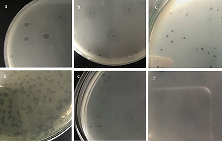
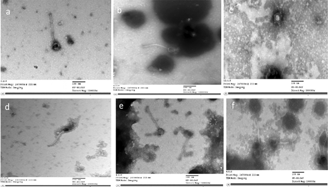
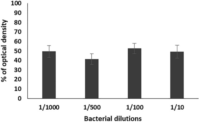
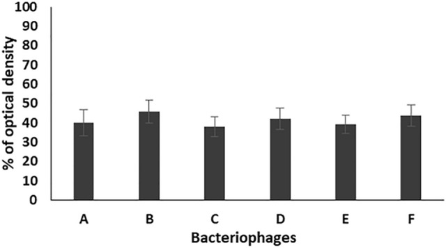
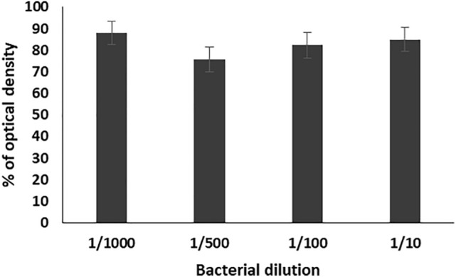
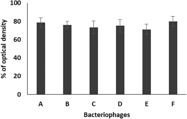
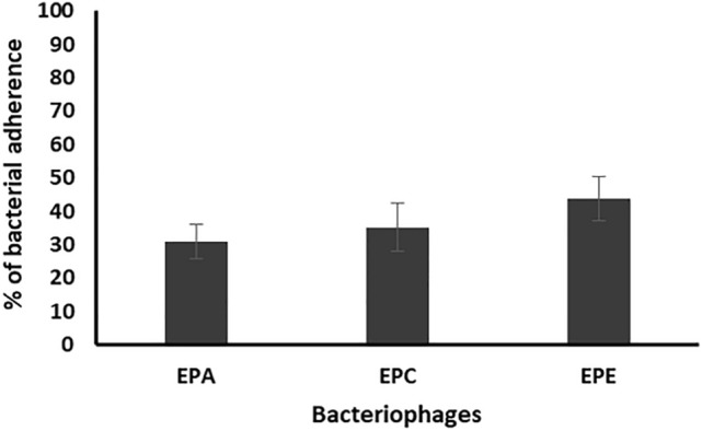
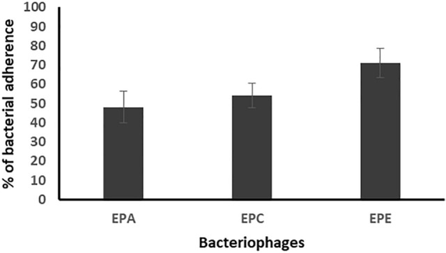
Strong biofilm-forming Enterococcus feacalis urinary tract pathogens (n = 35) were used to determine the lytic spectrum of six bacteriophages isolated from sewage samples. Only 17 Enterococcus feacalis isolates gave lytic zones with the tested bacteriophages from which five isolates were susceptible to all of them. The isolated enterococcal phages are characterized by wide range of thermal (30-90 °C) and pH (3-10) stability. They belong to order Caudovirales, from which four bacteriophages (EPA, EPB, EPD, EPF) belong to family Myoviridae and two (EPC, EPE) belong to family Siphoviridae. In addition, they have promising antibiofilm activity against the tested strong-forming biofilm E. faecalis isolates. The enterococcal phages reduced the formed and preformed biofilms to a range of 38.02-45.7% and 71.0-80.0%, respectively, as compared to the control. The same promising activities were obtained on studying the anti-adherent effect of the tested bacteriophages on the adherence of bacterial cells to the surface of urinary catheter segments. They reduced the number of adherent cells to a range of 30.8-43.8% and eradicated the pre-adherent cells to a range of 48.2-71.1%, as compared to the control. Overall, the obtained promising antibiofilm activity makes these phages good candidates for application in preventing and treating biofilm associated Enterococcus faecalis infections.


Conflict of interest statement

The authors declare no competing interests.

Figures

Figure 1
Figure 1
Plaques pictures of the 6 bacteriophages (EPA: F), as formed on Enterococcus double layer agar plates. (a) EPA: large plaques (diameter: up to 4 mm) with hallow zones. (b) EPB: medium plaques (diameter: 2–3 mm) with hallow zone. (c) EPC: medium plaques (diameter: 1–2 mm) without hallow zones. (d) EPD: large plaques (diameter: up to 4 mm) without hallow zone. (e) EPE: medium plaques (diameter: 1 mm) with hallow zone. (f) EPF: pinpoint plaques (diameter: < 1 mm) with hallow zone.
Figure 2
Figure 2
TEM images of the six different enterococcal bacteriophages (a) phage EPA, (b) phage EPB, (c) phage EPC, (d) phage EPD, (e) phage EPE, (f) phage EPF, with scale bar 100 nm.
Figure 3
Figure 3
Percentage of optical density of the formed biofilms of 17 enterococcal isolates, prepared in different dilutions (1/10, 1/100, 1/500, and 1/1000) from bacterial suspension adjusted to 0.5 MacFarland, in the presence of the six bacteriophages (EPA, EPB, EPC, EPD, EPE, EPF) as compared to the control. Each bar represents the mean of three independent measurements.
Figure 4
Figure 4
Percentage of optical density of the formed biofilms of 17 enterococcal isolates, prepared in 1/500 dilution from bacterial suspension adjusted to 0.5 MacFarland, in the presence of the six bacteriophages (A: EPA, B: EPB, C: EPC, D: EPD, E: EPE, F: EPF) as compared to the control. Each bar represents the mean of three independent measurements.
Figure 5
Figure 5
Percentage of the formed biofilms of 17 enterococcal isolates, prepared in different dilutions (1/10, 1/100, 1/500, and 1/1000) from bacterial suspension adjusted to 0.5 MacFarland, after treatment the preformed biofilms with the six bacteriophages (EPA, EPB, EPC, EPD, EPE, EPF) as compared to the control. Each bar represents the mean of three independent measurements.
Figure 6
Figure 6
Percentage of the formed biofilms of 17 enterococcal isolates, prepared in different dilutions (1/10, 1/100, 1/500, and 1/1000) from bacterial suspension adjusted to 0.5 MacFarland, after treatment the preformed biofilms with the six bacteriophages (A: EPA, B: EPB, C: EPC, D: EPD, E: EPE, F:EPF) as compared to the control. Each bar represents the mean of three independent measurements.
Figure 7
Figure 7
Percentage of bacterial adherence on the surface of catheters segments of 3 enterococcal isolates (EF104, EF134, and EF151), prepared in dilution 1/500 from bacterial suspension adjusted to 0.5 MacFarland, in the presence of the three bacteriophages (EPA, EPC, EPE) as compared to the control. Each bar represents the mean of three independent measurements.
Figure 8
Figure 8
Percentage of bacterial adherence on the surface of catheters segments of the 3 enterococcal isolates (EF104, EF134, and EF151), prepared in dilution 1/500 from bacterial suspension adjusted to 0.5 MacFarland, after treatment the preformed biofilms with the three bacteriophages (EPA, EPC, EPE) as compared to the control. Each bar represents the mean of three independent measurements.

References

    1. Turjeman A, et al. Risk factors for enterococcal urinary tract infections: A multinational, retrospective cohort study. Eur. J. Clin. Microbiol. Infect. Dis. 2021;40:2005–2010. doi: 10.1007/s10096-021-04207-4. - DOI - PubMed
    1. Abdelkareem MZ, Sayed M, Hassuna NA, Mahmoud MS, Abdelwahab SF. Multi-drug-resistant Enterococcus faecalis among Egyptian patients with urinary tract infection. J. Chemother. 2017;29:74–82. doi: 10.1080/1120009X.2016.1182358. - DOI - PubMed
    1. Stępień-Pyśniak D, Hauschild T, Kosikowska U, Dec M, Urban-Chmiel R. Biofilm formation capacity and presence of virulence factors among commensal Enterococcus spp. from wild birds. Sci. Rep. 2019;9:1–7. doi: 10.1038/s41598-019-47602-w. - DOI - PMC - PubMed
    1. Jamal M, et al. Bacterial biofilm and associated infections. J. Chin. Med. Assoc. 2018;81:7–11. doi: 10.1016/j.jcma.2017.07.012. - DOI - PubMed
    1. Oumer Y, Dadi BR, Seid M, Biresaw G, Manilal A. Catheter-associated urinary tract infection: Incidence, associated factors and drug resistance patterns of bacterial isolates in southern ethiopia. Infect. Drug Resist. 2021;14:2883. doi: 10.2147/IDR.S311229. - DOI - PMC - PubMed

Publication types