. 2023 Aug;14(4):589-607.
doi: 10.1007/s12975-022-01071-y. Epub 2022 Jul 29.

MicroRNA-193a-5p Rescues Ischemic Cerebral Injury by Restoring N2-Like Neutrophil Subsets

Affiliations

MicroRNA-193a-5p Rescues Ischemic Cerebral Injury by Restoring N2-Like Neutrophil Subsets

Ziping Han et al. Transl Stroke Res. 2023 Aug.

Abstract

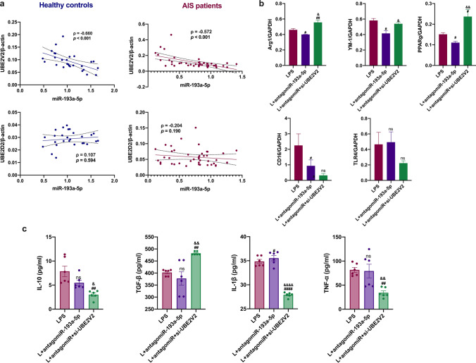
Circulating neutrophils are activated shortly after stroke and in turn affect the fate of ischemic brain tissue, and microRNAs (miRNA) participate in regulating neuroinflammation. We probed the role of neutrophilic miRNA in ischemic stroke. miR-193a-5p was decreased in circulating neutrophils of acute ischemic stroke (AIS) patients and healthy controls. In another set of AIS patients treated with recombinant tissue plasminogen activator, higher neutrophilic miR-193a-5p levels were associated with favorable outcomes at 3 months and non-symptomatic intracerebral hemorrhage. An experimental stroke model and human neutrophil-like HL-60 cells were further transfected with agomiR-193a-5p/antagomiR-193a-5p or ubiquitin-conjugating enzyme V2 (UBE2V2)-siRNA prior to model induction for in vivo and in vitro studies. Results of 2,3,5-triphenyl tetrazolium chloride staining and neurological function evaluations at post-experimental stroke showed that intravenous agomiR-193a-5p transfusion protected against ischemic cerebral injury in the acute stage and promoted neurological recovery in the subacute stage. This protective role was suggested to correlate with neutrophil N2 transformation based on the N2-like neutrophil proportions in the bone marrow, peripheral blood, and spleen of the experimental stroke model and the measurement of neutrophil phenotype-associated molecule levels. Mechanistically, analyses indicated that UBE2V2 might be a target of miR-193a-5p. Cerebral injury and neuroinflammation aggravated by miR-193a-5p inhibition were reversed by UBE2V2 silencing. In conclusion, miR-193a-5p protects against cerebral ischemic injury by restoring neutrophil N2 phenotype-associated neuroinflammation suppression, likely, in part, via UBE2V2 induction.

Keywords: Cerebral ischemic injury; N2-like neutrophil; UBE2V2; miR-193a-5p.


Conflict of interest statement

The authors declare no competing interests.

Figures

Fig. 1
Fig. 1
Expression of miR-193a-5p in circulating neutrophils of AIS patients. a Heatmap of the differentially expressed miRNA expression profile in ischemic stroke patients and controls using a miRNA sequencing; |fold-change|> 1.5, p < 0.05. b Validation of miR-193a-5p expression level in AIS patients (n = 43) and healthy controls (n = 27). c ROC analysis of miR-193a-5p in AIS patients and healthy controls. d Correlation analysis of the mRNA level of neutrophilic CD16 or CD206 and miR-193a-5p in neutrophils. AIS, acute ischemic stroke; AUC, area under ROC curve; CI, confidence interval; ROC, receiver operating characteristics. Bar charts reported in (b) show the median with interquartile range. ** p < 0.01 based on Man-Whitney U test (two-sided); the coefficient ρ (rho) was obtained via Spearman’s correlation analysis
Fig. 2
Fig. 2
Association between neutrophilic miR-193a-5p and outcomes of AIS patients receiving rtPA treatment. a Neutrophilic miR-193a-5p according to an mRS ≤ 2 or mRS > 2 at 3 months. ROC analysis of neutrophilic miR-193a-5p and outcome at 3 months. b Neutrophilic miR-193a-5p according to the non-sICH or sICH group. ROC analysis of neutrophilic miR-193a-5p and sICH. c Correlation analysis of CRP and NLR with neutrophilic miR-193a-5p. N = 128. AIS, acute ischemic stroke; ROC, receiver operating characteristics; AUC, area under ROC curve; rtPA, recombinant tissue plasminogen activator; CI, confident interval; CRP, C-reactive protein; mRS, modified Rankin Scale; NLR, neutrophil-to-lymphocyte ratio; sICH, symptomatic intracerebral hemorrhage. Bar charts reported in (ab) show the median with the interquartile range. *p < 0.05 based on the Man-Whitney U test (two-sided); the coefficient ρ (rho) was obtained via Spearman’s correlation analysis
Fig. 3
Fig. 3
Effect of agomiR-193a-5p on brain injury in experimental stroke at the acute stage. a Brief illustration of the in vivo experiments at the acute stage. b Representative transverse slices from TTC staining of sham, MCAO, and MCAO plus agomiR-193a-5p groups at 24 h post-MCAO. Quantitative analysis of the infarct volume in the MCAO and MCAO + agomiR-193a-5p groups. c ELISA evaluation of IL-10, TGF-β, TNF-α, and IL-1β levels in the mouse ipsilateral brain tissue homogenate. d RT-qPCR analysis of CD206, TGF-β, PPARγ, and TLR4 in the mouse ipsilateral brain tissue. Quantification of the relative value was based on the sham-operated control (set as 1). N = 6 per treatment arm. MCAO, transient middle cerebral artery occlusion; TTC, 2,3,5-triphenyltetrazolium chloride. Bar charts in (cd) show the mean with SEM. *p < 0.05, **p < 0.01, ***p < 0.001, ****p < 0.0001, significantly different from sham group. #p < 0.05, ##p < 0.01, ###p < 0.001, ####p < 0.0001, significantly different from MCAO group. ns, not significant
Fig. 4
Fig. 4
Effect of agomiR-193a-5p on functional recovery and brain injury in experimental stroke at the subacute stage. a Brief illustration of the in vivo experiments at the subacute stage. b Representative transverse slices from TTC staining of MCAO and MCAO + agomiR-193a-5p groups at 7 days post-MCAO. Quantitative analysis of the infarct volume in the MCAO and MCAO + agomiR-193a-5p groups. c Line chart of mouse body weights from pre-surgery to 1–7 days post-surgery. d Line chart of the mNSS, beam walking test score, time to contact/removal of the adhesive tape in the removal test for mice at pre-surgery, and 1, 3, 5, and 7 days post-surgery. N = 6 per treatment arm. MCAO, transient middle cerebral artery occlusion. TTC, 2,3,5-triphenyltetrazolium chloride; mNSS, mouse neurological severity score. Bar charts in (b − g) show the mean with the standard deviation. *p < 0.05, **p < 0.01, ***p < 0.001, ****p < 0.0001, significantly different from sham group. #p < 0.05, ##p < 0.01, ###p < 0.001, ####p < 0.0001, significantly different from MCAO group. ns, not significant
Fig. 5
Fig. 5
Flow cytometric analysis of the relative percentage of Ly6G+CD11b+CD206+ cells in the bone marrow, peripheral blood, and spleen at the acute stage of experimental stroke. N = 6 per treatment arm. Quantification of the relative percentage was based on the sham-operated control (set as 1). MCAO, transient middle cerebral artery occlusion. Bar charts show the mean with the SEM. *p < 0.05, **p < 0.01, significantly different from sham group. #p < 0.05, significantly different from MCAO. ns, not significant
Fig. 6
Fig. 6
Effect of agomiR-193a-5p on the human neutrophil-like cell line with inflammation induced by LPS. a Brief illustration of the in vitro experiments. b RT-qPCR analysis of miR-193a-5p, Arg1, YM1, CD16, PPARγ, and TLR4 in HL60 cells. c ELISA evaluation of TGF-β, TNF-α, IL-1β, and IL-10 levels in the supernatant of HL60 cells. Quantification of the relative value was based on the sham-operated control (set as 1). N = 6 per treatment arm. Bar charts reported in (bc) show the mean with the SEM. *p < 0.05, **p < 0.01, ***p < 0.001, ****p < 0.0001, significantly different from control. #p < 0.05, ##p < 0.01, ###p < 0.001, ####p < 0.0001, significantly different from LPS. ns, not significant
Fig. 7
Fig. 7
Prediction and validation of target genes of miR-193a-5p. a Biological functions (blue) and signaling (red) terms highlighted by GO and pathway analysis. b Prediction of putative target genes of miR-193a-5p. c Target gene validation by RT-qPCR in AIS patients (n = 43) and controls (n = 27). Analyzed by Man-Whitney U test (two-sided). d Target gene validation by RT-qPCR in HL60 cells (n = 6 per treatment arm). e Fluorescence in situ hybridization (FISH) analysis of miR-193a-5p (green), Ly6G (red), and UBE2V2 (purple) merged with DAPI (blue) present in C57BL/6 J mouse brain slices; bar = 20 μm. f RNA binding protein immunoprecipitation (RIP) of miR-193a-5p with UBE2V2 in HL60 cells and C57BL/6 J mouse brains (n = 4). AIS, acute ischemic stroke; MCAO, transient middle cerebral artery occlusion; NC, negative control. Bar charts reported in (c) show the median with the interquartile range; bar charts in (d, f) show the mean with the SEM. *p < 0.05, **p < 0.01, ***p < 0.001, ****p < 0.0001, significantly different from sham group. #p < 0.05, ##p < 0.01, ###p < 0.001, ####p < 0.0001, significantly different from MCAO group
Fig. 8
Fig. 8
Expression of UBE2V2 in AIS patients and UBE2V2 silencing in human neutrophil-like cells. a Correlation analysis of the miRNA level of UBE2V2 and UBE2D2 with miR-193a-5p in neutrophils of AIS patients (n = 43) and healthy controls (n = 27). b RT-qPCR analysis of Arg1, YM1, CD16, PPARγ, and TLR4 in HL60 cells; quantification of the relative value was based on the sham-operated control group (set as 1). c ELISA evaluation of TGF-β, TNF-α, IL-1β, and IL-10 in the supernatant of HL60 cells. N = 6 per treatment arm. The coefficient ρ (rho) was obtained via Spearman’s correlation analysis. AIS, acute ischemic stroke. Bar charts in (bc) show the mean with the SEM. #p < 0.05, ##p < 0.01, ###p < 0.001, ####p < 0.0001, significantly different from LPS group. &p < 0.05, &&p < 0.01, &&&p < 0.001, &&&&p < 0.0001, significantly different from LPS + antagomir-193a-5p group
Fig. 9
Fig. 9
Effect of UBE2V2 silencing on experimental stroke. a Representative transverse slices from TTC staining of MCAO, MCAO + antagomiR-193a-5p, and MCAO + antagomiR-193a-5p + UBE2V2-siRNA groups at 24 h post-MCAO. Quantitative analysis of the infarct volume in the MCAO, MCAO + antagomiR-193a-5p, and MCAO + antagomiR-193a-5p + UBE2V2-siRNA groups. b ELISA evaluation of IL-10, TGF-β, TNF-α, and IL-1β levels in the mouse ipsilateral brain tissue homogenate. d RT-qPCR analysis of CD206, TGF-β, PPARγ, and TLR4 in the mouse ipsilateral brain tissue. Quantification of relative values was based on the ham-operated control (set as 1). N = 6 per treatment arm. MCAO, transient middle cerebral artery occlusion. TTC, 2,3,5-triphenyltetrazolium chloride. Bar charts in (ac) show the mean with the SEM. #p < 0.05, ##p < 0.01, ###p < 0.001, ####p < 0.0001, significantly different from MCAO group. &p < 0.05, &&p < 0.01, &&&p < 0.001, &&&&p < 0.0001, significantly different from MCAO + antagomir-193a-5p group

References

    1. Macrez R, Ali C, Toutirais O, et al. Stroke and the immune system: from pathophysiology to new therapeutic strategies. Lancet Neurol. 2011;10:471–480. doi: 10.1016/S1474-4422(11)70066-7. - DOI - PubMed
    1. Zera KA, Buckwalter MS. The local and peripheral immune responses to stroke: implications for therapeutic development. Neurotherapeutics. 2020;17:414–435. doi: 10.1007/s13311-020-00844-3. - DOI - PMC - PubMed
    1. Weisenburger-Lile D, Dong Y, Yger M, et al. Harmful neutrophil subsets in patients with ischemic stroke: association with disease severity. Neurol Neuroimmunol Neuroinflamm. 2019;6:e571. doi: 10.1212/NXI.0000000000000571. - DOI - PMC - PubMed
    1. Elkins J, Veltkamp R, Montaner J, et al. Safety and efficacy of natalizumab in patients with acute ischaemic stroke (ACTION): a randomised, placebo-controlled, double-blind phase 2 trial. Lancet Neurol. 2017;16:217–226. doi: 10.1016/S1474-4422(16)30357-X. - DOI - PubMed
    1. Enlimomab Acute Stroke Trial Investigators. Use of anti-ICAM-1 therapy in ischemic stroke: results of the Enlimomab Acute Stroke Trial. Neurology. 2001;57:1428–1434. - PubMed

Publication types

LinkOut - more resources