. 2022 Jul 29;12(1):13031.
doi: 10.1038/s41598-022-16469-9.

Paternal age impairs in vitro embryo and in vivo fetal development in murine

Affiliations

Paternal age impairs in vitro embryo and in vivo fetal development in murine

Larissa Araújo Stábile et al. Sci Rep. .

Abstract

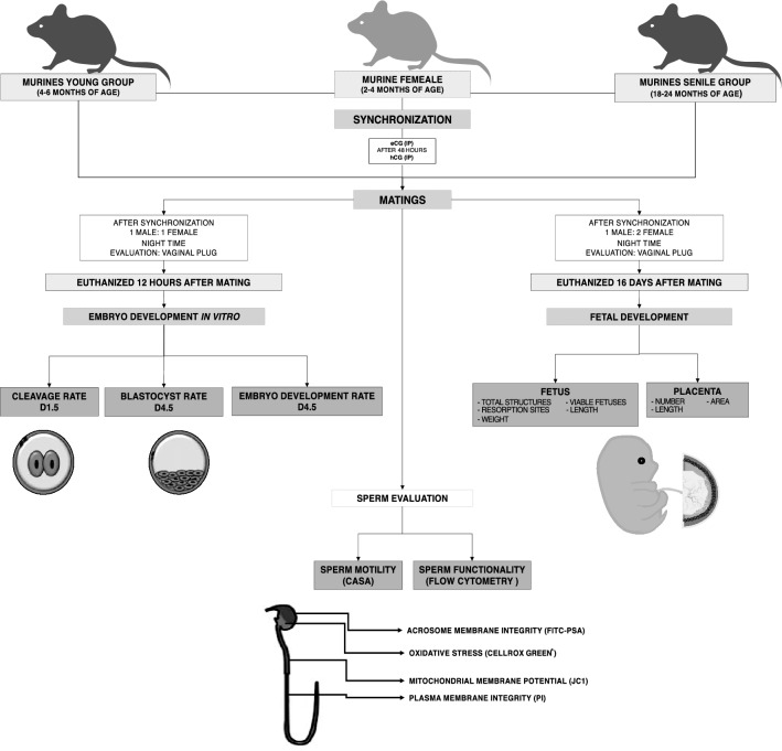
The association between advanced paternal age and impaired reproductive outcomes is still controversial. Several studies relate decrease in semen quality, impaired embryo/fetal development and offspring health to increased paternal age. However, some retrospective studies observed no alterations on both seminal status and reproductive outcomes in older men. Such inconsistency may be due to the influence of intrinsic and external factors, such as genetics, race, diet, social class, lifestyle and obvious ethical issues that may bias the assessment of reproductive status in humans. The use of the murine model enables prospective study and owes the establishment of homogeneous and controlled groups. This study aimed to evaluate the effect of paternal age on in vitro embryo development at 4.5 day post conception and on in vivo fetal development at 16 days of gestation. Murine females (2-4 months of age) were mated with young (4-6 months of age) or senile (18-24 months of age) males. We observed decreased in vitro cleavage, blastocyst, and embryo development rates; lighter and shorter fetuses in the senile compared to the young group. This study indicated that advanced paternal age negatively impacts subsequent embryo and fetal development.


Conflict of interest statement

The authors declare no competing interests.

Figures

Figure 1
Figure 1
Mating rate. Reproductive performance of young (n = 27) and senile (n = 29) groups. Different superscript letters in each bar represent p < 0.05, as indicated by T-test.
Figure 2
Figure 2
In vitro embryo development. Image and chart of cleaved embryos from the young and senile group (number of cleaved embryos/number of total structures × 100; a). Image and chart of blastocyst from the young and senile group (number of blastocysts/number of total structures × 100; b), and embryo development rates (number of blastocysts/number of cleaved embryos × 100; c) from young and senile groups. In the young group: 185 total structures (presumed zygotes) from 6 pregnant dams (30.8/pregnant dam), 122 cleaved embryos from 6 pregnant dams (20.3/pregnant dam), and 61 blastocysts from 6 pregnant dams (10.1/pregnant dam). In senile group: 107 total structures from 5 pregnant dams (21.4/pregnant dam), 27 cleaved embryos from 5 pregnant dams (5.4/pregnant dam) and 3 blastocysts from 5 pregnant dams (0.6/pregnant dam). The evaluations were performed in stereomicroscope (Olympus SZ61, Olympus®, Tokyo, Japan) at ×60 magnification. Different superscript letters in each bar represent p < 0.05, as indicated by statistical T-test.
Figure 3
Figure 3
Fetal development at 16 days of gestation. Fetuses length (a) and weight (b), and fetal: placental weight ratio (c) of young and senile groups. We considered the average of the litters per male, 14 litters as experimental unit (5 litters in the senile group and 9 litters in the young group). The photos were performed with a megapixel digital color camera (Olympus LC30, Olympus®, Munster, German) attached to a stereomicroscope (Olympus SZ61, Olympus®, Tokyo, Japan) at ×8 magnification. Different superscript letters in each bar represent p < 0.05, as indicated by statistical T-test.
Figure 4
Figure 4
Experimental design. Male murines divides into two groups, senile (18–24 months of age) and young (4–6 months of age), who mated with a group of murine females synchronized, for the realization of three experiments: embryo development in vitro, fetal development, and sperm evaluation.

References

    1. Kovac JR, et al. The effects of advanced paternal age on fertility. Asian J. Androl. 2013;15:723. doi: 10.1038/aja.2013.92. - DOI - PMC - PubMed
    1. Ramasamy R, Chiba K, Butler P, Lamb DJ. Male biological clock: A critical analysis of advanced paternal age. Fertil. Steril. 2015;103:1402–1406. doi: 10.1016/j.fertnstert.2015.03.011. - DOI - PMC - PubMed
    1. Qureshi AI, Saeed O, Malik AA, Suri MFK. Pregnancy in advanced age and the risk of stroke in postmenopausal women: Analysis of women’s health initiative study. Am. J. Obstet. Gynecol. 2017;216:409–e1. doi: 10.1016/j.ajog.2016.12.004. - DOI - PubMed
    1. Hassold T, Hunt P. Maternal age and chromosomally abnormal pregnancies: What we know and what we wish we knew. Curr. Opin. Pediatr. 2009;21:703. doi: 10.1097/MOP.0b013e328332c6ab. - DOI - PMC - PubMed
    1. Gill SK, et al. Association between maternal age and birth defects of unknown etiology—United States, 1997–2007. Birth Defects Res. Part A Clin. Mol. Teratol. 2012;94:1010–1018. doi: 10.1002/bdra.23049. - DOI - PMC - PubMed

Publication types