

Synchronous bilateral multifocal basal cell adenomas of the parotid gland-a case report
- PMID: 35906614
- PMCID: PMC9336062
- DOI: 10.1186/s12903-022-02339-3
Synchronous bilateral multifocal basal cell adenomas of the parotid gland-a case report
Abstract
Background: Bilateral parotid gland tumors account for up to 3% of cases. In this group, the vast majority are Warthin's tumors. However, bilateral presentations of other parotid gland tumor entities is also possible, an example of which is a basal cell adenoma (BCA). Bilateral BCA is extremely rare, which could cause misdiagnosing it as a Warthin tumor.
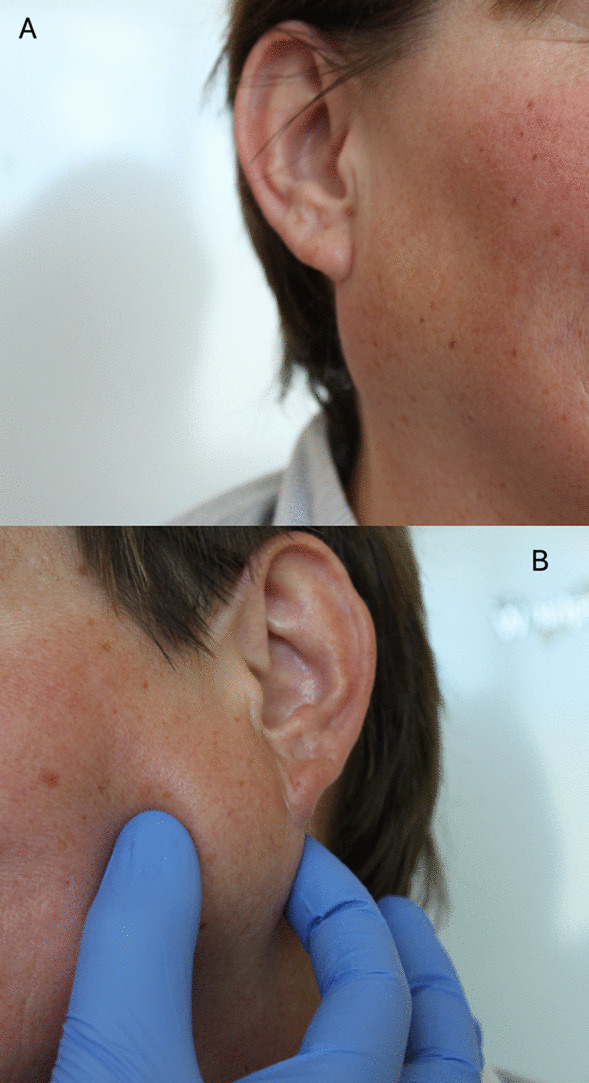
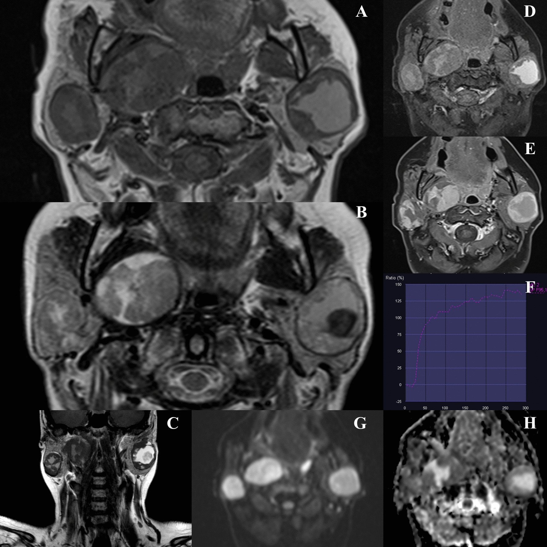
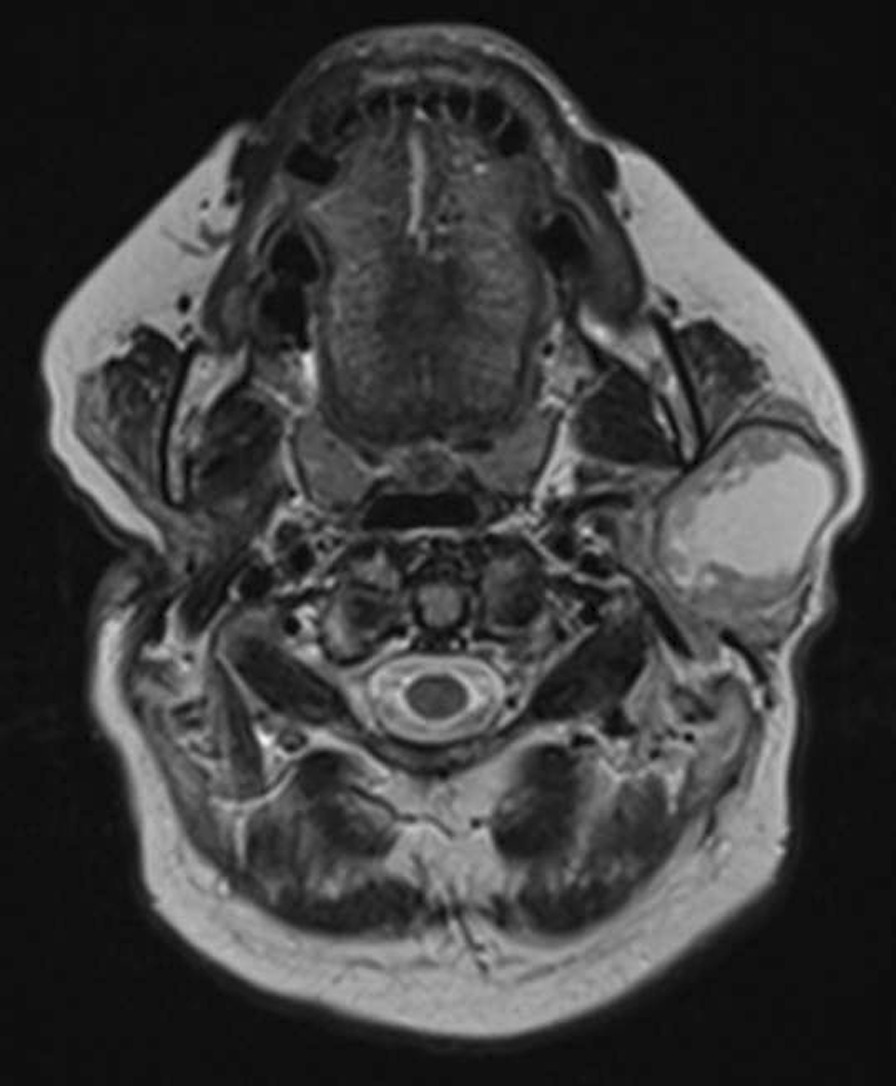
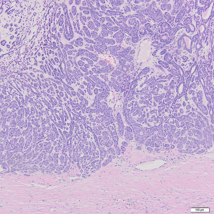
Case presentation: The current study reports the unique case of a 48-year-old woman who presented with a 6-month history of slowly growing masses located bilaterally in the parotid region, surgically treated with 5-year follow-up (no recurrence, normal facial nerve function). Magnetic resonance imaging (MRI) revealed three lesions: two in the superficial and deep lobes of the right parotid gland, and one in the superficial lobe of the left parotid gland. A total parotidectomy with facial nerve preservation was performed on the right side, and superficial parotidectomy on the left side 6 months later. Histopathological examination confirmed that all three tumors were BCAs. Molecular analysis didn't show any strong, potential of unknown clinical significance in the studied sample.
Conclusions: Multifocal bilateral lesions of the parotid gland are usually Warthin tumors. Detailed preoperative diagnostics including MRI and histopathological examination is essential to avoid misdiagnosing BCA and Warthin tumors. To our best knowledge, no case of synchronous bilateral multifocal basal cell adenomas of the parotid gland has been reported in English literature so far.
Keywords: Basal cell adenoma; Bilateral; Case report; Parotid gland; Synchronous.
© 2022. The Author(s).
Conflict of interest statement
The authors declare that they have no competing interests.
Figures
Similar articles
-
[Multiple bilateral lymphatic adenomas of the parotid glands].Otolaryngol Pol. 2006;60(2):139-42. Otolaryngol Pol. 2006. PMID: 16903327 Polish.
-
Uncommon Coexistence of Pleomorphic Adenoma and Warthin's Tumor in a Painfully Swollen Left Parotid Gland: A Surgical Case Report.Am J Case Rep. 2023 Nov 30;24:e940985. doi: 10.12659/AJCR.940985. Am J Case Rep. 2023. PMID: 38031394 Free PMC article.
-
Synchronous oncocytoma and Warthin's tumor in the ipsilateral parotid gland.Auris Nasus Larynx. 2004 Mar;31(1):73-8. doi: 10.1016/j.anl.2003.07.008. Auris Nasus Larynx. 2004. PMID: 15041058
-
Multifocal synchronous ipsilateral Warthin tumors: case report and review of the literature.Ear Nose Throat J. 2010 Sep;89(9):E1-3. doi: 10.1177/014556131008900901. Ear Nose Throat J. 2010. PMID: 20859854 Review.
-
[Bilateral adenolymphoma (Warthin's tumor) of the parotid. The anatomicoclinical, diagnostic and therapeutic aspects of 2 cases].Minerva Stomatol. 1994 Nov;43(11):535-42. Minerva Stomatol. 1994. PMID: 7739487 Review. Italian.
Cited by
-
Bilateral synchronous salivary gland tumors: report of three cases.Diagn Pathol. 2025 Jun 13;20(1):74. doi: 10.1186/s13000-025-01672-9. Diagn Pathol. 2025. PMID: 40514698 Free PMC article.
References
-
- Fonseca TC, Eisenberg ALA. Accuracy of intraoperative consultation in lesions of the salivary glands: analysis of 748 cases. J Bras Patol Med Lab. 2015;51:52–56.
-
- El-Naggar A, Chan J, Grandis J, et al., editors. World Health Organization classification of head and neck tumours. Lyon: IARC Press; 2017.
-
- Seifert G, Sobin LH. Histological typing of salivary gland tumors. In: WHO International Histological classification of Tumors, 2nd ed. Berlin: Springer; 1991. p. 245–247.
-
- Gnepp DR, Henley JD. Salivary and lacrimal glands. In: Gnepp DR, editor. Diagnostic surgical pathology of the head and neck 1. Philadelphia: Saunders; 2000. pp. 325–430.
Publication types
MeSH terms
LinkOut - more resources
Full Text Sources
