. 2022 Jul 29;17(1):296.
doi: 10.1186/s13023-022-02449-9.

Adjunct diagnostic value of radiological findings in mucopolysaccharidosis type IVa-related thoracic spinal abnormalities: a pilot study

Affiliations

Adjunct diagnostic value of radiological findings in mucopolysaccharidosis type IVa-related thoracic spinal abnormalities: a pilot study

Ya-Ting Jan et al. Orphanet J Rare Dis. .

Abstract

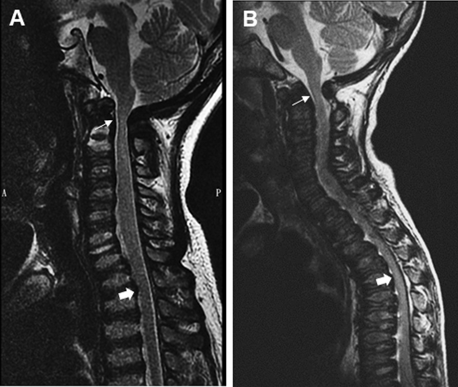
Background: In patients with mucopolysaccharidosis (MPS), systematic assessment and management of cervical instability, cervicomedullary and thoracolumbar junction spinal stenosis and spinal cord compression averts or arrests irreversible neurological damage, improving outcomes. However, few studies have assessed thoracic spinal involvement in MPS IVa patients. We aimed to evaluate thoracic spinal abnormalities in MPS IVa patients and identify associated image manifestations by CT and MRI study.

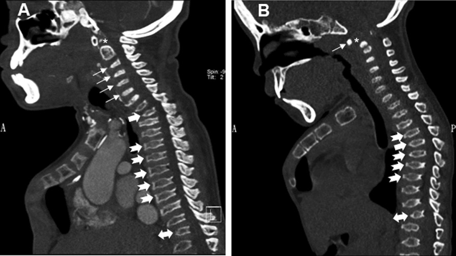
Results: Data of patients diagnosed and/or treated for MPS IVa at MacKay Memorial Hospital from January 2010 to December 2020 were extracted from medical records and evaluated retrospectively. Computed tomography (CT), plain radiography and magnetic resonance imaging (MRI) findings of MPS IVa-related spinal abnormalities were reviewed. Spine CT and plain radiography findings of 12 patients (6 males and 6 females with median age 7.5 years, range 1-28 years) revealed two subtypes of spinal abnormalities: thoracic kyphosis apex around T2 (subtype 1, n = 8) and thoracic kyphosis apex around T5 (subtype 2, n = 4). Spine CT and plain radiography clearly identified various degrees of thoracic kyphosis with apex around T2 or T5 in MPS IVa patients. Square-shaped to mild central beaking in middle thoracic vertebral bodies was observed in subtype 1 patients, while greater degrees of central beaking in middle thoracic vertebral bodies was observed in subtype 2 patients.

Conclusions: Spine CT findings clearly identify new radiological findings of thoracic kyphosis apex around T2 or T5 in MPS IVa patients. The degrees of central beaking at middle thoracic vertebral bodies may be a critical factor associated with different image presentations of thoracic kyphosis.

Keywords: Mucopolysaccharidosis; Spinal stenosis; Thoracic kyphosis; Thoracic vertebral body.


Conflict of interest statement

The authors declare that they have no competing interests.

Figures

Fig. 1
Fig. 1
Spinal CT manifestations in MPS IVa patients. Sagittal CT images grouped into subtypes 1 and 2 according to different levels of thoracic kyphosis apex. A MPS type IVa in a 26-year-old man (subtype 1). Sagittal reformatted spinal CT images show cervicothoracic kyphosis with apex around level of T2 (solid arrow), accompanied by square-shaped to mild central beaking of middle thoracic vertebral bodies (notched arrow). Mild thoracolumbar kyphosis with apex around level of T12 (double arrow), anterior central beaking of cervical (arrow) and visible lumbar vertebral bodies, and odontoid dysplasia (asterisk) are noted as well. B MPS type IVa in a 7-year-old girl (subtype 2). Sagittal reformatted CT images of the spine show middle thoracic kyphosis with apex around T5 (solid arrow), along with greater degrees of anterior central beaked thoracic vertebral bodies (notched arrow). Common spinal involvement of odontoid dysplasia (asterisk) with atlantoaxial instability (arrow) and associated spinal stenosis as well as thoracolumbar kyphosis (double arrow) in MPS IVa patients also well delineated by CT images
Fig. 2
Fig. 2
Spinal MRI images demonstration in MPS IVa patients. A MPS type IVa in a 14-year-old girl (subtype 1). Sagittal T2-weighted FSE MRI of the spine shows spinal canal narrowing around bony level of T2 (solid arrow) corresponding to CT manifestation of cervicothoracic kyphosis in addition to common spinal stenosis site around cervicomedullary junction (arrow). B MPS type IVa in a 5-year-old girl (subtype 2). T2-weighted FSE MRI of the spine in sagittal plane shows narrowing of the spinal canal more significant around bony level of T5 (solid arrow) related to CT reveals middle thoracic kyphosis. Atlantoaxial instability with mild cord myelopathy around cervicomedullary junction is also evident (arrow)

Similar articles

References

    1. EF N JM, et al. The mucopolysaccharidosis. In: Scriver CRBA. In: Scriver CR, Sly WS, Childs B, Beaudet AL, Valle D, Kinzler KW, et al., editors. The metabolic and molecular basis of inherited disease. 8. New York: McGraw-Hill; 2001.
    1. Lehman TJ, Miller N, Norquist B, Underhill L, Keutzer J. Diagnosis of the mucopolysaccharidoses. Rheumatology (Oxford) 2011;50(Suppl 5):v41–8. doi: 10.1093/rheumatology/ker390. - DOI - PubMed
    1. Rasalkar DD, Chu WC, Hui J, Chu CM, Paunipagar BK, Li CK. Pictorial review of mucopolysaccharidosis with emphasis on MRI features of brain and spine. Br J Radiol. 2011;84(1001):469–77. doi: 10.1259/bjr/59197814. - DOI - PMC - PubMed
    1. Northover H, Cowie RA, Wraith JE. Mucopolysaccharidosis type IVA (Morquio syndrome): a clinical review. J Inherit Metab Dis. 1996;19(3):357–65. doi: 10.1007/BF01799267. - DOI - PubMed
    1. Tomatsu S, Montano AM, Oikawa H, Smith M, Barrera L, Chinen Y, et al. Mucopolysaccharidosis type IVA (Morquio A disease): clinical review and current treatment. Curr Pharm Biotechnol. 2011;12(6):931–45. doi: 10.2174/138920111795542615. - DOI - PubMed

LinkOut - more resources