. 2022 Nov;88(5):2139-2156.
doi: 10.1002/mrm.29393. Epub 2022 Jul 30.

A joint linear reconstruction for multishot diffusion weighted non-Carr-Purcell-Meiboom-Gill fast spin echo with full signal

Affiliations

A joint linear reconstruction for multishot diffusion weighted non-Carr-Purcell-Meiboom-Gill fast spin echo with full signal

Philip K Lee et al. Magn Reson Med. 2022 Nov.

Abstract

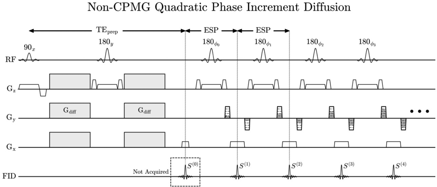
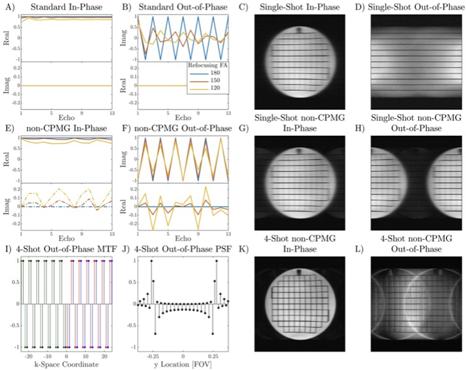
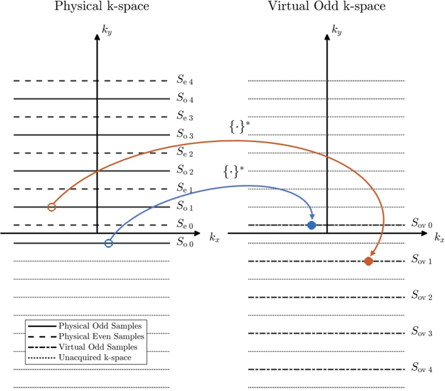
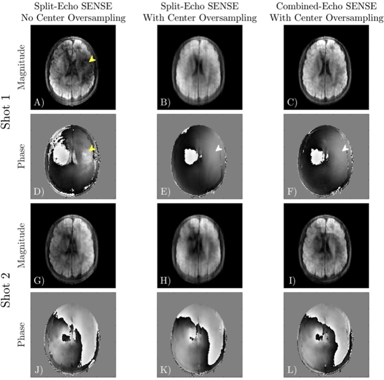
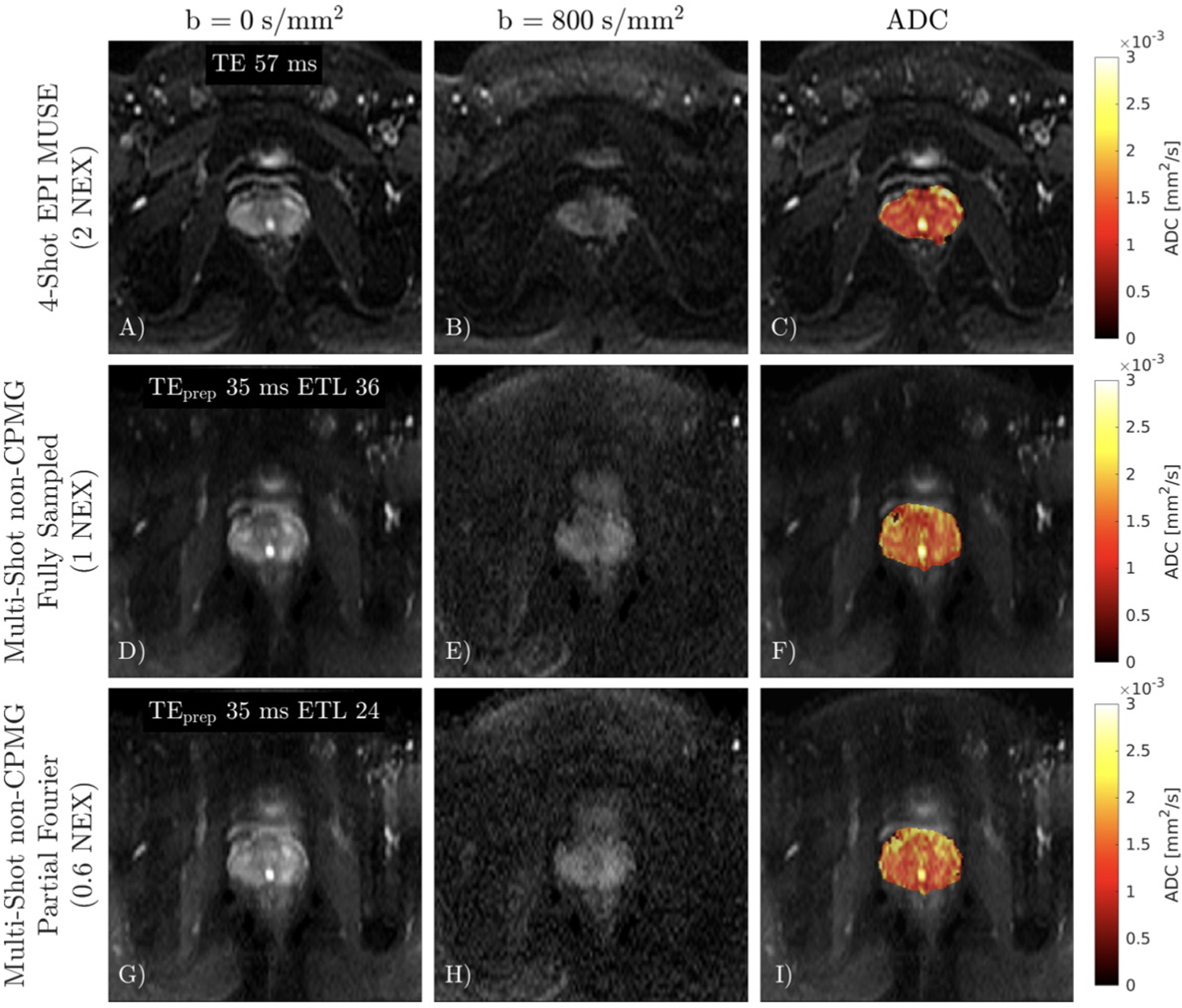
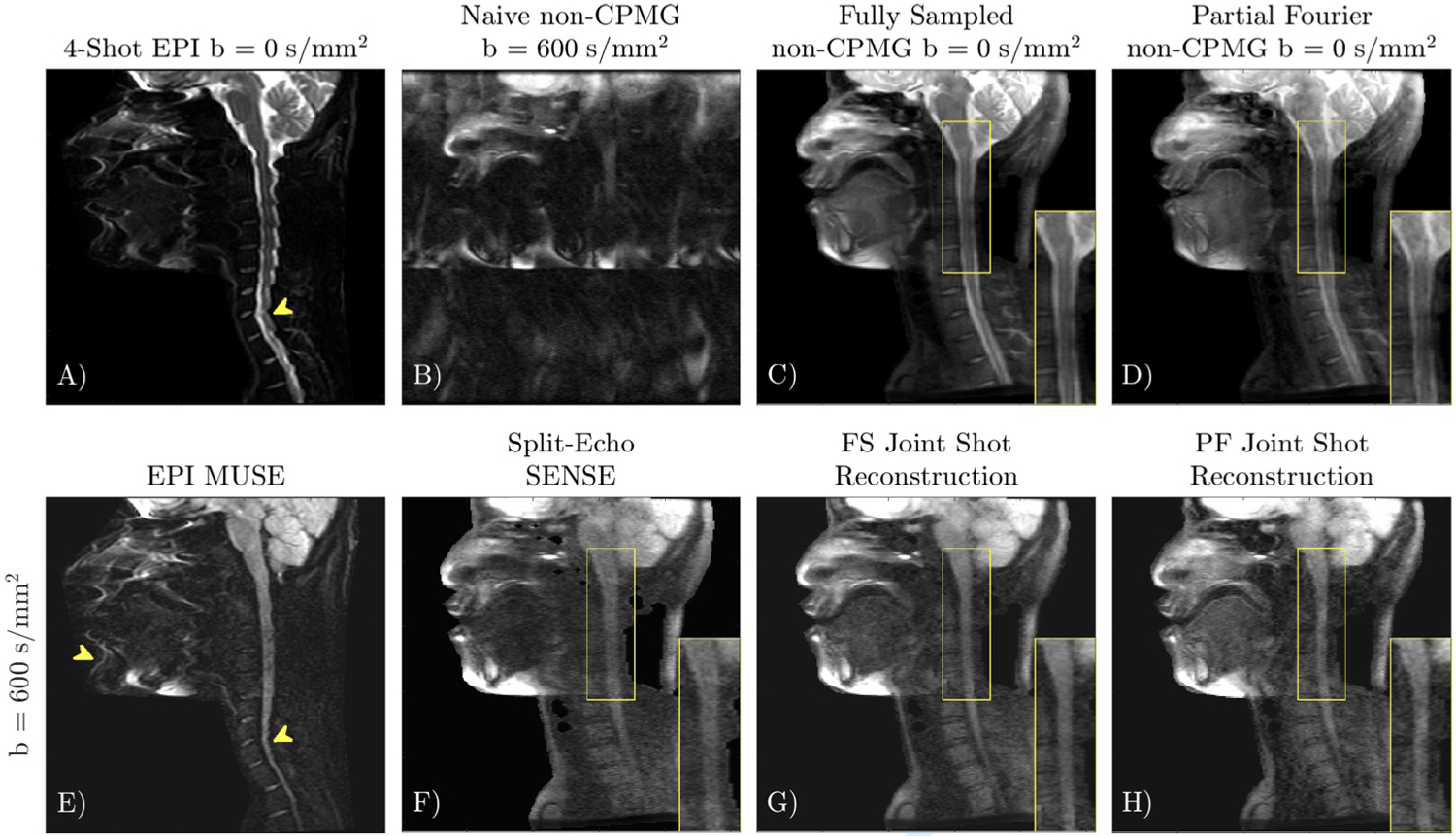
Purpose: Diffusion weighted Fast Spin Echo (DW-FSE) is a promising approach for distortionless DW imaging that is robust to system imperfections such as eddy currents and off-resonance. Due to non-Carr-Purcell-Meiboom-Gill (CPMG) magnetization, most DW-FSE sequences discard a large fraction of the signal ( 2 - 2 × $$ \sqrt{2}-2\times $$ ), reducing signal-to-noise ratio (SNR) efficiency compared to DW-EPI. The full FSE signal can be preserved by quadratically incrementing the transmit phase of the refocusing pulses, but this method of resolving non-CPMG magnetization has only been applied to single-shot DW-FSE due to challenges associated with image reconstruction. We present a joint linear reconstruction for multishot quadratic phase increment data that addresses these challenges and corrects ghosting from both shot-to-shot phase and intrashot signal oscillations. Multishot imaging reduces T2 blur and joint reconstruction of shots improves g-factor performance. A thorough analysis on the condition number of the proposed linear system is described.

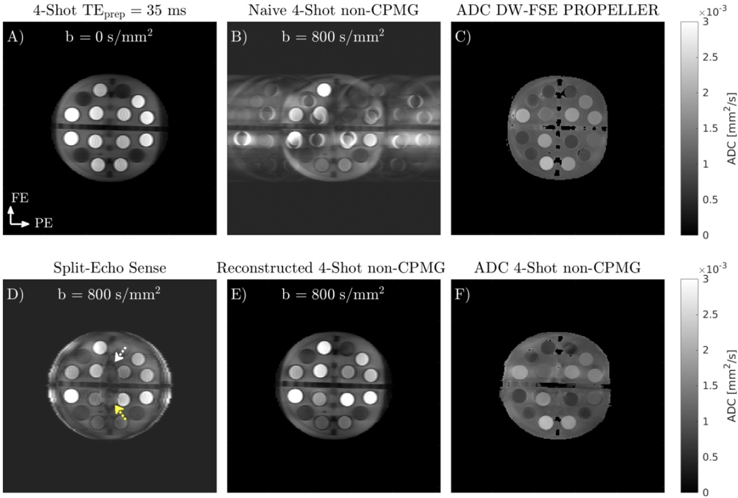
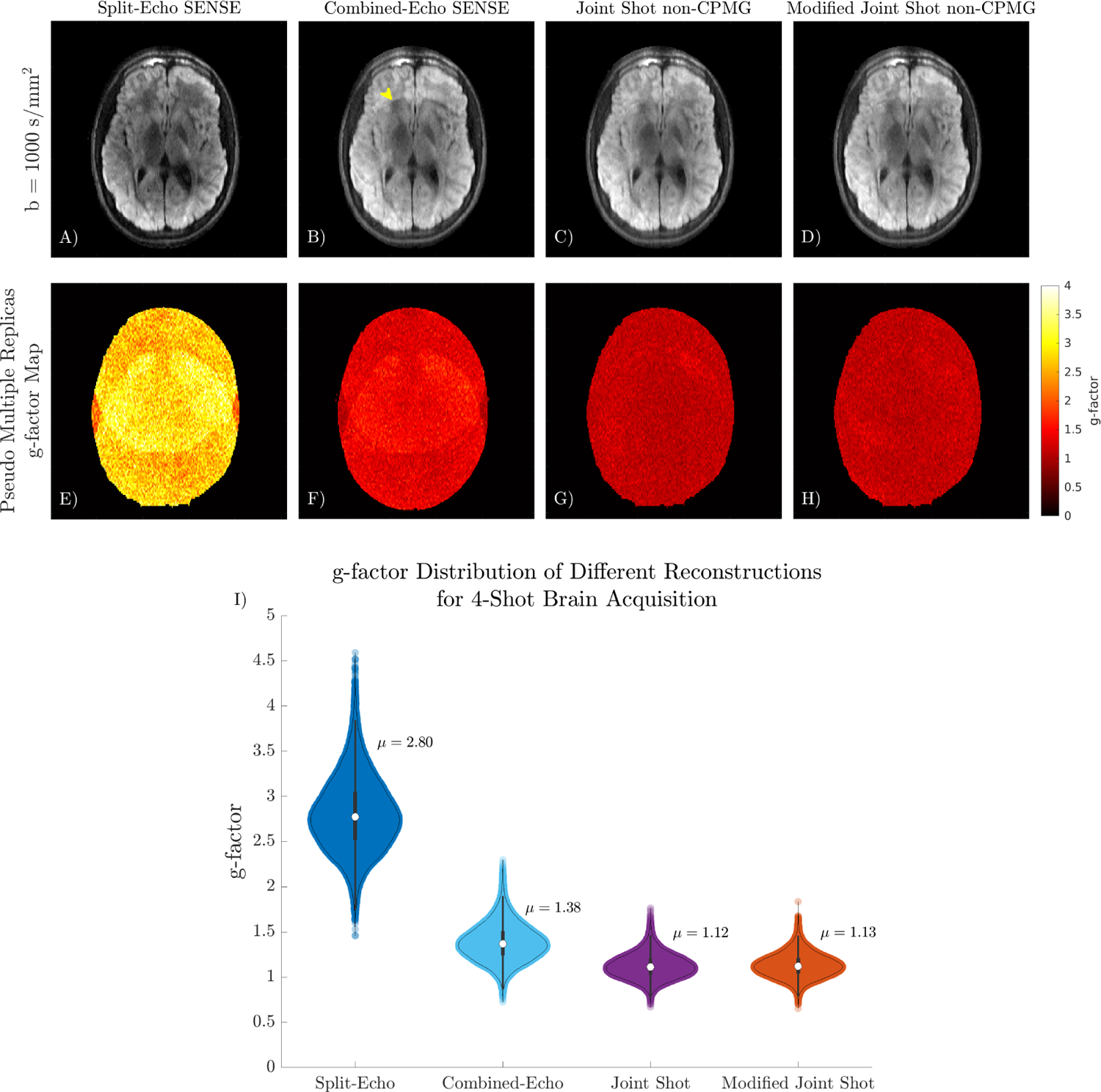
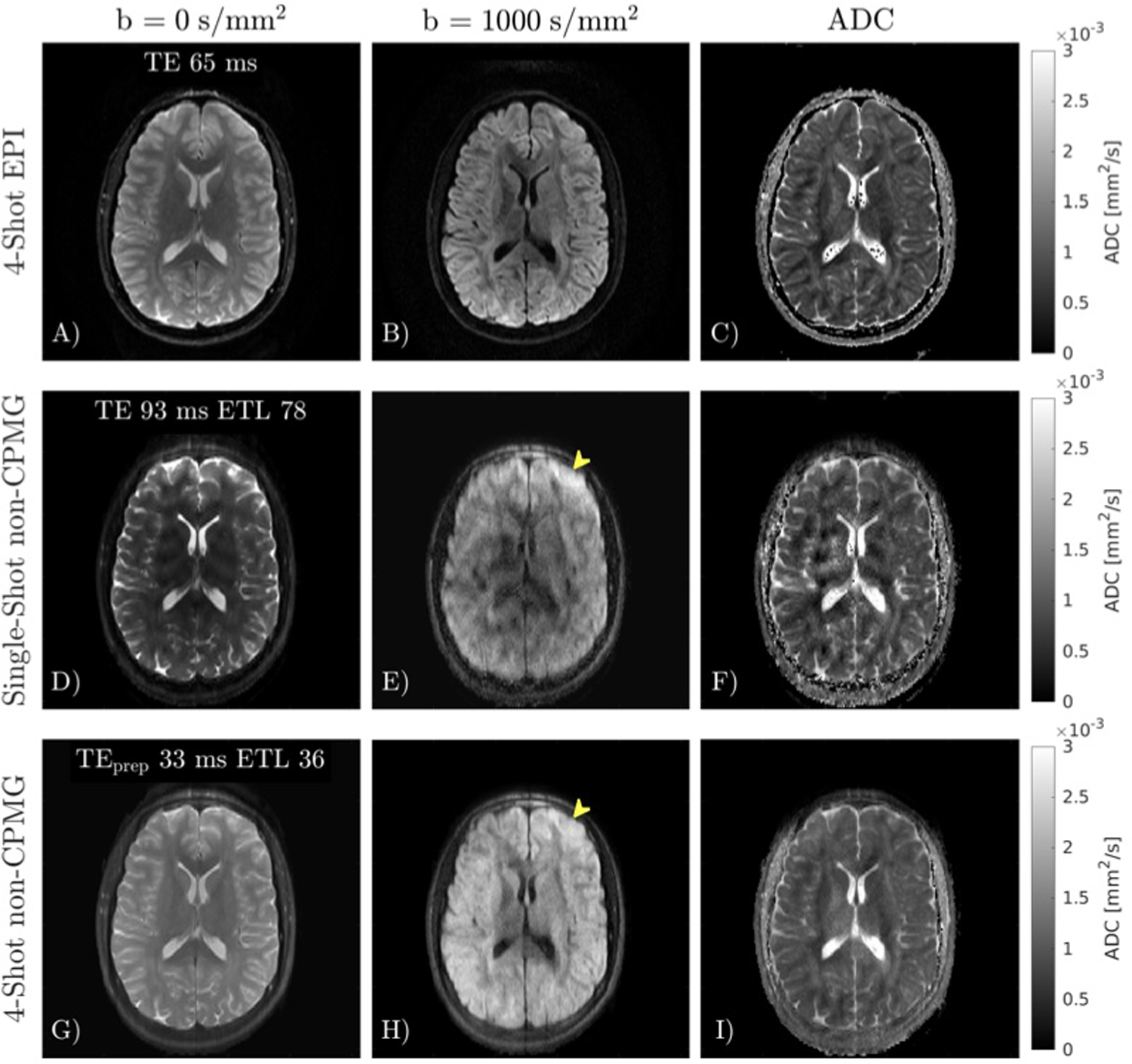
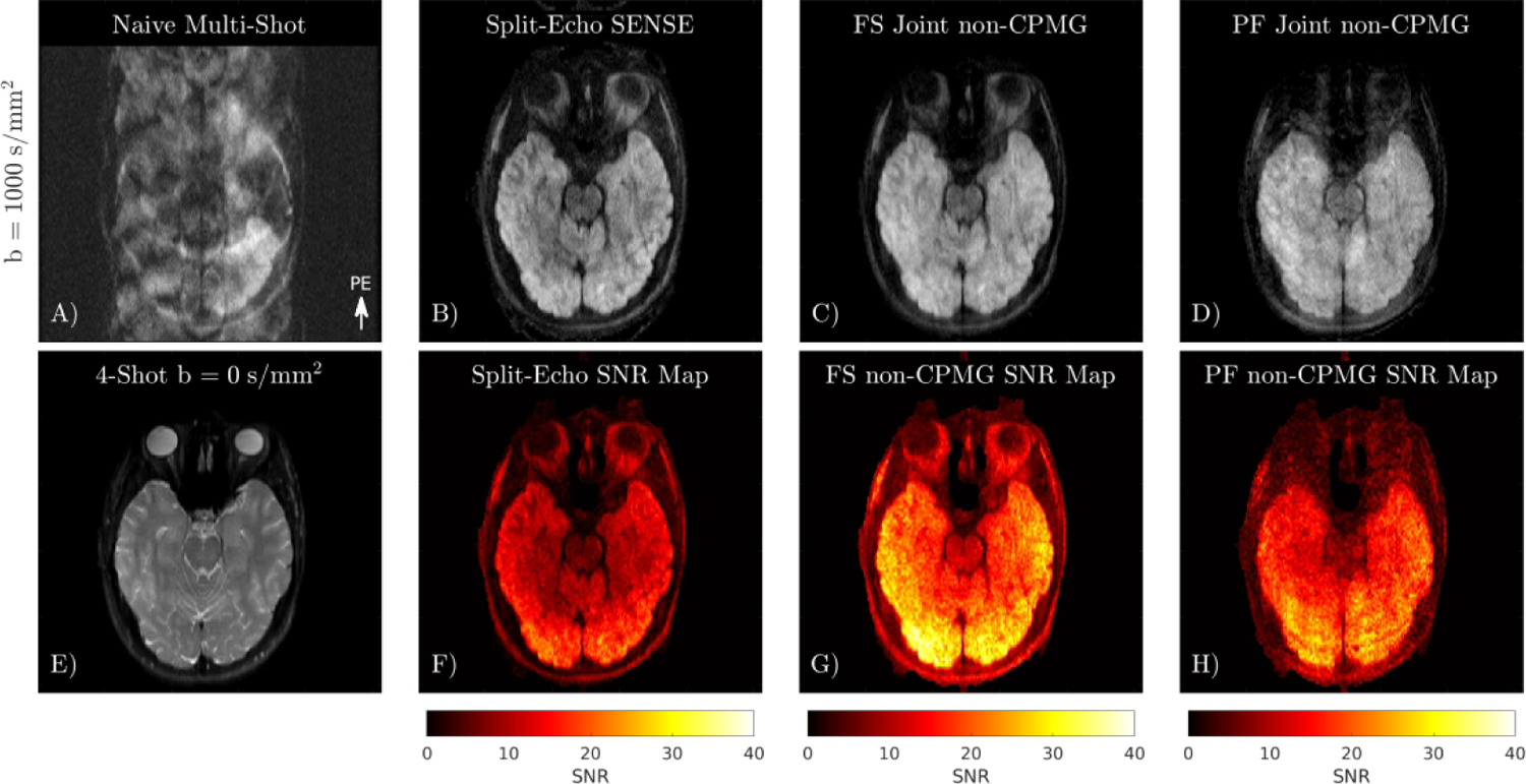
Methods: A joint multishot reconstruction is derived from the non-CPMG signal model. Multishot quadratic phase increment DW-FSE was tested in a standardized diffusion phantom and compared to single-shot DW-FSE and DW-EPI in vivo in the brain, cervical spine, and prostate. The pseudo multiple replica technique was applied to generate g-factor and SNR maps.

Results: The proposed joint shot reconstruction eliminates ghosting from shot-to-shot phase and intrashot oscillations. g-factor performance is improved compared to previously proposed reconstructions, permitting efficient multishot imaging. apparent diffusion coefficient estimates in phantom experiments and in vivo are comparable to those obtained with conventional methods.

Conclusion: Multi-shot non-CPMG DW-FSE data with full signal can be jointly reconstructed using a linear model.

Keywords: diffusion weighted imaging; g-factor; multiplexed sensitivity encoding; multishot fast spin echo; non-CPMG magnetization.


Figures

FIGURE 1
FIGURE 1
Stejskal-Tanner monopolar pulsed gradient spin echo with an FSE readout. The transmit phase of the first seven refocusing pulses are preset (referred to as the stabilization period), and were designed via optimization by Le Roux to enter the steady state. After the seven echo stabilization period, the transmit and receiver phase both increment quadratically.
FIGURE 2
FIGURE 2
Simulated signal evolution of the first thirteen echoes of CPMG and non-CPMG magnetization and its effects on image formation for two-sided center-out trajectories in a grid phantom with T2 ~ 300 ms, ETL 32. A-B) Signal modulation of in-phase and out-of-phase components without T2 decay for conventional CPMG. Out-of-phase magnetization has stable magnitude if the refocusing flip angle is 180°but will rapidly approach 0 if the flip angle deviates from 180°. C-D) In-phase and out-of-phase images obtained by offsetting the phase of the excitation pulse. Since the refocusing flip angle is not exactly 180°, out-of-phase magnetization rapidly decays, drastically reducing resolution. E-F) Quadratic phase increments maintain a stable echo train amplitude for out-of-phase magnetization if the refocusing flip angle is greater than 150°. G-H) For single-shot non-CPMG, the out-of-phase image is shifted by FOV/2 compared to the in-phase image (minor ghosts are visible due to slight flip angle or phase error). I-J) Center region of k-space modulation transfer function (MTF) and point spread function (PSF) for 4-shot out-of-phase condition. Different colors in the MTF represent different shots. The out-of-phase magnetization has FOV/2 replicas shifted by FOV/4 with multiple harmonics due to the rectangular k-space modulation. A discontinuity of the periodicity at the center of k-space creates an edge filter effect. K-L) Multi-shot in-phase and out-of-phase non-CPMG images.
FIGURE 3
FIGURE 3
Creation of virtual odd echo k-space from physical odd k-space samples by flipping about kx = 0 and ky = 0. {·}* denotes complex conjugation of the value.
FIGURE 4
FIGURE 4
A) 4-shot b = 0 s/mm2 image of ISMRM-NIST breast diffusion phantom. B) Naive combination of b = 800 s/mm2 data obtained using quadratic phase increment FSE readout has ghosts in the phase encode direction from the oscillating out-of-phase component. C) ADC map obtained with DW-FSE PROPELLER. D) Split-Echo SENSE, obtained by separately reconstructing odd and even lines from each shot. Yellow and white arrows indicate residual aliasing due to the high undersampling factor of each split shot. E) Joint reconstruction of all b = 800 s/mm2 shots using linear model and phase navigators estimated from data. F) Multi-shot non-CPMG ADC map.
FIGURE 5
FIGURE 5
Different phase navigator reconstructions for shot 1 (A-F) and shot 2 (G-L) of a 4-shot acquisition. Without center oversampling, a shot may miss the center of k-space, resulting in dropout and poor estimation of the phase (yellow arrow). Center oversampling improves phase estimation in this region (white arrow). Split-Echo SENSE and Combined-Echo SENSE with center oversampling obtain similar phase estimates.
FIGURE 6
FIGURE 6
A-D) Comparison of different linear reconstructions on 4-shot DW non-CPMG data. Split-Echo SENSE has low SNR in the region with large coil overlap due to the high acceleration factor. Some aliasing is present in the Combined-Echo reconstruction (yellow arrow). E-H) g-factor maps obtained using pseudo multiple replicas quantify the improved conditioning from jointly reconstructing all shots. I) Violin plot summarizing the distribution of g-factor in the slice.
FIGURE 7
FIGURE 7
In vivo comparison of 4-shot EPI MUSE, and single-shot and multi-shot quadratic phase increment FSE. A-C) 4-shot EPI reconstructed with MUSE. D-F) Single-shot FSE has considerable T2 blurring compared to EPI. G-H) Multi-shot FSE reduces T2 blur compared to single-shot FSE (yellow arrow). Despite reduced T2 blur, the apparent resolution of multi-shot FSE is still lower than EPI.
FIGURE 8
FIGURE 8
Comparison of different linear reconstructions on multi-shot non-CPMG diffusion data. A) Naive combination of 4-shot data exhibits severe ghosting from shot-to-shot phase, and aliasing of the out-of-phase component. B) Reconstruction of each shot split into even and odd lines. C) Joint reconstruction of shots using phase navigator. D) Joint reconstruction of partial Fourier data. E) 4-shot b = 0 s/mm2 image. F-H) SNR maps obtained using pseudo multiple replicas.
FIGURE 9
FIGURE 9
A-C) 4-shot DW-EPI with 2 NEX reconstructed with MUSE. D-F) Fully sampled non-CPMG image. There is no apparent ghosting in the multi-shot non-CPMG images. G-I) Partial Fourier non-CPMG reconstruction has lower SNR compared to the fully sampled acquisition.
FIGURE 10
FIGURE 10
Application of multi-shot quadratic phase increment FSE in the cervical spine. 4-shot DW-EPI (A, E) has distortion due to off-resonance (yellow arrows). Quadratic phase increment DW-FSE (F-H) exhibits no distortion. Split-Echo SENSE (F) can correct ghosts from shot-to-shot phase and intra-echo train oscillations, but has low SNR (inset) and magnitude bias. Joint reconstruction of shots (G) improves SNR. The non-CPMG reconstruction applied to prospectively acquired partial Fourier data (H), has similar resolution to the fully sampled acquisition (G).

Similar articles

Cited by

References

    1. Chawla S, Kim S, Wang S, Poptani H. Diffusion-weighted imaging in head and neck cancers. Future Oncology. 2009; 7: 959–975 - PMC - PubMed
    1. Tyagi N, Cloutier M, Zakian K, Deasy JO, Hunt M, Rimner A. Diffusion-weighted MRI of the lung at 3T evaluated using echo-planar-based and single-shot turbo spin-echo-based acquisition techniques for radiotherapy applications. J Appl Clin Med Phys. 2019; 20: 284–292. - PMC - PubMed
    1. Czarniecki M, Caglic I, Grist JT, Gill AB, Lorenc K, Slough RA, Priest AN, Barrett T. Role of PROPELLER-DWI of the prostate in reducing distortion and artefact from total hip replacement metalwork. European Journal of Radiology. 2018; 102: 213–219 - PubMed
    1. Rosenkrantz AB, Taneja SS. Use of Reduced Field-of-View Acquisition to Improve Prostate Cancer Visualization on Diffusion-Weighted Magnetic Resonance Imaging in the Presence of Hip Implants: Report of 2 Cases. Current Problems in Diagnostic Radiology. 2018; 2: 125–127 - PubMed
    1. Zegg M, Spicher A, Pfausler B, Nowosielski M, Krappinger D. Bilateral medial medullary syndrome following anterior screw fixation of type 2 odontoid fracture, a case report of two patients. Spinal Cord Series and Cases. 2021; 7: 101. - PMC - PubMed

Publication types