. 2022 Dec;148(12):3485-3499.
doi: 10.1007/s00432-022-04128-5. Epub 2022 Jul 30.

Methyltransferase-like 3 facilitates lung cancer progression by accelerating m6A methylation-mediated primary miR-663 processing and impeding SOCS6 expression

Affiliations

Methyltransferase-like 3 facilitates lung cancer progression by accelerating m6A methylation-mediated primary miR-663 processing and impeding SOCS6 expression

Shengshu Li et al. J Cancer Res Clin Oncol. 2022 Dec.

Abstract

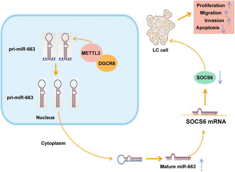
Objective: Lung cancer (LC) remains a threatening health issue worldwide. Methyltransferase-like protein 3 (METTL3) is imperative in carcinogenesis via m6A modification of microRNAs (miRNAs). This study estimated the effect of METTL3 in LC by regulating m6A methylation-mediated pri-miR-663 processing.

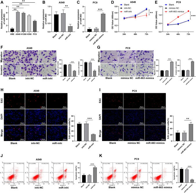
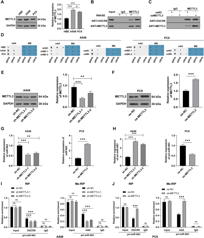
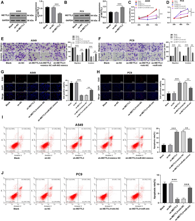
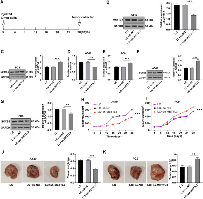
Methods: miR-663 expression in 4 LC cell lines and normal HBE cells was determined using RT-qPCR. A549 and PC9 LC cells selected for in vitro studies were transfected with miR-663 mimics or inhibitor. Cell viability, migration, invasion, proliferation, and apoptosis were detected by CCK-8, Transwell, EdU, and flow cytometry assays. The downstream target genes and binding sites of miR-663 were predicted via Starbase database and validated by dual-luciferase assay. LC cells were delivered with oe-METTL3/sh-METTL3. Crosslinking between METTL3 and DGCR8 was verified by co-immunoprecipitation. Levels of m6A, miR-663, and pri-miR-663 were measured by m6A dot blot assay and RT-qPCR. m6A modification of pri-miR-663 was verified by Me-RIP assay. Finally, the effects of METTL3 in vivo were ascertained by tumor xenograft in nude mice.

Results: miR-663 was upregulated in LC cells, and miR-663 overexpression promoted cell proliferation, migration, invasion, and inhibited apoptosis, but miR-663 knockdown exerted the opposite effects. miR-663 repressed SOCS6 expression. SOCS6 overexpression annulled the promotion of miR-663 on LC cell growth. METTL3 bound to DGCR8, and METTL3 silencing elevated the levels of pri-miR-663 and m6A methylation-modified pri-miR-663, and suppressed miR-663 maturation and miR-663 expression. METTL3 facilitated tumor growth in mice through the miR-663/SOCS6 axis.

Conclusion: METTL3 promotes LC progression by accelerating m6A methylation-mediated pri-miR-663 processing and repressing SOCS6.

Keywords: Lung cancer; Methyltransferase-like 3; Migration and invasion; Primary miR-663; SOCS6; Tumor formation assay; m6A methylation.


Conflict of interest statement

The authors declare that they have no conflict of interest.

Figures

Fig. 1
Fig. 1
miR-663 promoted proliferation, migration, and invasion of LC cells. ac miR-663 expression detected using RT-qPCR; d, e viability of A549 and PC9 cells assessed by CCK-8 assay; f, g migration and invasion of A549 and PC9 cells evaluated through Transwell assays; h, i cell proliferation analyzed via EdU method; j, k cell apoptosis detected by flow cytometry. Cell experiment was conducted three times and data were expressed as mean ± SD. One-way ANOVA was used for comparisons among multiple groups, followed by Tukey's test. **p < 0.01, ***p < 0.001
Fig. 2
Fig. 2
miR-663 inhibited SOCS6 expression. a Starbase database predicted the binding relationship between miR-663 and SOCS6; b dual-luciferase assay verified the targeted binding of miR-663 and SOCS6; ce WB measured SOCS6 expression. Cell experimentation was conducted three times, and data were exhibited as mean ± SD. One-way ANOVA was employed for comparisons among multiple groups, followed by Tukey's test. **p < 0.01, ***p < 0.001, ns indicated p > 0.05
Fig. 3
Fig. 3
Overexpression of SOCS6 partially annulled the promotion of miR-663 on LC cell growth. a, b SOCS6 expression was determined using WB; c cell viability detected using CCK-8 method; d, e cell migration and invasion estimated by Transwell assays; f, g cell proliferation assessed via EdU assay; h, i cell apoptosis analyzed through flow cytometry. Cell experiment was conducted three times, and data were presented as mean ± SD. One-way ANOVA was employed for comparisons among multiple groups, followed by Tukey's test. *p < 0.05, **p < 0.01, ***p < 0.001
Fig. 4
Fig. 4
METTL3-mediated m6A methylation regulated the maturation of miR-663 in LC cells. A549 and PC9 cells were, respectively, transfected with sh-METTL3 or oe-METTL3. a WB assay was employed to detect METTL3 expression; b, c Co-IP was used to verify the crosslinking between METTL3 and DGCR8; d m6A dot blot assay was conducted to determine m6A level, with MB stain as loading control; e, f WB assay measured METTL3 expression; g, h RT-qPCR determined levels of miR-663 and pri-miR-663; i, j RIP and Me-RIP assays were introduced to verify the m6A modification of pri-miR-663. Cell experimentation was conducted three times and data were displayed as mean ± SD. One-way ANOVA was adopted for comparisons among multiple groups, followed by Tukey's test. **p < 0.01, ***p < 0.001, nsp > 0.05. Compared with A549 cells, #p < 0.05
Fig. 5
Fig. 5
METTL3 facilitated LC cell growth by regulating miR-663. a, b METTL3 expression was measured using WB; c, d cell viability detected using CCK-8 method; e, f cell migration and invasion assessed by Transwell assays; g, h cell proliferation evaluated via EdU assay; i, j cell apoptosis analyzed through flow cytometry. Cell experiment was conducted three times and data were expressed as mean ± SD. One-way ANOVA was employed for comparisons among multiple groups, followed by Tukey's test.*p < 0.05, **p < 0.01, ***p < 0.001
Fig. 6
Fig. 6
METTL3 promoted tumorigenesis and growth in mice via the miR-663/SOCS6 axis. a Schematic diagram of tumor formation assay in vivo; b, c WB detected METTL3 expression; d, e RT-qPCR determined miR-663 expression; f, g WB measured SOCS6 expression; h, i changes of tumor volume was recorded; j, k representative images and weight of tumors. Data were exhibited as mean ± SD. One-way ANOVA was performed for comparisons among multiple groups, followed by Tukey's test. **p < 0.01, ***p < 0.001
Fig. 7
Fig. 7
Mechanism of METTL3 in LC development by regulating the miR-663/SOCS6 axis

Similar articles

Cited by

References

    1. Alarcon CR, Lee H, Goodarzi H et al (2015) N6-methyladenosine marks primary microRNAs for processing. Nature 519:482–485 - PMC - PubMed
    1. Barbieri I, Tzelepis K, Pandolfini L et al (2017) Promoter-bound METTL3 maintains myeloid leukaemia by m(6)A-dependent translation control. Nature 552:126–131 - PMC - PubMed
    1. Bi X, Lv X, Liu D et al (2021) METTL3-mediated maturation of miR-126-5p promotes ovarian cancer progression via PTEN-mediated PI3K/Akt/mTOR pathway. Cancer Gene Ther 28:335–349 - PubMed
    1. Chen Y, Min L, Ren C et al (2017) miRNA-148a serves as a prognostic factor and suppresses migration and invasion through Wnt1 in non-small cell lung cancer. PLoS ONE 12:e0171751 - PMC - PubMed
    1. Chen M, Wei L, Law CT et al (2018) RNA N6-methyladenosine methyltransferase-like 3 promotes liver cancer progression through YTHDF2-dependent posttranscriptional silencing of SOCS2. Hepatology 67:2254–2270 - PubMed

LinkOut - more resources