. 2022 Jul 28;14(14):5925-5945.
doi: 10.18632/aging.204194. Epub 2022 Jul 28.

miR-21 upregulation exacerbates pressure overload-induced cardiac hypertrophy in aged hearts

Affiliations

miR-21 upregulation exacerbates pressure overload-induced cardiac hypertrophy in aged hearts

Wei-Ting Chang et al. Aging (Albany NY). .

Abstract

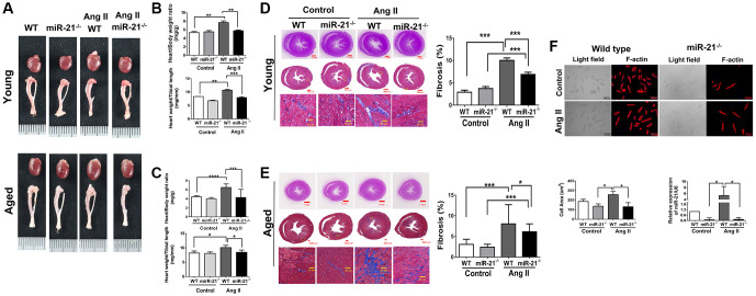
Young and aging hearts undergo different remodeling post pressure overload, but the regulator that determines responses to pressure overload at different ages remains unknown. With an angiotensin II (Ang II)-induced hypertensive model, miR-21 knockout mice (miR-21-/-) were observed regarding the effects of miR-21 on hypertension-induced cardiac remodeling in young (12 week-old) and old (50 week-old) mice. Although the aged heart represented a more significant hypertrophy and was associated with a higher expression of miR-21, Ang II-induced cardiac hypertrophy was attenuated in miR-21-/- mice. Upon results of cardiac-specific arrays in miR-21-overexpressing cardiomyocytes, we found a significant downregulation of S100a8. In both in vitro and in vivo models, miR-21/S100a8/NF-κB/NFAT pathway was observed to be associated with pressure overload-induced hypertrophic remodeling in aged hearts. To further investigate whether circulating miR-21 could be a biomarker reflecting the aged associated cardiac remodeling, we prospectively collected clinical and echocardiographic information of patients at young (<65 y/o) and old ages (≥65 y/o) with and without hypertension. Among 108 patients, aged subjects presented with a significantly higher expression of circulating miR-21, which was positively correlated with left ventricular wall thickness. Collectively, miR-21 was associated with a prominently hypertrophic response in aged hearts under pressure overload. Further studies should focus on therapeutic potentials of miR-21.

Keywords: aging; cardiac hypertrophy; hypertension; miR-21; pressure overload.


Conflict of interest statement

CONFLICTS OF INTEREST: The authors declare no conflicts of interest related to this study.

Figures

Figure 1
Figure 1
Angiotensin II (Ang II)-induced cardiac hypertrophy and fibrosis, especially in aged mice. (A) Echocardiography measurements are shown in young and aged mice with or without Ang II. Systolic and diastolic blood pressures recorded. Echocardiographic measurements of intraventricular septal thickness at diastole (IVSd) and ejection fraction and fractional shortening. (B) Quantitative analysis of heart weight/tibia length. (C) Representative sections and amplified images of the highlighted area of hearts stained with Masson's trichrome for fibrosis detection (blue); scale bars, 30 μm (left panel). Quantification of cardiac fibrosis in the indicated groups of rats (right panel). Expression of (D) circulating and (E) heart tissue expression of miR-21 in mice. Data are expressed using mean ± standard deviation (S.D.). *P < 0.05, **P < 0.01, ***P < 0.001 for difference from each group (N = 6–12).
Figure 2
Figure 2
Angiotensin II (Ang II)-induced more cardiac hypertrophy and miR-21 expression in primarily isolated aged cardiomyocytes than in primary young cardiomyocytes. (A) The cultured neonatal rat cardiomyocytes for 14 days displayed cardiomyocytes senescence. SA-β-gal staining results for cardiomyocytes of neonatal rats. Blue precipitation in the cytoplasm was observed in the senescent cells. Percentage of β-gal-positive cardiomyocytes was increased in cultured cardiomyocytes for 14 days. (B) Telomere length expression in cardiomyocytes of neonatal rats. (C) The expression of cell senescence-associated protein in cultured neonatal rat cardiomyocytes was detected by qRT-PCR. (D) The levels of miR-21 and cardiac injury-associated genes in cultured neonatal rat cardiomyocytes were detected by qRT-PCR. (E) Immunofluorescence assay of F-actin was performed to identify the cell area in each group. Bar charts showing the individual cardiomyocyte cell areas. (F) The levels of miR-21 in cultured young and aged rat cardiomyocytes with and without treatment of Ang II detected by qRT-PCR. (G) Primarily isolated young and aged cardiomyocytes were transfected with a miR-21 mimic or inhibitor for 24 hours. Representative merged images of F-actin immunofluorescence staining of cardiomyocytes. Overexpression of miR-21 enhanced Ang II-induced cardiac hypertrophy, especially in primarily isolated aged cardiomyocytes. *P < 0.05, **P < 0.01, and ***P < 0.001 for difference from each group (N = 6–8).
Figure 3
Figure 3
miR-21 knockout (miR-21−/−) protects against angiotensin II (Ang II)-induced cardiac alterations in both young and aged mice. (A) The study design. (B) Sequential echocardiography measurements are shown in wild-type (WT) and miR-21−/− mice with or without exposure to Ang II. Echocardiographic measurements of (C) fractional shortening (FS), (D) interventricular septum (IVSd), and (E) left ventricular internal diameter in diastole (LVIDd) are shown for each group. *P < 0.05, **P < 0.01, and ***P < 0.001 for difference from each group (N = 6–8).
Figure 4
Figure 4
miR-21 knockout (miR-21−/−) decreased angiotensin II (Ang II)-induced cardiac hypertrophy and fibrosis in both young and aged mice. (A) Representative images of harvested hearts. Quantitative analysis of heart weight/body weight and heart weight/tibia length in wild type (WT) or miR-21−/− of (B) young and (C) aged mice. In WT or miR-21−/− of (D) young and (E) aged mice, representative sections of hearts stained with Masson's trichrome for fibrosis detection (blue); scale bars, 30 μm (left panel). Quantification of cardiac fibrosis (right panel). (F) miR-21−/− decreased Ang II-induced increased cardiac hypertrophy miR-21 expression in primary mouse cardiomyocyte. The representative merged images of light field and F-actin immunofluorescence staining for primary cardiomyocyte isolated from WT and miR-21−/− of young mice. The cell area was measured 100 random cells in each group. The expression of miR-21 was measured by qRT-PCR in each group. *P < 0.05, **P < 0.01, and ***P < 0.001 for difference from each group. (N = 6–8).
Figure 5
Figure 5
The treatment of miR-21 antagomir mitigated angiotensin II (Ang II)-induced cardiac hypertrophy, especially in the aged mice. (A) The study design investigating the effects of miR-21 antagomir in young (12 week-old) and aged mice(50 week-old) of Ang II-induced pressure overload. The sequential changes of (B) systolic, (C) diastolic blood pressures, echocardiography derived (D) intraventricular septal thickness at diastole (IVSd), and (E) fractional shortening (FS) in young and old mice treated with miR-21 antagomir or not under Ang II-induced pressure overload. (F) The comparison of harvested hearts in mice of control, Ang II, miR-21 antagomir and Ang II+ miR-21 antagomir. *P < 0.05 for difference from each group (N = 4–6).
Figure 6
Figure 6
miR-21 decreased s100a8 expression in primarily isolated cardiomyocytes. (A) Heat map of cluster analysis showed dynamic changes of cardiac specific genes after miR-21 mimic treatment compared with the control. The levels of S100a8, Nr3C2 and NKX2.5 were measured by qRT-PCR in primary cardiomyocytes transfected with miR-21 mimic and inhibitor for 24 hours. (B) The protein expressions of S100a8, NFκB, calcineurin, and NFAT were measured by Western blot in primary cardiomyocytes. (C) The relative expression level of each protein was quantified by densitometry. miR-21 knockout (miR-21−/−) increased S100a8 and decreased NFκB, calcineurin, and NFAT expression in heart tissue of mice after angiotensin II (Ang II) treatment. The representative expressions and quantifications of S100a8, NFκB, calcineurin, and NFAT in wild type (WT) or miR-21−/− of mice were detected by Western blot. *P < 0.05 for difference from each group. (N = 4–6).
Figure 7
Figure 7
High expression of circulating miR-21 in aged hypertensive subjects. (A) Circulating miR-21 expression in normotensive young, normotensive old, hypertensive young and hypertensive old subjects. (B) The linear correlation between intraventricular septal thickness at diastole (IVSd) and circulating miR-21 expression in hypertensive subjects. **P < 0.01 for difference from each group.
Figure 8
Figure 8
The summary of miR-21 regulation in the cardiac hypertrophy under pressure overload.

Similar articles

Cited by

References

    1. Triposkiadis F, Xanthopoulos A, Butler J. Cardiovascular Aging and Heart Failure: JACC Review Topic of the Week. J Am Coll Cardiol. 2019; 74:804–13. 10.1016/j.jacc.2019.06.053 - DOI - PubMed
    1. Azad N, Lemay G. Management of chronic heart failure in the older population. J Geriatr Cardiol. 2014; 11:329–37. 10.11909/j.issn.1671-5411.2014.04.008 - DOI - PMC - PubMed
    1. Husaini BA, Mensah GA, Sawyer D, Cain VA, Samad Z, Hull PC, Levine RS, Sampson UK. Race, sex, and age differences in heart failure-related hospitalizations in a southern state: implications for prevention. Circ Heart Fail. 2011; 4:161–9. 10.1161/CIRCHEARTFAILURE.110.958306 - DOI - PMC - PubMed
    1. Obokata M, Reddy YNV, Borlaug BA. Diastolic Dysfunction and Heart Failure With Preserved Ejection Fraction: Understanding Mechanisms by Using Noninvasive Methods. JACC Cardiovasc Imaging. 2020; 13:245–57. 10.1016/j.jcmg.2018.12.034 - DOI - PMC - PubMed
    1. Gradman AH, Alfayoumi F. From left ventricular hypertrophy to congestive heart failure: management of hypertensive heart disease. Prog Cardiovasc Dis. 2006; 48:326–41. 10.1016/j.pcad.2006.02.001 - DOI - PubMed

Publication types